Acta Med. Nagasaki 43: 19-25
Mechanisms for the Occurrence of Three Uniparental Disomies Associated with Abnormal Phenotypes
Osamu MIYOSHI
Department of Human Genetics, Nagasaki University School of Medicine, Nagasaki
Results of a molecular-genetic study on the mechanism of uniparental disomy (UPD) in three individuals are reported.
Case 1 was a physically normal adult whose Rh blood-type showed mosaicism of two phenotypes, D+ (or D/D geno- type) and D- (or d/d genotype), while his father and mother were a D/d heterozygote and a D/D homozygote re- spectively. Allele-typing of his peripheral blood leukocytes and buccal membrane cells using polymorphic DNA markers on chromosome 1 revealed both paternal and maternal al- leles, but demonstrated paternal uniparental transmissions of alleles in the monoclonal B-lymphocytes and in hair-root cells from various body regions. The results indicate that he had two cell lines each with paternal UPD1 and mater- nal UPD1. Only a plausible mechanism for the mosaicism includes abnormal segregation at first mitosis, where both chromatids of each homologs 1 migrated together to the same direction, resulting in two daughter cells having D/D and d/d genotypes. This sort of cell division has hith- erto been undescribed in man. Case 2 was a Silver-Russell syndrome patient with a mosaic 46, XX / 47, XX, +r (7) karyotype. Allele-typing with chromosome 7 markers re- vealed that she inherited maternal uniparental alleles at telomeric regions of the chromosome but biparental alleles at the centromeric region, the result indicating that the two normal chromosomes 7 were of maternal UPD and the ring chromosome 7 was of paternal origin. A likely mechanism for her UPD7 is "monosomy duplication", followed by so- matic loss of the ring chromosome. The finding also indi- cates that the putative SRS locus can be ruled out from the centromeric region, 7p13-qll. Case 3 had intrauterine growth retardation and a 45, XY, i (14) karyotype. Allele- typing revealed maternal uniparental transmissions of al- leles at both centromeric and telomeric regions of chromo- some 14, but showed biparental alleles at other regions. The results indicate that the isochromosome was of maternal Address Correspondence :
Osamu Miyoshi, M.D., Department of Human Genetics, Nagasaki University School of Medicine, Sakamoto 1-12-4,
Nagasaki 852-8523, Japan
UPD and may have arisen through "gametic complementa-
tion" mechanism.
Key words: uniparental disomy (UPD); allele-typing; genotyping;
Rh-phenotypic mosaicism; Silver-Russell syndrome;
intrauterine growth retardation; mechanism
Introduction
In 1980, Engel" first introduced a concept of uniparental disomy (UPD). Normally, each member of homologous chromosomes originates from each parent,
whereas UPD is a phenomenon where both homolo- gous chromosomes come from a single parent. UPD is subdivided into two classes: contribution of both homologs of a parent to a child is heterodisomy; and double transmission of one parental homolog to a child is isodisomy1-". Recent development of molecular-
genetic analysis with polymorphic DNA markers has made it possible to ascertain the origin and mecha- nism of UPD. Dinucleotide (CA) repeat polymorphisms
are useful multiallelic markers for this purpose'). UPD is not extremely rare in man and has been observed for chromosomes 1, 2, 4, 6-11, 13-16, 20-22, and X, and
the majority of persons with UPD for a particular chromosome manifested abnormal phenotypes'-'). One of the explanations of the parent-of-origin specific phe-
notype is genomic imprinting that is defined as parent-of-origin dependent differential expression of a gene, i.e., only one parental allele is expressed (active)
and the other allele is imprinted (silent)'). When chil- dren inherit UPD chromosomes in which no active al- leles exist, they may have abnormal phenotypes, such as growth retardation and / or congenital malforma- tions. UPD may arise through either of the following mechanisms (Fig. 1)'3) : (a) fusion of a nullisomic gamete with a gamete disomic for the same chromo- some (gametic complementation); (b) loss of one
member of trisomic chromosomes in a zygote at an early mitotic division (trisomy rescue); or (c) duplica- tion of a monosomic chromosome in a zygote
(monosomy duplication). "Gametic complementation"
may result in partial (or segmental) UPD, depending on meiotic cross-overs, and "trisomy rescue" may re- make a normal zygote in 2/3 of cases and complete or segmental UPD in 1/3; and "monosomy duplication"
leads to complete isodisomy. All of these events can save life from harmful effects of the abnormal karyotype. Postzygotic events are also possible for the latter two mechanisms, resulting in somatic mosaicism.
This paper deals with a molecular-genetic study on the mechanisms of three different UPD chromosomes in three individuals with unusual phenotypes.
Materials and Methods
Case 1
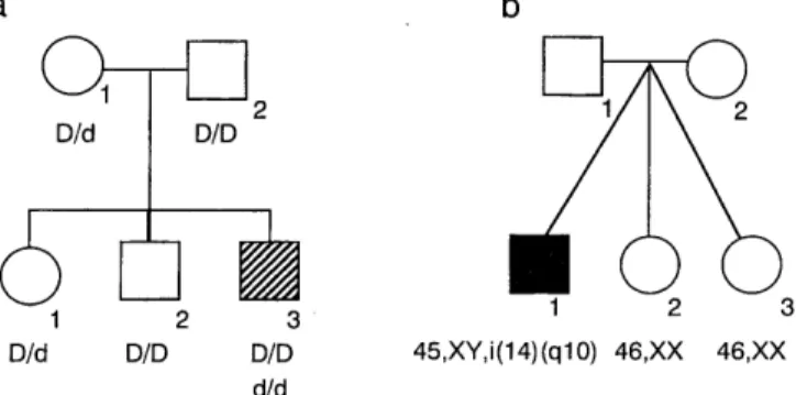
The proband was a 36-year-old Japanese man who visited West Tokyo Red-Cross Blood Center for a blood-donor volunteer in 1985. He was physically and mentally normal, and his healthy parents were not consanguineous. Two elder siblings were also healthy
(Fig. 1). When the proband's blood types were deter- mined, the Rh phenotype was found to be unusual.
Flow cytometric analysis for RhD antigen density pro- file demonstrated that he had two cell lines each with the Rh phenotype D+ (or genotype D/D) and with D-
(or genotype d/d), respectively (Fig. 1), suggesting a chimera for the Rh blood type. Family analysis re- vealed that his father was a D / d heterozygote, the mother a D/ D homozygote, an elder sister a D/ d heterozygote, and an elder brother a D/D homozygote.
Cytogenetic studies on 100 metaphase cells from cul- tured lymphocytes of the proband revealed a mosaic 46, XY [71] / 46, XY, lqh+, lqh+ [29] karyotype, and
a b
1 2 EI-N-0 1 2
6 1
1D/d D/D 1 6
2 3 1 2 3D/d D/D D/D 45,XY,i(14)(g10) 46,XX 46,XX d/d
Fig 1. Family trees of Case 1 (a, 11-3) and Case 2 (b, II-1).
Letters below individual symbols implicate genotypes of the RhD gene (a) and karyotypes (b).
his parents had 46, XY, lqh+ and 46, XX, lqh+
karyotypes, respectively. The B-cell of the proband was transformed with Epstein-Barr virus (EBV) to lymphoblastoid cell lines and then they were cloned into monoclonal cell lines. Monoclonality of these cells was confirmed with the lqh+ heteromorphism.
Case 2
The patient was a 20-month-old Japanese girl, who was born at 34 gestational weeks by caesarean section because of premature rupture of the membrane, intrau- terine growth retardation (IUGR) and fetal distress, with a birthweight of 1,020 g (-3.0 SD) and length of 36 cm (-4.4 SD). Physical examination revealed the following abnormalities: triangular face, hypertelorism, micrognathia, clinodactyly of the fifth fingers, asym- metry of the trunk and limbs, patent ductus arteriosus (PDA ), and right recurrent nerve paralysis. Re- examination at age 18 months revealed marked growth and developmental retardation with a length of 68.5 cm (-4.7 SD), weight of 5,900 g (-3.5 SD), OFC of 44.5 cm (-1.4 SD) and chest circumference of 39.0 cm (-3.7 SD). The muscle was hypotrophic and bone mineral density showed a low level for her age. These clinical manifestations led to the diagnosis of Silver- Russell syndrome. Chromosome analysis on 45 meta- phase cells from her lymphocytes revealed a mosaic 46, XX [30] /47, XX,+r(?) [ 15] karyotype. Whole chro- mosome painting using a chromosome 7-specific probe pool (WCP probe, VYSIS, UK) demonstrated that 22 (73%) of 30 cells analyzed had two normal chromo- somes 7, and 8 (27%) a ring chromosome 7 in addi- tion to the normal homologs 7. Thus, the karyotype of the proband was finally identified as mos 46, XX/47, XX,+r (7).
Case 3
The patient, a 9-month-old Japanese boy, was one of triplets born to unrelated, healthy parents (Fig. 2). At 29 gestational weeks, the patient was delivered by caesarean section with a weight of 634 g (small for dates) and length of 31 cm. Two other members of the triplets, two girls, were healthy and both weighed 1,110 g, being appropriate for dates. Physical examina- tion revealed the following abnormalities: dolichocephaly with frontal bossing, low-set ears, short neck, small hands and feet, overlapping fingers and PDA. A skele- tal survey showed no abnormality. Re-examination at 259 days revealed growth and developmental retarda- tion with a length of 59.3 cm, weight of 4,210 g and OFC of 41.0 cm. Chromosome analysis on his
D151596
Father
Mother
Case 1 PBL
Hair-root
by 100 105 110 115 120 125
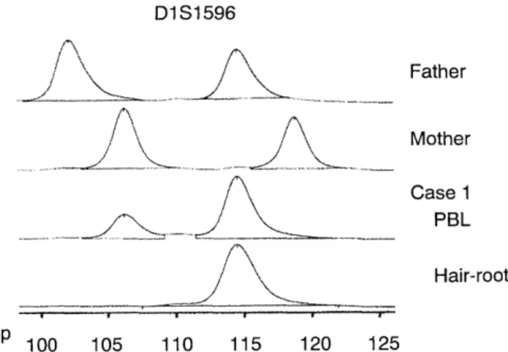
Fig 2. Electrophoretic patterns of CA-repeat polymorphic al- leles at the D1S1596 locus in the family of Case 1.
lymphocytes revealed a 45, XY, i (14) (q10) karyotype.
Karyotypes of the other two triplet-members and the parents were all normal. To know the zygosity of the triplets, quinacrine fluorescence Q (QFQ)-banded chro- mosomal heteromorphisms on chromosomes 3 and 4 and acrocentric chromosomes were analyzed as de- scribed previously". The result demonstrated that the triplets were trizygotic, because the infants had differ- ent karyotypes or different QFQ polymorphic patterns (data not shown).
CA-repeat polymorphic marker analysis
Since UPD for chromosomes 1, 7, and 14 was sus- pected in Cases 1, 2, and 3, respectively, genotypes of the probands and their respective parents were ana- lyzed as follows: Parent-child transmission modes of al- leles were traced using dinucleotide (CA)- and/or tetranucleotide-repeat polymorphic markers which were located on respective chromosomes (Table 1), ac- cording to published genetic maps"'. Markers on other chromosomes were also used as controls to con- firm paternity as well as biparental transmission. Sets of oligonucleotides were synthesized for marker loci according to their base sequences4'; one of each pair was labeled with fluorescence dye (Cy-5) and the other of the pair unlabeled, and used them as forward and reverse primers for polymerase chain reaction (PCR)-based DNA amplification. Genomic DNA was extracted with DNA-extraction kit (WAKO Chemical, Tokyo, Japan) from peripheral blood leukocytes (PBL) of Cases 1, 2 and 3, and of their parents. In addition, in Case 1, five monoclonal EBV-transformed cell lines, a total of 50 pieces of hairs and/or hair-root cells from the scalp, elbow, axillary and shin, and buccal mem- brane cells were used as the source of genomic DNA.
PCR was cycled 30 times under the following condi- tions: denaturation at 95°C, annealing at 55°C and ex- tension at 72°C, each for 30 sec in a mixture contain- ing 50mM KCI, 20mM Tris-HC1 (pH 8.5), 1.5mM MgCI2, 200mM each of dNTP and 0.5U AmpliTaq (Perkin Elmer, USA). PCR products were electro- phoresed on automated sequencer (ALFexpressTM , Pharmacia Biotech, Sweden), electrophoretic patterns were analyzed with computer software, Fragment Manager" (Pharmacia Biotech), and allele-types of the family members were finally determined, as described elsewhere'-"'. Informative marker locus was defined when parental alleles are differentiated from those of their spouses.
Results
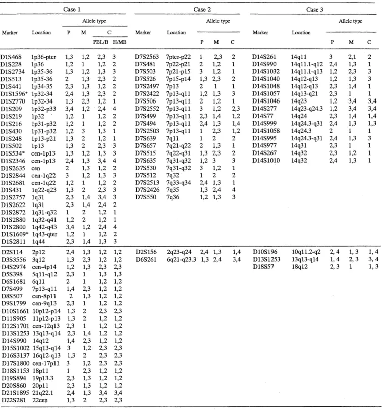
Genotype analysis in Case 1 revealed that 24 polymorphic marker loci on chromosome 1 were infor- mative (Table 1). All these markers showed heterozy- gous patterns in PBL and buccal membrane cells, while only a paternally derived allele was detected at these loci in all of the 5 monoclonal B-cell lines and 50 pieces of hair-root cells from various body regions (Table 1). The results suggested biparental inheritance of the alleles in PBL and buccal membrane cells, but indicated paternal uniparental isodisomy (UPD) in the monoclonal B-lymphocytes and the hair-root cells.
Transmission patterns of alleles from other chromo- somes were consistent with biparental inheritance and confirmed the paternity.
Likewise, genotyping of PBL in Case 2 revealed only a maternal allele at 16 informative loci from telomeric regions of chromosome 7 (Table 1). On the other hand, she inherited biparental alleles at 4 centromeric loci (D7S2552, D7S499, D7S494 and D7S2503), encom- passing a 7p l 3-q l l region. The results suggested that two normal-looking homologues 7 were maternal isodisomy, and the ring chromosome 7 were of pater- nal origin. Biparental inheritance and paternity were confirmed at two loci on chromosomes 2 and 6.
In Case 3, 15 polymorphic loci on the chromosome 14 were informative. At all these loci, the patient lacked any of the paternal alleles but inherited one or two maternal alleles (Table 1). Based on the fact that he had an isochromosome 14, i.e., mirror-image of the chromosome, he must have two copies of one- maternal-allele at 4 centromeric and at 5 telomeric loci. On the other hand, he inherited both of the ma- ternal alleles at the remaining 4 intermediate loci (D14S1046, D14S277, D14S77 and D14S999). The re- sults indicated that both centromeric and telomeric
Table 1. Polymorphic allele types at informative loci in Cases 1, 2 and 3 and their parents
Case 1 Case 2 Case 3
Allele type Allele type Allele type
Marker Location P M C Marker Location Marker Location
PBLJB H/MB P M C P M C
D1S468 1p36-pter 1,3 1,2 2,3 3 D7S2563 7pter-p22 1 2,3 2 D14S261 14q11 3 2,1 2
D1S228 1p36 1,2 1 1,2 2 D7S481 7p22-p21 2 1,2 1 D14S990 14g11.1-q12 2,4 1,3 1
D1S2734 lp35-36 1,3 1,2 1,3 3 D7S503 7p21-p15 3 1,2 1 D14S1032 14g11.1-q13 1,2 2,3 3 D1S513 1p35-36 2 1,3 2,3 2 D7S526 7p15-p14 1,3 2,3 2 D14S1040 14g12-q13 1,2 1,3 3
D1S441 1p34-35 2,3 1,3 1,2 2 D7S2497 7p13 2 1 1 D14S1048 14g12-q13 2,3 1,4 1
D1S1596* lp32-34 2,4 1,3 2,3 2 D7S2422 7p13-qll 1,2 1,3 3 D14S1057 14g13-q21 2,3 1 1 D1S2770 lp32-34 1,3 2,3 1,2 1 D7S506 7p13-q11 2 1,2 1 D14S1046 14q23 1,2 3,4 3,4 D1S209 1p32-p33 3,4 1,2 2,4 4 D7S2552 7p13-q11 3 1,2 2,3 D14S277 14q23-q24.3 1,2 3,4 3,4
D1S219 1p32 ' 1,2 1 1,2 2 D7S499 7p13-qll 2,3 1,4 1,2 D14S77 14q24 2,3 1,4 1,4
D1S216 lp3l-p32 1,2 1 1,2 2 D7S494 7p13-qll 2,4 1,3 1,4 D14S999 14g24.3-q31 2,4 1,3 1,3
D1S430 lp3l-p32 1,2 3 1,3 1 D7S2503 7p13-qll 1 2,3 1,2 D14S1058 14q24.3 2 1 1
D1S248 1p13-p21 1,3 2 1,2 1 D7S639 7q11 1 2 2 D14S995 14g24.3-q31 2,4 1,3 3
D1S502 1pl3 1,3 2 2,3 3 D7S657 7g21-q22 2 1,3 1 D14S977 14g31 2,3 1 1
D1S534* cen-1p13 1,3 1,2 1,3 3 D7S515 7g22-q31 1,3 2,3 2 D14S267 14q32 2,3 1,2 1 D1S2346 cen-1p13 2,4 1,3 3,4 4 D7S635 7g31-q32 1,2 3 3 D14S1010 14g32 2,4 1,3 1
D1S2635 cen 2 1,3 1,2 2 D7S530 7g31-q32 3 1,2 1
D1S2844 cen-1822 3 1,2 1,3 3 D7S512 7q32 1 2 2
D1S2681 cen-1g22 1,2 1 1,2 2 D7S2513 7q33-q34 2,4 1,3 1 D1S431 1g22-q23 1,3 2 2,3 3 D7S2426 7q35 1,3 2,4 4
D1S2757 1q31 2,3 1,4 3,4 3 D7S550 7q36 1,2 1,3 3
D1S2622 1q31 2,3 1,4 2,4 2 D 1 S 2872 1 q31-q3 2 1 2 1,2 1 D1S2880 1g32-q41 1,2 2 1,2 1 D1S2800 1g42-q43 3,4 1,2 2,4 4 DlS1609* 1g43-qter 1,2 1 1,2 2 D1S2811 1q44 2,3 1,4 1,3 3
D2S114 2p12 2,4 1,3 1,2 1,2 D2S156 2q23-q24 2,4 1,3 1,4 D10S196 10gl1.2-q2 2,4 1,3 1,4 D3S3556 3q12 1,3 2,3 1,2 1,2 D6S261 6g21-g23.3 1,3 2,4 3,4 D13S1253 13g13-q14 1,4 2,3 3,4
D4S2974 cen-4p14 1,2 1,3 2,3 2,3 D18S57 18g12 2,3 1 1,3
D5S398 5g11-q12 2,3 1 1,3 1,3 D6S 1681 6q11 2 1 1,2 1,2 D7S499 7p13-ql l 1,4 2,3 1,2 1,2 D8S507 cen-8p11 2 1,3 1,2 1,2 D9S 1799 cen-9q13 2,3 1 1,2 1,2 D10S1661 1Op12-p14 1,3 2 2,3 2,3 D11S905 11p12-p13 1,3 2 1,2 1,2 D12S1701 cen-12q13 2,3 1 1,2 1,2 D13S1253 13g13-q14 2,3 1,4 1,2 1,2 D14S990 14q12 1,4 2,3 1,2 1,2 D15S1002 15g13-q14 3 1,2 2,3 2,3 D16S3137 16g12-q13 1,3 2 2,3 2,3 D17S1800 cen-17p11 3 1,2 2,3 2,3 D18S1153 18pl l 1 2,3 1,2 1,2 D19S894 19pl3.3 2,3 1,3 1,2 1,2 D20S860 20pl l 2,3 1,3 1,2 1,2 D21S1895 21q22.1 2,4 1,3 3,4 3,4 D22S281 22cen 1,3 2 2,3 2,3
P, M, C, PBL, B, H, MB implicate father, mother, child, peripheral blood leukocytes, buccal membrane cells, hair-root cells, and monoclonal
B-lymphocytes, respectively.
Markers with asterisks are tetra-nucleotide repeats.
regions of chromosome 14 were of isodisomy, whereas the other region is heterodisomic (segmental UPD).
Biparental inheritance and paternity were confirmed with markers from chromosomes 10, 13 and 18.
Discussion
Case 1 was coincidentally found by flow cytometric analysis for RhD antigen to have unusual Rh
Osamu Miyoshi : Mechanisms of Uniparental Disomy in Man phenotypes composed of D+ and D- bearing cells, i.e., a mosaic for the Rh phenotype with the D/D genotype and the d/d genotype (Fig. 1). Since his father was a D/d heterozygote and mother a D/D homozygote, and the RhD gene is located at 1p36-p34, it was assumed that the d/d cells in Case 1 reflect UPD for chromo- some 1 (UPD1) that was derived from his father.
Chromosomal heteromorphism, lqh+, that was ob- served in one cell line supported the presumption.
Allele-typing of the proband's PBL with polymorphic loci on the chromosome 1 revealed biparental trans- missions of alleles. This transmission mode may be due to the mosaicism of this tissue. Integral analysis of allele-curves (electrophoretic patterns, Fig. 2) revealed that the ratio of the paternal allele to the maternal al- lele was 4:1, probably reflecting their proportions in PBL (data not shown). On the other hand, analysis of the same alleles in the monoclonal B-cells and the hair- root cells from various skin areas demonstrated the paternal uniparental inheritance. Since each hair and /or its root-cells is derived from a single cell, this tis- sue may have represented monoclonality with paternal UPD1. In addition, the finding that all the 50 hair cells
examined showed the same transmission pattern may indicate that the skin tissue originates from one or a few ancestral cells. In contrast, another ectodermal tis- sue, the buccal membrane cell, was composed of two different cells, as was PBL, suggesting that these tis- sues were derived from at least two ancestral cells.
The phenomenon that one individual has two cell lines
each with maternal or paternal UPD1 seems very un- usual and has not been known in the literature. Only a following mechanism is plausible to explain the find- ing: the zygote may have resulted from the normal fertilization of a "D" allele-bearing ovum by a "d"
allele-bearing sperm, followed by abnormal segregation at the first cleavage division, where both chromatids of a homologous chromosome 1 may have migrated together to the same direction with opposite-directed migration of chromatids of the counterpart chromo- some, resulting in two daughter cells having D/D and d/d genotypes (Fig. 3). The hair-root cells belong to the same "d/d" cell lineage, while PBL and buccal membrane cells are the mixture of "D/D" and "d/d"
cells. The abnormal cell division at first mitosis, a similar manner to the first meiotic division, has hith- erto been undescribed in man. Two previously re- ported individuals with UPD1 had only one cell line with either maternal or paternal UPD 1-121 . However, the author was recently informed about another indi- vidual who is a mosaic for the Rh phenotype as was Case 1, indicating that such segregation may not be very rare. Further analysis is needed in this second case.
Allele-typing of Case 2 detected only one maternal allele at 16 telomeric loci on the chromosome 7, but revealed biparental alleles at 4 centromeric loci (Table
1). Since each of her cell lines, 46, XX and 47,XX,+r(7), contains two normal chromosomes 7, these transmission patterns indicated that she inherited
gametic trisomy monosomy Case 1 Case 2 Case 3
complementation rescue duplication
Q O DO .,, d 0 © o0
monosomy crossing-over
duplication at MI
~O QO O ®Dd chromatid loss 00® of ring ( UH
T - i 1 replication isochromosome
DD dd formation
O O oo O abnormal~® ~0 (1]U
segregation
gametic
DD dd complementation
00 00 O oo O
DD dd dd
b PBL/buccal cells other cells examined C d a Fig 3. Possible mechanisms for UPD in man (a) and those for UPD in Case 1 (b), Case 2 (c) and Case 3 (d).
one maternal-allele in duplicate at the telomeric loci, and one copy of paternal-allele in addition to two cop- ies of maternal-allele at the centromeric loci. This indi- cates that the two normal chromosomes 7 were of ma- ternal isodisomy (UPD), and the ring chromosome 7 was of paternal origin. There was no evidence of recombinations on chromosome 7 in the maternal meiosis. From these findings, "monosomy duplication"
is the most likely mechanism for her UPD7 (Fig. 3), although monosomy was restricted to distal regions of chromosome 7. The patient may have arisen as a 46,XX,r(7) zygote (partial monosomy for most part of chromosome 7), where the normal chromosome 7 and the r(7) were of maternal and paternal origin, respec- tively. Then, duplication of the maternally-derived chromosome 7 may have occurred at an early postzygotic cell division, followed by death of the par- tially monosomic cells and survival of the UPD cells because of resistibility against cell selection"-"), result- ing in a 47, XX, upd (7),+r (7) karyotype. Finally, the ring chromosome 7 may have been lost in a subset of cells during somatic cell divisions due to ring chromo- some fragility 16'. Maternal UPD7 has been identified in 17 individuals including our patient1-24'. Of the 17 in- dividuals, 13 had a common phenotype reminiscent of Silver-Russell syndrome (SRS). It has been suggested that there is at least one maternally imprinted gene on chromosome 7 that controls intrauterine and postnatal growth17 2°. 24>. If the putative SRS gene is maternally imprinted (paternally expressed), it can be ruled out from the centromeric region, 7p13-qll, that corre- sponds to the retaining extent of the paternally- derived ring chromosome in Case 2.
UPD with an isochromosome without its normal chromosome member, as in Case 3, can arise through either of the 3 different mechanisms described above25' Allele-typing of Case 2 demonstrated maternal isodisomy for the centromeric and telomeric regions and heterodisomy for the middle region of chromosome 14 (Table 1). This indicates that meiotic recombinations had occurred between the maternal homologs 14, and thus prefers "gametic complementation" as the mecha- nism (Fig. 3). Non-mosaicism in Case 3 denied any postzygotic events. Maternal UPD for chromosome 14 has been identified in 12 individuals including Case 326-s6'. All but two such individuals had a com- mon phenotype, such as growth and developmental re- tardation, neonatal hypotonia, small hands and feet, and precocious puberty. On the other hand, paternal UPD for chromosome 14 is associated with different manifestations"-"). Thus, the phenotype characteristic for the maternal UPD is most likely due to lack of paternally-derived allele(s) through genomic imprinting
mechanism.
Acknowledgements
I express my gratitude to Professor Norio Niikawa for his helpful comments and suggestions.
References
1) Engel E: A new genetic concept: Uniparental disomy and its poten- tial effect, isodisomy. Am J Med Genet 6: 137-143, 1980 2) Ledbetter DH, Engel E: Uniparental disomy in humans: develop-
ment of an imprinting map and its implications for prenatal diag-
nosis. Hum Mol Genet 4: 1757-1764, 1995
3) Spence JE, Perciaccanate RG, Greig GM, Willard HF, Ledbetter DH, Hejtmancik JF, Pollack MS, O'Brien WE, Beaudet AL: Uniparental
disomy as a mechanism for human genetic disease. Am J Hum Genet 42: 217-226, 1988
4) Dib C, Faur S, Fizames C, Samson D, Drouot N, Vignal N, Millasseau P, Marc S, Hazan J, Seboun E, Lathrop M, Gyapay G,
Morissette J, Weissenbach J: A comprehensive genetic map of the human genome based on 5,264 microsatellites. "The Genethon
Human Genetic Linkage Map". Nature 380: 152-154, 1996 5) Niikawa N: Genomic imprinting and its relevance to genetic dis-
eases. Jpn J Hum Genet 41: 351-362, 1996
6) Abe K, Harada N, Itch T, Hirakawa 0, Niikawa N: Trisomy 13/
trisomy 18 mosaicism in an infant. Clin Genet 50: 300-303, 1996 7) The Cooperative Human Linkage Center, CHLC: Integrated Maps:
http://www.chlc.org
8) Bruns GA, Matise TC, Weith A: Report of the committee on the genetic constitution of chromosome 1. In: Cuticchia AJ,
Chipperfield MA, Foster PA, eds. Human Gene Mapping 1995.
Baltimore: Johns Hopkins Univ Press, 165-233, 1995
9) Miyoshi 0, Hayashi S, Fujimoto M, Tomita H, Sohda M, Niikawa N: Maternal uniparental disomy for chromosome 14 in a boy with
intrauterine growth retardation. J Hum Genet 43, 138-142, 1998 10) Miyoshi 0, Kondoh T, Taneda H, Otsuka K, Matsumoto T, Niikawa
N: 47,XX,upd (7)mat,+r(7)pat/ 46,XX,upd(7)mat mosaicism in a
girl with Silver-Russell syndrome (SRS): Possible exclusion of the
putative SRS gene from a 7p13-ql1 region. J Med Genet (in press) 11) Pulkkinen L, Bullrich F, Czarnecki P, Weiss L, Uitto J: Maternal
uniparental disomy of chromosome I with reduction to
homozygosity of the LAMB3 locus in a patient with Herlitz
junctional epidermolysis bullosa. Am J Hum Genet 61: 611-619,
1997
12) Gelb BD, Willner JP, Dunn TM, Kardon NB, Verloes A, Poncin J, Desnick RJ: Paternal uniparental disomy for chromosome 1 re- vealed by molecular analysis of a patient with pycnodysostosis.
Am J Hum Genet 62: 848-854, 1998
13) Petersen MB, Bartsch 0, Adelsberger PA, Mikkelsen M, Schwinger E, Antonarakis SE: Uniparental isodisomy due to duplication of
chromosome 21 occurring in somatic cells monosomic for chromo-
some 21. Genomics 13: 269-274, 1992
14) Bartsch 0, Petersen MB, Stuhlmann 1, Mau G, Frantzen M, Schwinger E, Antonarakis SE, Mikkelsen: "Compensatory" uniparental
disomy of chromosome 21 in two cases. J Med Genet 31: 534-540,
1994
15) Niikawa N, Kajii T : The origin of mosaic Down syndrome: Four cases with chromosomal markers. Am J Hum Genet 36: 123-130,
1984
16) Kosztolnyi G : Does "ring syndrome" exist? Analysis of 207 case reports on patients with a ring autosome. Hum Genet 75: 174-179,
1987
17) Voss R, Ben-Simon E, Avital A, Godfrey S, Zlotogora J, Dagan J, Tikochinski Y, Hillel J: Isodisomy of chromosome 7 in a patient
with cystic fibrosis. Could uniparental disomy be common in
humans? Am J Hum Genet 45: 373-380, 1989
18) Shuman C, Weksberg R, Nedelcu R, Northey A, Scherer S:
Chromosome 7 uniparental disomy in Silver-Russell syndrome. Am J Hum Genet 59: A648, 1996
19) Spotila LD, Sereda L, Prockop DJ : Partial isodisomy for maternal chromosome 7 and short stature in an individual with a mutation
at the COLIA2 locus. Am J Hum Genet 51: 396-1405, 1992 20) Kotzot D, Schmitt S, Bernasconi F, Robinson WP, Lurie IW, Ilyina
H, Mdhes K, Hamel BCJ, Otten BJ, Hergersberg M, Werder E,
Schoenle E and Schinzel A: Uniparental disomy 7 in Silver-Russell
syndrome and primordial growth retardation. Hum Mol Genet 4:
583-587, 1995
21) Langlois S, Yong SL, Wilson RD, Kwong LC, Kalousek DK:
Prenatal and postnatal growth failure associated with maternal
heterodisomy for chromosome 7. J Med Genet 32: 871-875, 1995 22) Eggermann T, Wollmann HA, Kuner R, Eggermann K, Enders H,
Kaiser P, Ranke MB: Molecular studies in 37 Silver-Russell syn- drome patients: Frequency and etiology of uniparental disomy.
Hum Genet 100: 415-419, 1997
23) Preece MA, Price SM, Davies V, Clough L, Stanier P, Trembath RC, Moore GE: Maternal uniparental disomy 7 in Silver-Russell syn-
drome. J Med Genet 34: 6-9, 1997
24) Cuisset L, LeStunff C, Dupont JM, Vasseur C, Cartigny M, Despert F, Delpech M, Bougnere P, Jeanpierre M: PEG1 expression in ma-
ternal uniparental disomy 7. Ann Genet 40: 211-215, 1997 25) Robinson WP, Bernasconi F, Basaran S, Yuksel-Apak M, Neri G,
Serville F, Balicek P, Haluza R, Farah LMS, Schinzel AA: A so- matic origin of homologous Robertsonian translocations and
isochromosomes. Am J Hum Genet 54: 290-302, 1994
26) Temple IK, Cockwell A, Hassold T, Pettay D, Jacobs P: Maternal uniparental disomy for chromosome 14. J Med Genet 28: 511-514,
1991
27) Pentao L, Lewis RA, Ledbetter DH, Patel PI, Lupski JR: Maternal uniparental isodisomy of chromosome 14: Association with
autosomal recessive rod monochromasy. Am J Hum Genet 50: 690-
699, 1992
28) Antonarakis SE, Blouin JL, Maher Joseph, Avramopoulos D, Thomas G, Talbot CCJr: Maternal uniparental disomy for human
chromosome 14, due to loss of a chromosome 14 from somatic
cells with t(13;14) trisomy 14. Am J Hum Genet 52: 1145-1152, 1993
29) Healey S, Powell F, Battersby M, Chenevix-Trench G, McGill J:
Distinct phenotype in maternal uniparental disomy of chromosome
14. Am J Med Genet 51: 147-149, 1994
30) Morichon-Delvallez N, Segues B, Pinson MP, Brub D, Domrnergues
M, Aubry MC, Cessot F, Lyonnet S, Munnich A, Vekemans M:
Maternal uniparental disomy for chromosome 14 by secondary
nondisjunction of an initial trisomy. Am J Hum Genet 55: A2224,
1994
31) Sirchia SM, DeAdnreis C, Pariani S, Grimoldi MG, Molinari A, Buscaglia M, Simoni G: Chromosome 14 maternal uniparental
disomy in the euploid cell line of a fetus with mosaic 46,XX/
47,XX,+ 14 karyotype. Hum Genet 94: 355-358, 1994
32) Papenhausen PR, Mueller OT, Johnson VP, Sutcliffe M, Diamond TM, Kousseff BG: Uniparental disomy of chromosome 14 in two
cases: An abnormal child and a normal adult. Am J Med Genet 59:
271-275, 1995
33) Tomkins DJ, Roux AF, Waye J, Freeman VCP, Cox DW, Whelan DT: Maternal uniparental isodisomy of human chromosome 14 as-
sociated with a paternal t(13g14q) and precocious puberty. Eur J
Hum Genet 4: 153-159, 1994
34) Coviello DA, Panucci E, Mantero MM, Perfumo C, Guelfi M, Borronco C, Dagna-Bricarelli F: Maternal uniparental disomy for
chromosome 14. Am J Hum Genet 57: 111A, 1995
35) Splitt MP, Goodship JA: Another case of maternal uniparental disomy chromosome 14 syndrome. Am J Med Genet 72: 239-240,
1997
36) Wang JC, Passage MB, Yen PH, Shapiro LJ, Mohandas TK:
Uniparental heterodisomy for chromosome 14 in a phenotypically
abnormal familial balanced 13/ 14 Robertsonian translocation car- rier. Am J Hum Genet 48: 1069-1074, 1991
37) Walter CA, Shaffer LG, Kaye CI, Huff RW, Ghidoni PD, McCaskill C, McFarland MB, Moore CM: Short limb dwarfism and
hypertrophic cardiomyopathy in a patient with paternal isodisomy
14: 45,XY,idic(14) (pl l ). Am J Med Genet 65: 259-265, 1996 38) Cotter PD, Kaffe S, McCurdy LD, Jhaveri M, Willner JP,
Hirschhorn K: Paternal uniparental disomy for chromosome 14 A
case report and review. Am J Med Genet 70 74-79, 1997