• 検索結果がありません。

Changes in the Transmembrane Action Potential's Pattern of the Specialized Fiber in Guinea Pig Atria During Anaphylaxis in Vitro

N/A
N/A
Protected

Academic year: 2021

シェア "Changes in the Transmembrane Action Potential's Pattern of the Specialized Fiber in Guinea Pig Atria During Anaphylaxis in Vitro"

Copied!
11
0
0

読み込み中.... (全文を見る)

全文

(1)

Acta med. nagasaki. 9 : 29 - 39

Changes in the Transmembrane Action Potential's Pattern of the Specialized Fiber in Guinea Pig Atria During Anaphylaxis in Vitro

Masayuki KIDO*

Department of Pharmacology,

Nagasaki University

School of Medicine, Nagasaki, Japan

Received for publication, June 5, 1964

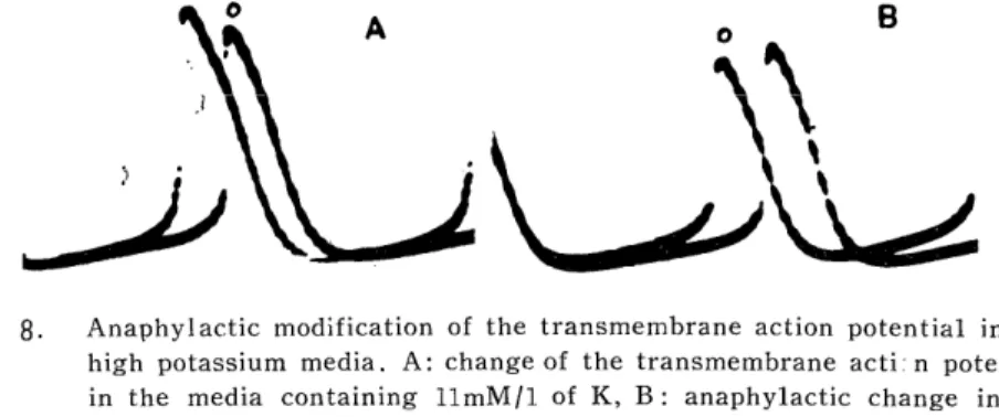
Although it was reported from our laboratory that the duration in the transmembrane A. P. of the proper atrial fiber is prolonged by anaphylaxis, no study on the changes in the action potential of the specialized fiber have been reported. So the present study was attempted to search changes in the membrane function of the pacemaker, using the ultramicroelectrode technique. Anaphylaxis causes an acceleration of the rhythm with the increased slope of the prepotential and without any changes in its height, so the beat acceleration may depend on the activities of ionic movement during diastole but not on the membrane excitability. And no specific change was observed in anaphylaxis in the high K-media. The A. P. pattern of the specialized fiber in anaphylaxis apparently differs from that caused by histamine, acetylcholine and serotonin, while it is very similar to adrenaline. But adequate dose of DCI gave no influence on anaphylactic manifestations. A possible role of these drugs on anaphylactic reactions may be excluded through changes in the pattern of the action potential and the attitude of anaphylaxis against DCI.

It has been reported from our laboratory14) that adding of antigen to an auricle preparation taken from a sensitized guinea pig causes an augmentation of the contractile force and an acceleration of the rate of beat. And the reaction was confirmed as an anaphylactic manifestation occurring in the atrial tissue, through facts that the reaction occurs specifically in sensitized atria while never in the atria once exposed to antigen.

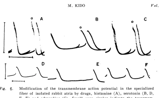
K. GREEFF4) has reported that in the auricle anaphylaxis some of antihistaminics depress the cardiac acceleration, but extensive studies by F. KIHARA10) have shown that antihistaminics did not depress the anaphylactic reaction in the auricle preparation. KIHARA pointed out that histamine may not be a substance principally responsible for the

*木 戸 正 之

(2)

30 M. KIDO Vol. 9.

anaphylactic reaction in the guinea pig atria. On the other hand, in foregoing papers from our laboratory11)15) it was discussed that acetyl- choline, noradrenaline and serotonin seemed to play little role in the auricle anaphylaxis through differences in their mode of action, their attitude to respective antagonists and their contents in the cardiac tissue.

Recently Y. NAKAZAWA and A. UENO" found that there was a specific change in transmembrane action potentials (A. P.) of the proper atrial fibers during the anaphylactic reaction.

In the above reports the mode of changes in the contractile force in the anaphylactic reaction was researched but the rhythm-accelerating effect observed simultaneously has not been stressed. The contractile force of the heart is altered by changing the beating rate, which is already known as the stair-case phenomena'"' or the interval-strength relationship'-'. M. MATAYOSHI13) concluded in his paper that anaphy- lactically induced rhythm acceleration itself may minimize augmentation of the contractile force. T. K&JIYAMA4' reported that the rhythm acceler- ating effect in the atrial anaphylaxis was not influenced by premedi- cation of corticosteroids despite its force augmenting effect is suppressed markedly. K. GREEFF4' also pointed out that inhibition of the atrial anaphylaxis by antihistaminics was found only in its force augmentation.

Above facts clearly indicate that each of the rhythm and the contractile force may respond independently to drugs, although a change of the rhythm can modify the contractile force. So some differences in changes of the pattern of the transmembrane A. P. induced by anaphylaxis could be expected between those of the proper atrial fiber and the pace-

maker fiber. Our present attempt is to search a basic mechanism to cause anaphylaxis through changes occurred in the action potential pattern of a single fiber in the pacemaker area.

Methods and Materials

Atria taken from male and female guinea pigs weighing 200 to 300g were mainly used in the experiment. Immediately after removing from an exsanguinated guinea pig, a sinus node belonging with an atrium was put into a horizontal acryl resin bath which contains 80 ml, 28 to 32°C nutrient solution, aeroated with oxygen containing carbon dioxide at 5 % level, and the atrial piece was pinned horizontally on a cork block in the bath. The nutrient solution was composed of NaCI 0.9%, KC1 0.042%, CaCI2 0.024%, glucose 0.1% and NaHCO;z 0.05%. Rabbit atria were also used in a part of the experiment.

The transmembrane A. P. of the fiber in the area described below

was led by single fiber stinging with a suspension intracellular micro-

electrode having an electric resistance of 15 to 50 MQ. Observation and

recording were carried out by a long recording camera with film speed

of 2 cm/sec attached to a cathode ray oscilloscope and by an ink writing

(3)

1964 ANAPHYLAXIS AND PACEMAKER ACTION POTENTIALS 31 recorder for monitoring time orientation through a low input high impedance amplifier (Nihonkoden Co. MZ - 3 A) and DC amplifier.

Microelectrode stinging was made via epicardium or, via endocardium on a fiber in the region along the crista terminalis between the caval orfices, according to the maps by T. C. WEST'" and by A. P. DE CARVALHO3). This region corresponds to the sinoatrial ring bundle parallel to the crista terminalis. In normal atria, when an action potential constantly had the amplitude over 60 mV, the height of the prepotential over 5 mV and no hump, the electrode was regarded to be successfully inserted into the pacemaker fiber.

A slow depolarization occurring in diastole in the fiber of the sinus venous, which was found by BOZLER", has been called as the pre- potential.

Anaphylactic sensitizations were carried out passively by injecting intravenously anti-egg-albumin rabbit serum having 1 x 128 antibody titer (0.3 ml for 100 g body weight) 18 to 24 hours prior to the experi- ment, and actively by intraperitoneal injections of 0.2% egg-albumin (0.5 ml) for 1 to 3 times every 4 to 7 days 2 weeks prior to the experi- ment. The anti-egg-albumin sera were obtained from rabbits sensitized by repeated injections of egg-albumin, the -method of sensitization in detail was described elsewhere12' .

Results

1. The contour of the transmembrane A. P. of the specialized fiber of the guinea pig.

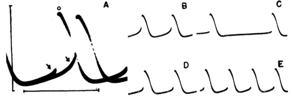
Contours of the transmembrane A. P. taken from the specialized fibers mentioned above were intermediate in appearance between those shown in Fig. 1A and Fig. 1C. A contour, having steeper and higher prepotential and smoother transition to the upstroke of the action potential, is considered as that of the true pacemaker (Fig. 1A), and a contour, having more similarities to the action potential pattern of the proper atrial fiber, as that of the latent pacemaker (Fig. IC). In the specialized fibers the amplitude of the action potential was lower and its duration was longer than in the proper atrial fibers. Average value of the transmembrane A. P. of specialized fibers of guinea pigs from calibrated 30 cases ases were appeared 82 ± 10 mV for the height of the action potential, 14 : 7 mV for the height of the prepotential and 300 + 70 msec for the duration of the action potential.

2. Changes in the contour of the transmembrane A. P. during anaphylaxis.

Acceleration of the beat was caused from 40 to 60 sec after adding

antigen to a sensitized atrium, reaching 55 ± 25 % (Table 1) at 4 to 5

minutes and then gradually returned to the initial rate 20 to 30 minutes

later. During the acceleration, the slope of the prepotential was increased

(4)

32 M. KIDO Vol. 9.

Fig. 1. The transmembrane action potentials recorded from the specialized fiber of isolated guinea pig atria, in true pacemaker (A), proper atrial fiber

(B), and latent pacemaker (C). Abbreviations of PD, PH and TD in

(D) show the duration of the prepotential, the height of the piepo

l

tential and the duration of the action potential respectively.

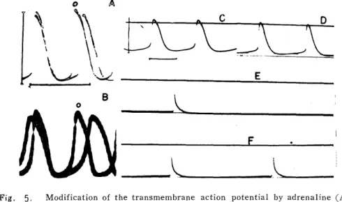
markedly, but the height of the prepotential was almost unchanged (Fig. 2C). The illustration in Fig. 3 shows time courses of changes of the slope and height of the prepotential and the beat rate in 5 cases.

The slope of the prepotential increased along with increase of the rate, while the height of the prepotential was almost unchanged. Therefore, anaphylactically induced beat acceleration may depend on the increased slope of the prepotential, but not on the height of the prepotential or the membrane excitability.

The duration of the action potential was shortened slightly by

Table 1.

Responses of the specialized fibers in guinea pig atria to some agents

Mean'

duration

*15*

± Serotonin

n prepotential pre

of ~o increase ini nitial rateagents

beats/mipotential beat rate slope I height action potential

Anaphylaxis 125 ± 35 55 ± 25 115 ± 65 - 5 ±-15 ± 10*

Histamine 120 ± 245

_

± 15 -35 ± 15

5120 ± 2

_ _

35 ± 10 -10 _+. 10

IAd

renaline 110 ± 25 50 ± 15 95 ± 50 - 5 ± 25 -15 ± 5

25 25 +5 652

-

5 35 +10 5 +25 -

+ Eeach value represents mean of at least 4 experiments Minus means decrease

~`* Minus means shortening

(5)

1964 ANAPHYLAXIS AND PACEMAKER ACTION POTENTIALS 33

Fig. 2. Modification of the action potential by anaphylaxis and histamine. The control action potential was superimposed on the action potential modi-

fied by anaphylaxis (A, C) and by histamine (B, D). Upper figures show shortening of the action potential duration (A, B), the lower

changes in the slope and the height of the prepotential (C, D). Small

open circles indicate the transmembrane action potential after adding

antigen or histamine.

Fig. 3. Time courses of changes in the beat rate, the height of the prepotential and the slope of the prepotential, A -A : the slope of the prepoten-

tial, 0-0 : the beat rate, ®-® : the height of the prepotential.

anaphylaxis at almost all points in the repolarization phase (Fig. 2A).

The above change in the duration differed from that, of prolongation, observed in the proper atrial fiber during anaphylaxis.

3. Modification of the contour of the transmembrane A, P. by

some drugs,

(6)

34 M. KIDO Vol. 9, Effects of some drugs, which possess possibility to play a role in anaphylactic manifestations, on the transmembrane A. P. of the special- ized fiber was observed and compared with that of anaphylaxis.

a) Histamine

Histamine hydrochloride (lpg/ml) caused an increase in the rate of beat 20 to 50 seconds after adding and the beat was increased 65 ± 25 % from the initial rate (Table 1) 3 to 4 minutes later. In that time,

the slope of the prepotential was increased moderately as in the anaphy- latic change, but the height of the prepotential was decreased markedly in contrast with anaphylaxis (Fig. 2D). The duration of the action potential was more shortened by histamine than in anaphylaxis (Fig.

2B). In rabbit atria, 1 dug/ml of histamine caused same effects on the beat rate and the action potential of the specialized fiber as in guinea pig atria (Fig. 6A).

b) Serotonin

Adding of serotonin creatinine sulfate (10,ug/ml) reduced the rate of beat 40 to 60 seconds after administration and the reduction was followed by an increase in 100 to 120 seconds later, while only an increase was caused by 1,eg/ml of serotonin. The maximum increase was 35 ::1-- 10 % of the control rate by either doses (Table 1) . The rate increasing effect was apparently less than that of anaphylaxis and histamine. Serotonin has a dual effect, initially depressed and then accelerated, on the beat rate, although the effect may vary with its dose. When the rate was reduced by serotonin, the prepotential was decreased both in the slope and the height. However, only the slope of the prepotential was recovered and the height of the prepotential remained still lowered, when the rate was increased. And slight shorten- ing of the repolarization was observed (Fig. 4). It should be mentioned that shortening by the rate change may occur only slightly, since an increase of the rate caused by serotonin is less than that by histamine

or anaphylaxis. In rabbit atria, 2pg/ml of serotonin caused a similar

Fig. 4. Modification of the transmembrane action potential by serotonin 10 Ng/ml. The superimposed figure A was made from B and E. A small

open circle indicates the curve from E. B : before, C, D & E : after

administraion of serotonin.

(7)

1964 ANAPHYLAXIS AND PACEMAKER ACTION POTENTIALS 35 dual effect on the beat rate and the action potential pattern to that in guinea pig atria (Fig. 6. B, D, E & F).

c) Adrenaline

40 to 60 seconds after adding adrenaline hydrochloride (1,ug/ml), the rate of beat began to increase and reached 150 ± 15% of the initial rate 3 to 5 minutes after medication (Table 1). Modification of the contour of the transmembrane A. P. by adrenaline was most similar to that by anaphylaxis out of drugs used in the experiment (Fig. 5 A).

d) Acetylcholine

Acetylcholine chloride in a concentration of 2,ug/ml caused abruptly a marked decrease of the rate which recovered very slowly. The slope and the height of the prepotontial were markedly diminished, so special features of the specialized fiber in the contour of the transmembrane A. P. were almost lost. The maximum diastolic potential was increas- ed moderately by adding acetylcholine. The spike of the action potential was distinguished because of remarkable shortening of the duration and more rapid upstroke in the rising phase. These changes caused by ace- tylcholine were obviously different from those in anaphylaxis (Fig. 5.

C, D, E & F).

4. Anaphylaxis in isolated guinea pig atria pretreated with dichloro- isoproterenol (DO).

About 10 minutes after administration of 10-' of DCI, adding of 10"7 of adrenaline did not change the beat rate and the contour of the trans- membrane A. P. of the specialized fibers, while the dose caused an

Fig. 5. Modification of the transmembrane action potential by adrenaline (A), acetylcholine (C, D, E, F) and anaphylaxis in the presence of DCI (B).

Small open circles indicate the transmembrane action potentials after

adding adrenaline or antigen. Acetylcholine was applied lied between C

and D.

(8)

36 M. KIDO Vol. 9.

Fig. 6. Modification of the transmembrane action potential in the specialized fiber of isolated rabbit atria by drugs, histamine (A), serotonin (B, D,

E, F) and adrenaline (C). Small open circles indicate the transmem-

brane action potentials after adding the drugs. D, E and F show the

dual effect of serotonin on the beat rate, D : before, E & F : after

administration of serotonin.

acceleration of the beat rate and some changes in contour of transmem- brane A. P. in the atria untreated with DCI. On the other hand, adding of antigen to a sensitized atria in the bath containing same concentration of DCI caused an increase both in the beat rate (55%) and in the slope of the prepotential (75 %). The height of the pre- potential was slightly decreased but the change was not significant. In short, anaphylactically induced changes were not modified by DCI (Fig.

5. B).

5. Effects of increased potassium concentration in the nutrient solution.

a) Effects of high potassium media on the normal sinus node.

When the normal nutrient solution was replaced with high K-solution which contains 2 times of potassium (11 mM) of the normal solution the height of the prepotential was increased up to about 1.2 times of

Fig. 7. The transmembrane action potential in the high potassium (22 mM/l ) media, upper : the transmembrane action potential of the specialized

fiber. lower: that of the proper atrial fiber. A : control, B : high

potassium.

(9)

1964 ANAPHYLAXIS AND PACEMAKER ACTION POTENTIALS 37

Fig. 8. Anaphylactic modification of the transmembrane action potential in the high potassium media. A: change of the transmembrane acti: n potential

in the media containing 11mM/1 of K, B : anaphylactic change in the

media. Small open circles indicate the action potentials after increasing

KCI and adding antigen.

the original, but the slope of the prepotential was not influenced.

whereas the rate was slowed slightly (Fig. 8A). In the nutrient solution containing K in 22 mM per liter, the rate of beat was gradually reduced and eventually arrested. During the time course, an. increase of the height of the prepotential, a slight decrease of the slope and a smoother transition to the rising phase were observed initially, then the trans- membrane A. P. lost its propagated potential and eventually became a state of repeating of prepotentials. On the other hand, the membrane potential simultaneously taken from the proper atrial fiber showed no action potential corresponding to the prepotential without the propagated potential (Fig. 7). Above facts clearly indicate that the high potassium media cause a marked decrease of excitability.

b) Anaphylaxis in the high potassium media.

Added antigen to a sensitized atrium in the high K nutrient solution containing 11 mM of potassium per liter, the rate of beat and the slope of the prepotential were increased, but the height of the prepotential was not influenced (Fig. 8B).

Discussion

Although the rhythm acceleration with the increase of the contractile force in the atria during anaphylaxis in vitro was reported from our laboratory, foregoing papers concerning the heart anaphylaxis dealt mainly with its force enhancing effect. Whereas, the author made an attempt to investigate the mode of the anaphylactically induced rhythm change in the atrium.

In the pattern of the transmembrane A. P. led from the specialized

fiber in the atria using an ultramicroglasselectrode technique, a diastolic

slow depolarization preceding to rapid depolarization, so called prepoten-

tial, is considered as a fundamental factor to develope the automacity

in the heart. According to WEIDMANN'0) there are three factors altering

(10)

38 M. KIDO Vol. 9.

automatic rhythm, that is, the maximum diastolic potential, the thresh- old potential and the slope of the prepotential. The terms can be substantially replaced by ionic concentration gradient across the mem- brane, excitability of the membrane and activity of ionic transport.

Namely these factors are assumed as essential factors controlling the rhythm. Many investigators')")") emphasized that the slope of the prepotential may be caused by changes of activities of ionic transport, especially by gradually decreased movement of potassium ions. So concentration change of potassium ion in the nutrient solution may give some changes in the configuration of the prepotential and the beat rate.

In the present study, high concentration of potassium ions in the nutrient solution decreases the slope of the prepotential and increases its height. These phenomena are appeared as essential factors partici- pating in the slowed rhythm in high K media.

In contrast anaphylaxis causes an acceleration of the rhythm which is found parallel to the slope of the prepotential, while no significant change is observed in the height of the prepotential, meaning no change in the threshold to induce propagated A. P. So the acceleration of the beat during anaphylaxis is not related to excitability but to ionic move- ment in diastolic state. Though it was already reported from our la- boratory') that the anaphylactic reaction can be provoked even in the high potassium media, similar anaphylactic changes in the rhythm and the contour of the action potentials are also induced in the high potassium media, that is to say, anaphylactic changes are riot modified by altering concentration of potassium ions in the nutrient media. This fact sug-

gests that potassium ion may not play an essential role to cause anaphy- lactic manifestations.

It is well known that, in general, in the excitable membrane a change of frequency of excitation affects the duration of the action potential'. And a recently appearing paper from our laboratory'") have pointed out that the duration in the action potential of the proper atrial fiber was modified several ways by anaphylaxis in spontaneously beating atria, while prolongation of the duration was always observed in elec- trically driven atria. That is to say, prolonging effect of anaphylaxis can be reduced by shortening-effect of anaphylactic rhythm acceleration as a physiological variable. In the specialized fiber, the duration of the propagated action potential was always shortened during anaphylaxis, but the shortening was less than that by histamine and serotonin.

Relatively slight shortening of the duration in the propagated action potential of the specialized fiber during anaphylaxis may suggest that anaphylactic reaction itself acts to prolong the duration as in the proper atrial fiber. KATSH & MARSHALL" also reported that anaphylaxis prolongs the duration of the action potential in the uterine smooth muscl fibers.

So it is very probable that anaphylactic reaction prolongs the repolari-

zation process of the cell membrane.

(11)

1964 ANAPHYLAXIS AND PACEMAKER ACTION POTENTIALS 39 Of some chemicals accepted as mediators to cause several symptoms in the anaphylaxis, histamine is reported" to play a similar reaction to anaphylaxis but serotonin and adrenaline to induce no change in the a: tion potential of the proper atrial fibers. On the other hand, in the specialized fiber, adrenaline causes same changes in the action potential as anaphylaxis, but histamine, serotonin and acetylcholine induces different changes from those caused by anaphylaxis. But rhythm accel- eration as an anaphylactic manifestation is also provoked even in atria under presence of DCI, while action of adrenaline is completely abol- ished. Through the findings in the contour of the action potential in the specialized fiber mentioned above, an essential cause of anaphylactic manifestation is not concerned with above chemicals accepted as mediators up to date, but probably may be with antigen-antibody re-

action itself took place in the cell membrane.

ACKNOWLEDGEMENT. The author is gratefull to professor Dr. Y. NAKAZAWA and assistant professor Dr. A. UENO for their helpfull comments and

suggestions.

REFERENCES

1) BLINKS, J. R. and KocH-WESEZ, J. : J. Pharmacol. Exptl. Therap. 134 : 373 (1961).

2) BOZLER, E. : Am. J. Physiol. 138: 273 (1942).

3) de CARVALLHO, A. P., de MELLO, W, C. and HOFFMAN, B. F. : Am. J.

Physio l . 196 : 483 (1959).

4) GREEFF, K., BENFEY, B. G. and BDKELMANN, A. : Arch. f. exp. Path. u, Pharmakol. 236 : 421 (1959).

5) HOFFMAN, B. F, and CRANEFIELD, P. F. : "Electrophysiology of the heart"

51, McGraw-Hill, New York. (1960).

6) HOFFMAN, B. F. and CRANEFIELD, P. F. : "Electrophysiology of the heart"

127, MacGraw-Hill, New York. (1960).

7) IRIE, H. : Nisshin Igaku 47 : 527 (1960) (Japanese).

8) KAJIYAMA, T. : This journal 6 74 (1961).

9) KATSH, S, and MARSHALL, J. M. : Am. J. Physiol. 196 : 39 (1959).

10) KIHARA, F. : Nagasaki Igaku Zassi 34 : 1612 (1959) (Japanese).

11) KIHARA, F. Nisshin Igaku 47 : 383 (1960) (Japanese).

12) KIMURA, Y. : "Allergy" 609, Kanahara, Tokyo. (1957) (Japanese), 13) MATAYOSHI, M. : Nagasaki Igaku Zassi 39 : 234 (1964) (Japanese).

14) NAKAZAWA, Y., SAKAI, O. and KOBATA, S. : Nisshin Igaku 45 : 147 (1958) (Japanese).

15) NAKAZAWA, Y., UENO, A. and ODA, T. : Fol is pharmacol. japon. 56 : 173 (1960) (Japanese).

16) NIEDERGERK, R. : J. Physiol. 134 : 569 (1956).

17) NOBLE, D. J. Physiol. 160 : 317 (1962).

18) TRAUTWEIN, W. : Pharmacol. Rev 15 (1963).

19) UENO, A., MURAKAMI, K. and NAKAZAWA, Y. : Acta med. nagasaki. 8 : 41 (1964).

20) WEIDMANN, S. "Elektrophysiologie der Herzmuskelfasern" : 75, Huber, Bern (1956).

21) WEST, T. C. : J. Pharmacol. Exptl. Therap, 115 : 283 (1955).

Fig.  1.  The  transmembrane  action  potentials  recorded  from  the  specialized  fiber            of  isolated guinea  pig  atria, in  true  pacemaker (A),  proper  atrial fiber
Fig.  2.  Modification  of  the  action  potential  by  anaphylaxis  and  histamine.  The            control action potential was  superimposed on  the  action  potential
Fig.  4.  Modification  of  the  transmembrane  action  potential  by  serotonin  10            Ng/ml
Fig.  5.  Modification  of  the  transmembrane  action  potential  by  adrenaline  (A),           acetylcholine (C,  D,  E,  F)  and  anaphylaxis in  the  presence of  DCI  (B)
+3

参照

関連したドキュメント