Acta med. nagasaki. 8 : 41-48
Changes in Membrane Potentials and Mechanical Responses
of Guinea Pig Atria During Anaphylaxis in Vitro*1
Akira. UENO, Keisuke MURAKAMI
and Yoshiro NAKAZAWA*2
Department of Pharmacology,
Nagasaki University School of Medicine, Nagasaki Japan
Received for publication, January 15, 1964
The anaphylactic reaction in the isolated atria of sensitized guinea pigs is principally to cause acceleration of cardiac activities as already reported. The present attempt was made to observe changes in transmem- brane action potentials (AP) and mechanical responses of the atria during the anaphylaxis. Four types of changes in AP were found in spontaneously beating atria that is prolongation of AP at all points of the repolarization, prolongation of the duration with proceeding of repolarization, shortening of the duration at first half, and no change at all in AP pattern. However the change in electrically driven left atria was only a parallel prolongation of the duration to the control in repolarization phase. On the other hand, an increase in the rate shortend the duration of AP in the normal prepara- tion electrically driven. The essential change of the contour of AP in the anaphylactic reaction is the prolongation of AP in repolarization phase.
Histamine induced a similar reaction in electrical and mechanical responses to the anaphylactic reaction.
It has been reported by Y. NAKAZAWA 5.8.9) and K. GREEFF1) that the anaphylactic reaction in the auricle of guinea pigs previously sensitized was acceleration of the cardiac movement, i.e. positive inotropic and chronotropic response. While a test of tissue anaphylaxis has been usually carried out with ileal and uterine segments, the
anaphylactic reaction in the heart preparations occurs more dynami- cally and has more calculative factors than that with smooth muscle organs whose reaction in only muscle contraction. Both the cardiac muscle and innervation differ from smooth muscle organs, and have the considerable physiological specificity, so it is significant to analyse pharmacodynamically the mechanism of anaphylaxis provocated in the tissue.
*1 Supported by a research grant (# 710429) from the Ministry of Education
*2上 野 昭 ,村 上 圭 介,中 沢与四郎
42 A. UENO, K. MURAKAMI, Y. NAKAZAWA Vol. 8.
On the other hand, intracellular microelectrode technic brought by LING & GERALD was applied for cardiac muscle cell by HOFFMAN2) and
others and electrophysiology of the heart has been greatly progressed.
Transmembrane action potentials of single cell by means of an intra- cellular microelectrode provides an accurate and sensitive index of the
electrical changes associated with activities of excitable tissue, and those have been applied for the research of pharmacodynamic actions of various drugs.
S. KATSH and J. M. MARSHALL' reported a study about electrical and mechanical responses of uterine smooth muscle during anaphylaxis in vitro, in which a good correlation between membrane potential, rate of spike discharge and fluctuation in tension was observed. However, no report of transmembrane action potentials of the cardiac muscle cell following the anaphylactic provocation was uncovered. As the provoca-
tion is carried out after the auricle preparation used for the experiment were repeatedly washed by nutrient fluid, a site in which antigen
interacts with the antibody series must be the cell.
There is a possibility that the interaction of antigen and plasma membrane might be as a trigger of dynamic phenomena of the heart anaphylaxis. Therefore, it is of our great interest of what electrical changes occurs at the transmembrane potentials and mechanical response of the cardiac muscle, and what correlation exists between the potentials and the tension of the muscle.
MATERIALS AND METHODS
About one hundred male and female guinea pigs weighing 180 to 300g were used in the experiment. Antigenic sensitization of the guinea pig was passively carried out by intravenous injection of anti-egg- albumin rabbit serum, having 1 x 128 to 1 x 256 antibody title (0.3m1 per 100g body weight), 24 hours prior to the experiment. The method of the sensitization are described in details elsewhere. The atrium was removed immediately from exsanguinated guinea pigs and placed in Ringer solution (NaCI, 0.9%; KC1, 0.042%; CaCL, 0.024%; glucose, 0.1%; NaHCO3 0.05%). And a right auricle of the atrium was pinned horizontally on the cork block in the bath and left one was joined to a unbonded strain gage transducer (Shinkoh UL-10-120) for recording the contractile tension of the atrium in connection with strain amplifier (Shinkoh DS6-P). This is sensitive enough to record a tension as small as 10mg and to respond lineally up to 130 c/s.
The bath was made by acryl resin and specially designed for the
experiment in the light of the method Of MATSUDA et all', total fluid
content is 50 ml and bath temperature is 32°C. The nutrient fluid was
actively circulated through a stream of bubbles of oxygen containing
carbon dioxide at 5 % level.
1963 ANAPHYLAXIS ON TRANSMEMBRANE ACTION POTENTIALS 43 The ultramicroelectrode was made from a capillary tube 1 to 1.5 mm in diameter prepared from Telex glass (Toshiba) with aid of Andrews type puller and filled with 3 M KC1 solution by means of ethanol-water-KC1 replacements. The microelectrode of 15 to 55 M2 was chosen for the experiment, and used tip of this as a suspension
electrode.
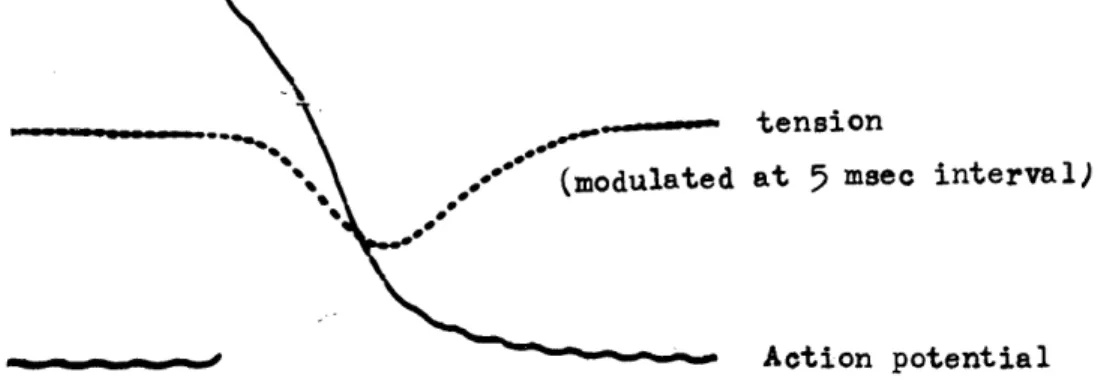
The transmembrane action potential was led from a location near by top of the pinned right auricle. And this has been amplified by means of a conventional cathode follower input amplifier (Nihon Koden MZ 3 A) and D.C. amplifier for observation of cathode ray oscilloscope and ink writting record. A dual beam oscilloscope was used to observe transmembrane potentials and contractile tension simultaneously and the tension beam was modulated at 5 or 10 msec interval for time orientation.
The plateau of the tension originally indicates zero potential.
RESULTS
1. Normal contour of transmembrane action potentials of proper atrial fibers of the guinea pig
Compared the contour of transmembrane action potentials of the proper atrial fibers with its mechanical activities, contraction of the fibers appeared at a gentle sloping point of the time course of spike potential, this is correspond to beginning of `Phase 2'. Critical period of the tension in contraction was equal to one third or half in repolarization phase and muscle fibers reached relaxation state when the potential returned to the resting one (Fig. 1). Intracellular resting and action potentials of spontaneously beating atria of 70 cases of guinea pigs which survived in 32°C Ringer were found most frequently in -85 mV for diastolic and 104 mV for hight of action potentials, so the overshoot was 19 mV. Total duration of the action potentials were varied from 80 msec to 150 msec. These values practically accord with higher bound of mammalian atria with reported by several investigators.
Fig. 1 Transmembrane action potential pattern and tension curve of the guinea pig atrium,
44 A. UENO, K. MURAKAMI, Y. NAKAZAWA Vol. 8.
2. Modification of action potential pattern by anaphylactic reaction
At 20 to 50 sec after the auricle preparation was exposed to antigen (2mg% in the bath solution) an increase in the tension and the rate occurs, which continued 10 to 20 min and then returned to a state before the exposure. No reaction was observed in non-sensitized atria and nothing or very weak in the preparations once provoked by antigen exposure, that is desensitization.
Fig. 2 Changes in transmembrane action potential during anaphylaxis.
Arrows indicate direction of the change.
Change in transmembrane action potentials in spontaneonsly beating atria during anaphylaxis could be devided into four types of group as shown in Fig. 2, a) slight prolongation of action potential (APD) at almost all points of the repolarization phase (type a), b) prolonging APD with proceeding of repolarization (type b), c) prolongation of APD in the latter half of repolarization phase while it in the first half was shortend (type c), d) no change at all in AP pattern despite there were marked response in the mechanical activities (typed). However, in the electrically driven (2c/s) left atria only a parallel prolongation of the duration to the control in repolarization phase was constantly observed overall, this is likely to type a) of the change in spontaneously beating atria. Illustration in Fig.3 shows a relationship existed between tension change and duration change in 50% and 90% repolarized state.
3. Change of rate and contour of action potential during the heart anaphylaxis
A difference in the results between the spontaneously beating atria
and the electrically driven left atria suggests a possibility that an
increase the rate affects the contour of the action potential. In order
to search a relation between the rate of frequency and contour of
1963 ANAPHYLAXIS ON TRANSMEMBRANE ACTION POTENTIALS 45