EED regulates epithelial‑mesenchymal
transition of cancer cells induced by TGF‑β
著者 ドゥラムスレン オクチャブリ
著者別表示 Dulamsuren Oktyabri journal or
publication title
博士論文本文Full year 2015‑09‑28
学位授与番号 13301甲第4285号
学位名 博士(医学)
学位授与年月日 2015‑09‑28
URL http://hdl.handle.net/2297/44641
doi: 10.1016/j.bbrc.2014.09.082
EED regulates epithelial-mesenchymal transition of cancer cells induced by TGF-β.
1
2
3
Dulamsuren Oktyabri†, Shoichiro Tange†, Minoru Terashima, Akihiko Ishimura, Takeshi
4
Suzuki1
5
6
Division of Functional Genomics, Cancer Research Institute, Kanazawa University,
7
Kanazawa 920-1192, Ishikawa, Japan
8
9
† These authors contributed equally to this study
10
1 To whom correspondence should be addressed
11
12
Takeshi Suzuki, Division of Functional Genomics, Cancer Research Institute, Kanazawa
13
University, Kakuma-machi, Kanazawa 920-1192, Ishikawa, Japan
14
Phone: +81-76-264-6741; Fax: +81-76-234-4502; E-mail: [email protected]
15
16
Keywords
1
Epithelial-mesenchymal transition ; Histone methylation ; Transcriptional regulation ;
2
microRNA ; Cancer progression
3
4
Abbreviations
5
EMT, epithelial-mesenchymal transition; TGF-β, Transforming Growth Factor-beta;
6
miR-200, microRNA-200; ChIP, chromatin immunoprecipitation; PRC2, Polycomb
7
repressive complex-2; H3K27me3, histone H3 trimethylated Lys27; GAPDH,
8
glyceraldehyde-3-phosphate dehydrogenase; IgG, immunoglobulin G; shRNA, small
9
hairpin RNA.
10
11
12
Abstract
1
Histone methylation is involved in various biological and pathological processes including
2
cancer development. In this study, we found that EED, a component of Polycomb
3
repressive complex-2 (PRC2) that catalyzes methylation of lysine 27 of histone H3
4
(H3K27), was involved in epithelial-mesenchymal transition (EMT) of cancer cells induced
5
by Transforming Growth Factor-beta (TGF-β). The expression of EED was increased
6
during TGF-β-induced EMT and knockdown of EED inhibited TGF-β-induced
7
morphological conversion of the cells associated with EMT. EED knockdown antagonized
8
TGF-β-dependent expression changes of EMT-related genes such as CDH1, ZEB1, ZEB2
9
and microRNA-200 (miR-200) family. Chromatin immunoprecipitation assays showed that
10
EED was implicated in TGF-β-induced transcriptional repression of CDH1 and miR-200
11
family genes through the regulation of histone H3 methylation and EZH2 occupancies on
12
their regulatory regions. Our study demonstrated a novel role of EED, which regulates
13
PRC2 activity and histone methylation during TGF-β-induced EMT of cancer cells.
14
15
16
1. Introduction
1
Lysine (K) methylation on the N-terminal tail of histone H3 (K4, K9, K27 and K36) plays
2
critical roles in transcriptional regulation and maintenance of genome integrity [1,2].
3
Methylation of H3K4 has been associated with active transcription whereas methylation of
4
H3K9 and H3K27 are repressive marks of chromatin. Methylation is regulated by two
5
classes of enzymes: histone lysine methyltransferases (KMTs) and lysine demethylases
6
(KDMs). Recent studies have revealed that the deregulation of these enzymes may
7
contribute to the developmental defects and the pathogenesis of human diseases including
8
cancer [1,2,3].
9
Using retroviral insertional mutagenesis in mice, we have identified hundreds of
10
candidate cancer genes including many genes encoding KMTs and KDMs [4,5].
11
Previously, we reported that KDM5B, an H3K4 demethylase, down-regulated the
12
expression of KAT5 and CD82 genes to increase cell invasion [6] and repressed the
13
expression of microRNA-200 (miR-200) family, thereby promoting epithelial-mesenchymal
14
transition (EMT) of cancer cells [7]. Thus our studies indicated that histone
15
methyl-modifying enzymes were involved not only in tumor initiation but also in tumor
16
progression.
1
Tumor progression has been associated with the activation of the EMT program that is
2
induced by extrinsic signals such as Transforming Growth Factor-beta (TGF-β) [8,9]. EMT
3
is characterized by the changes in epithelial and mesenchymal gene expression. Especially,
4
the down-regulation of E-cadherin is essential for EMT. Many studies on the mechanisms
5
for E-cadherin repression demonstrated that several transcriptional repressors such as ZEB1
6
and ZEB2 are involved in the complex network that control EMT [10]. The plasticity of
7
EMT suggests that epigenetic regulation such as DNA methylation, histone modification
8
and microRNA may be involved in EMT program [11]. There are several papers including
9
our study demonstrating the connection between E-cadherin repression and the function of
10
histone methyl-modifying enzymes [7,12,13]. However, the role of histone methylation in
11
EMT is just beginning to be disclosed.
12
Polycomb repressive complex-2 (PRC2) is known to regulate important gene expression
13
patterns during development [14]. PRC2 is composed of essential core subunits, EZH2,
14
SUZ12, EED, RBBP4 and RBBP7. The conserved SET domain of EZH2 contains the
15
active site for catalysis of histone H3K27 methylation, a mark correlated with gene
16
repression. Deregulation of PRC2 enzyme activity is thought to contribute to a number of
1
human diseases including cancer. It was reported that EZH2 was overexpressed in
2
metastatic cancer, and the expression levels were associated with tumor progression
3
[15,16]. Overexpression of EZH2 in immortalized mammary epithelial cells was shown to
4
promote cell invasion through transcriptional repression of CDH1/E-cadherin gene [12],
5
suggesting that EZH2 might drive malignant progression through an EMT program.
6
However, the role of PRC2 core components during cancer progression including EMT
7
remains unknown.
8
In this study we investigated the function of EED, one of the PRC2 core components,
9
during TGF-β-induced EMT of cancer cells. We found that TGF-β-dependent expression
10
changes of EMT-related genes were inhibited by EED knockdown and enhanced by EED
11
overexpression. Mechanistic investigations suggested that EED was involved in
12
TGF-β-induced transcriptional repression of CDH1 and miR-200 family genes through the
13
modulation of EZH2 recruitment and histone H3 methylation on the chromatin.
14
15
16
2. Materials and methods
1
2.1. Plasmids and Cell culture
2
The small hairpin RNA (shRNA)-expressing retrovirus vectors were constructed as
3
described previously [6]. The sequences of the oligonucleotides are listed in Supplementary
4
Table S1. We confirmed that EED transcripts were down-regulated with the infection of
5
both EED shRNA-expressing retroviruses even in the presence of TGF-β by quantitative
6
RT-PCR (QRT-PCR) (Supplementary Fig. S1). We also confirmed that both EED shRNAs
7
caused the same effects in our EMT studies (Supplementary Fig. S2), and thus we
8
presented the data of EED shRNA#1 as a representative result. Mouse EED cDNA was
9
tagged with FLAG-6xHis-tag, and then cloned into pDON-5 Neo plasmid (Takara) to
10
produce retroviruses expressing EED.
11
A549 human lung cancer cell line and HT29 colon cancer cell line were maintained as
12
described previously [7]. For EMT induction, the cells were treated with 1 to 5 ng/ml of
13
TGF-β (R&D Systems) for 24 to 72 hours. The production and the infection procedures of
14
shRNA or cDNA-expressing retroviruses were described previously [6].
15
16
2.2. Quantitative PCR
1
RNA preparation and QRT-PCR analysis were performed as described previously [6].
2
PCR data were normalized with respect to control human GAPDH expression. The
3
averages from at least three independent experiments are shown with the standard
4
deviations. P-values were calculated between control and the samples using Student’s
5
t-test. Primers used for the QPCR were described previously [6,7] and listed in
6
Supplementary Table S1. The primers for human EED could also detect mouse EED
7
transcripts.
8
For microRNA quantification, TaqMan MicroRNA Assays (Applied Biosystems) for
9
miR-200a (#000502) and miR-200c (#002300) were used. All data were normalized with
10
respect to RNU6B (#001093) expression.
11
12
2.3. Immunoblotting, cell staining and immuno-fluorescence assay
13
Immunoblotting was performed as described previously [7]. Anti-E-cadherin (#610181,
14
BD Transduction Lab), anti-ZEB1 (#3396, Cell Signaling), anti-ZEB2 (#61096, Active
15
Motif), anti-phosphorylated SMAD3 (ab51451, Abcam) and anti-GAPDH (6C5, Millipore)
16
antibodies were used. To detect cell morphological changes, A549 or HT29 cells were
1
stained with 0.4 % crystal violet (Waldeck). To allow direct fluorescence of actin
2
cytoskeleton, the cells were stained with 0.25µM tetramethylrhodamine isothiocyanate
3
(TRITC)-conjugated phalloidin (Sigma). For indirect immunofluorescence, the specimens
4
were incubated with anti-E-cadherin antibody and treated with Alexa546-conjugated
5
anti-mouse IgG antibody (Invitrogen). Nuclei were visualized with
6
4’,6-diamidino-2-phenylindole (DAPI).
7
8
2.4. Chromatin immunoprecipitation (ChIP) assays
9
ChIP experiments were performed as previously described [6,17]. The cross-linked
10
chromatins were immunopreciptated with mouse antibody (anti-H3K27me3,
11
anti-H3K4me3 [17], anti-EZH2 (#17-662, Millipore) and anti-FLAG (M2, #F1804,
12
Sigma)). The enrichment of the specific amplified region was analyzed by QPCR and
13
percentage enrichment of each histone modification over input chromatin DNA was shown.
14
Primers used for QPCR correspond to the region a of CDH1 gene, region b of
15
miR-200b/a/429 gene and region b of miR-200c/141 gene, respectively, as described
16
previously [7].
1
2
3. Results
1
3.1. EED is involved in TGF-β-induced EMT of cancer cells.
2
To investigate the involvement of PRC2 enzyme in EMT, we first examined the
3
changes in gene expression of PRC2 core members (EZH2, SUZ12, EED, RBBP4 and
4
RBBP7) during TGF-β-induced EMT process. We used a lung cancer cell line, A549,
5
because it shows rapid and clear morphological changes during EMT caused by TGF-β
6
treatment [18]. A549 cells were treated without or with TGF-β, and RNAs were extracted
7
from the cells and transcribed to cDNAs. Quantitative RT-PCR (QRT-PCR) indicated that
8
only EED expression was clearly increased by TGF-β treatment and that the expression of
9
other PRC2 components did not show any significant changes (Fig. 1A).
10
To elucidate the function of EED in EMT, we examined whether knockdown of EED
11
would influence the EMT process induced by TGF-β. A549 cells were infected with the
12
control retrovirus or the retrovirus expressing EED shRNA, and the infected cells were
13
treated with or without TGF-β. After TGF-β treatment, the control cells were dispersed,
14
elongated and assumed a fibroblast-like appearance associated with EMT (Fig. 1B). EED
15
knockdown itself did not change cell shapes significantly, but inhibited morphological
16
changes of the cells induced by TGF-β (Fig. 1B), suggesting that EED knockdown might
1
antagonize TGF-β-induced EMT. To confirm this, we performed immunofluorescence
2
assay for A549 cells using an antibody against E-cadherin, an epithelial cell marker.
3
Untreated control A549 cells showed heterogeneous E-cadherin staining, and this staining
4
was almost lost in TGF-β-treated cells (Fig. 1C) as described previously [18]. EED
5
knockdown induced slightly stronger staining of E-cadherin on the cell membrane
6
compared to control cells, and this staining was similarly detected after TGF-β treatment
7
(Fig. 1C). These results suggested that the epithelial properties might be strengthened by
8
EED knockdown and maintained even after TGF-β treatment. We also confirmed that actin
9
reorganization, which was induced during TGF-β-induced EMT, was not observed in EED
10
knockdown cells even after TGF-β treatment (Supplementary Fig. S3). We next examined
11
whether EED knockdown would cause similar effects on another EMT model. We used a
12
human colon cancer cell line, HT29, because it responds to TGF-β for EMT [7]. QRT-PCR
13
revealed that EED expression was also induced by TGF-β in HT29 cells (Supplementary
14
Fig. S4). TGF-β treatment induced morphological changes, disappearance of E-cadherin
15
staining and formation of actin stress fiber in HT29 cells (Supplementary Fig. S5).
16
However, EED knockdown could antagonize these TGF-β-induced phenotypes
1
(Supplementary Fig. S5). Altogether, these results indicated that knockdown of EED
2
counteracted TGF-β-induced morphological changes and cytoskeletal rearrangements of
3
the cells characteristic of EMT.
4
5
3.2. Knockdown of EED affected the changes in expression of EMT-related genes induced
6
by TGF-β.
7
EMT is characterized by the changes in epithelial and mesenchymal marker gene
8
expression [8]. Thus we analyzed the expression of an epithelial marker,
9
CDH1/E-cadherin, and a mesenchymal marker, FN1/Fibronectin, in the EED knockdown
10
cells. QRT-PCR showed that TGF-β decreased the expression of CDH1/E-cadherin mRNA
11
in A549 cells (Fig. 2A) as previously reported [18]. EED knockdown increased CDH1
12
expression, and inhibited the repression of CDH1 induced by TGF-β (Fig. 2A), which was
13
consistent with immunofluorescence for E-cadherin (Fig. 1C). For FN1/Fibronectin whose
14
expression was up-regulated by TGF-β, EED knockdown cancelled the effect of TGF-β
15
(Fig. 2B). These results suggested that EED knockdown inhibited the gene expression
16
program of TGF-β-induced EMT in A549 cells.
1
During EMT process, it has been reported that the expression of E-cadherin is regulated
2
by the transcriptional repressors such as ZEB1 and ZEB2 [10]. Thus we analyzed the
3
expression of ZEB family transcription repressors in the EED knockdown cells. TGF-β
4
treatment up-regulated the expression of ZEB1 and ZEB2 in A549 cells (Fig. 2C and 2D).
5
EED knockdown slightly decreased the expression of ZEB1 and ZEB2, but the reduction
6
was statistically not significant. Instead, EED knockdown inhibited the TGF-β-dependent
7
increase of both transcripts (Fig. 2C and 2D). This finding led us to investigate the
8
possibility that the effect could be due to the regulation of miR-200 family of microRNAs.
9
The miR-200 family has been reported to target and inhibit ZEB1 and ZEB2 specifically
10
during the EMT process [19,20]. We examined whether EED knockdown would affect the
11
expression of two representative miRNAs, miR-200a and miR-200c. Consistent with the
12
previous reports [19], TGF-β treatment resulted in the decreased expression of miR-200a
13
and miR-200c in A549 cells (Fig. 2E and 2F). EED knockdown increased their expression,
14
and inhibited the down-regulation of both microRNAs induced by TGF-β (Fig. 2E and 2F).
15
We also analyzed the changes in protein expression for some of the EMT-related gene
16
products in A549 cells. EED knockdown cancelled TGF-β-dependent reduction of
1
E-cadherin protein and increase of ZEB1 and ZEB2 proteins (Fig. 2G), which enabled us to
2
confirm the QRT-PCR results. Next we tried to examine whether TGF-β signaling pathway
3
would be impaired or not in the EED knockdown cells by detecting the phosphorylated
4
SMAD3 proteins after TGF-β treatment [9]. As shown in Fig. 2G, the phosphorylated
5
SMAD3 proteins were induced by TGF-β and their levels were similar in control cells and
6
EED knockdown cells. This result indicated that activation of the downstream SMAD3
7
transcription factor by TGF-β signal would not be impaired by EED knockdown.
8
Moreover, we confirmed the effects of EED knockdown in the regulation of the
9
EMT-related genes in another cancer cell line, HT29. QRT-PCR showed that EED
10
knockdown similarly inhibited TGF-β-dependent changes of CDH1/E-cadherin,
11
FN1/Fibronectin, ZEB1, miR-200a and miR-200c expression in HT29 cells (Supplementary
12
Fig. S6). These results together suggested that EED might be directly involved in
13
TGF-β-dependent transcriptional regulation of EMT-related genes in cancer cells.
14
15
3.3. EED is implicated in the transcriptional regulation of CDH1 and microRNA-200 family
16
gene by TGF-β through the conversion of histone H3 methylation.
1
EED is a core component of PRC2 enzyme complex that catalyzes methylation of K27
2
residue of histone H3 for transcriptional repression. Thus we examined the methylated
3
status of histone H3 on the regulatory regions of CDH1 and miR-200 family genes, which
4
were transcriptionally repressed during TGF-β-induced EMT, by ChIP assays. Genetically,
5
the miR-200 family is grouped in two polycistronic units: miR-200b/200a/429 and
6
miR-200c/141 [21]. Following immunoprecipitation, the primer sets positioned upstream
7
from the transcription start sites of CDH1 gene and two microRNA clusters were used in
8
quantitative PCR [7].
9
We first analyzed the transcriptionally repressive tri-methylated H3K27 (H3K27me3)
10
status. On the regulatory regions of CDH1, miR-200b/200a/429 and miR-200c/141 genes,
11
the levels of H3K27me3 were significantly increased after TGF-β treatment (Fig. 3), which
12
was correlated with the transcriptional repression of these genes. On the other hand,
13
transcriptionally active H3K4me3 marks decreased substantially on these regulatory
14
regions by TGF-β (Fig. 3). More importantly, we observed the enhanced occupancies of
15
EZH2, a catalytic subunit of PRC2, on these regulatory regions after TGF-β treatment (Fig.
16
3). These results suggested that TGF-β-induced recruitment of EZH2 on the regulatory
1
regions might be responsible for the transcriptional repression. EED knockdown caused the
2
decrease of H3K27me3, the increase of H3K4me3 and the decrease of EZH2 occupancies
3
on these regions compared to control cells (Fig. 3), which was consistent with the observed
4
increase of their expression (Fig. 2A, 2E and 2F). Moreover, EED knockdown inhibited
5
TGF-β-induced changes of histone methylation and EZH2 occupancies on these regions
6
(Fig. 3). These results suggested that endogenous EED was involved in TGF-β-induced
7
transcriptional repression of CDH1 and miR-200 family genes during EMT and that its
8
function was associated with the regulation of recruitment of EZH2 on the chromatin for
9
histone methylation.
10
11
3.4. Over-expression of EED activated TGF-β-dependent transcriptional regulation leading
12
to EMT.
13
To extend our understanding for the regulation of EMT by EED, we examined the effects
14
of EED over-expression in the cells. EED over-expression was confirmed by QRT-PCR
15
and Western blot, and the level of over-expressed EED was not significantly changed
16
before and after TGF-β treatment (Supplementary Fig. S7). Then we examined the
1
expression of CDH1/E-cadherin, FN1/Fibronectin, ZEB1, ZEB2, miR-200a and miR-200c
2
in the EED over-expressing cells. EED over-expression itself did not show any significant
3
changes in the expression of EMT-related genes, but enhanced the effects of TGF-β in the
4
expression of EMT-related genes (Fig. 4). In the EED over-expressing cells, the expression
5
of CDH1, miR-200a and miR-200c was repressed more by TGF-β (Fig. 4A, 4E and 4F), and
6
the expression of FN1, ZEB1 and ZEB2 was activated more by TGF-β (Fig. 4B, 4C and
7
4D). These results indicated that over-expression of EED potentiated TGF-β-dependent
8
transcriptional regulation during EMT process.
9
Next we tried to examine whether EED over-expression would affect the methylated
10
status of histone H3 and the occupancies of EZH2 on the regulatory regions of CDH1,
11
miR-200b/200a/429 and miR-200c/141 genes by ChIP. On these regulatory regions, EED
12
over-expression itself did not change the levels of H3K27me3 and H3K4me3 significantly,
13
but enhanced the effects of TGF-β on both modifications (Fig. 4G and Supplementary Fig.
14
S8), which was correlated well with the expression levels of CDH1 and miR-200 family
15
genes. Furthermore, EED over-expression itself had little effect in EZH2 occupancies, but
16
augmented EZH2 recruitment on these regulatory regions after TGF-β treatment. We could
1
also detect the increased recruitment of FLAG-tagged EED proteins on the regions only in
2
the presence of TGF-β (Fig. 4G and Supplementary Fig. S8). These results suggested that
3
EED was directly involved in the recruitment of EZH2 on the regulatory regions of CDH1,
4
miR-200b/200a/429 and miR-200c/141 genes for transcriptional repression, which was
5
highly dependent on TGF-β signal.
6
7
4. Discussion
1
In this study, we found that knockdown of EED antagonized TGF-β-induced EMT by
2
inhibiting TGF-β-dependent expression changes of EMT-related genes such as CDH1, ZEB
3
family and miR-200 family. On the other hand, overexpression of EED was shown to
4
enhance the effects of TGF-β in their expression. ChIP analyses revealed that EED caused
5
the increase of EZH2 occupancies and histone H3K27 methylation on the regulatory
6
regions of CDH1 and miR-200 family genes in the presence of TGF-β, thereby leading to
7
TGF-β-dependent transcriptional repression. Our study uncovers a novel role of EED, one
8
of the core components of PRC2, in TGF-β-induced EMT of cancer cells.
9
Methylation of histone H3K27 is an important modification implicated in development,
10
stemness and cancer [14]. The PRC2 complex regulates H3K27 methylation for gene
11
silencing and EZH2 is its enzymatic component. Over-expression of EZH2 was involved in
12
tumor initiation and progression [16]. It was reported that EZH2 could repress the
13
expression of CDH1/E-cadherin and miR-200 family genes possibly through the regulation
14
of H3K27 methylation [12,22]. Moreover, a genome-wide profiling of histone methylation
15
during EMT revealed strong correlations between the dynamic changes of histone
16
methylations and gene expression [23]. For certain target genes that have both
1
modifications of H3K27me3 and H3K4me3, the level of transcription was dependent on the
2
relative intensities of repressive H3K27me3 and active H3K4me3 marks. These previous
3
findings were consistent with our results of ChIP experiments: TGF-β induced the increase
4
of EZH2 occupancies and H3K27me3 and the decrease of H3K4me3 on the regulatory
5
regions of CDH1, miR-200b/200a/429 and miR-200c/141 genes, resulting in transcriptional
6
repression, but EED knockdown antagonized TGF-β-induced changes of EZH2
7
occupancies and histone methylations, consequently inhibiting TGF-β-dependent
8
transcriptional repression. Therefore, this study validated the importance of H3K27
9
methylation during EMT program and demonstrated a novel mode of regulation for H3K27
10
methylation and PRC2 function mediated by EED.
11
Among the PRC2 core components, EED was found to be only one member whose
12
expression was induced during TGF-β-induced EMT. We demonstrated that EED
13
knockdown itself could increase the expression of CDH1, miR-200a and miR-200c possibly
14
through the decrease of EZH2 occupancies and repressive H3K27me3 on their regulatory
15
regions. These results suggest that endogenous EED is directly involved in the regulation of
16
H3K27 methylation for the expression of EMT-related genes. Furthermore, EED
1
knockdown inhibited TGF-β-induced EMT, indicating that EED is required for
2
TGF-β-induced EMT of cancer cells. On the other hand, EED over-expression itself had
3
little effect in the levels of EZH2 occupancies and H3K27 methylation on the target genes,
4
but potentiated the effects of TGF-β in the expression of EMT-related genes through PRC2
5
recruitment. These results suggested that some additional factors and/or signals induced by
6
TGF-β might be required for EED to regulate the recruitment and activation of PRC2
7
complex on the target genes. Currently, the activation mechanisms of EED and PRC2
8
during TGF-β-induced EMT remain elusive. Further studies will be required to extend our
9
understanding.
10
Increasing evidence indicates that deregulation of enzymes and cofactors engaged in
11
histone methylation has been associated with the initiation and progression of many human
12
cancers [1,2,3]. Over-expression of EZH2 has been found in a number of tumors and
13
correlated with poor prognosis [15,16]. However, there are few reports demonstrating the
14
relationship between other PRC2 components and cancer development. A recent paper
15
reported that up-regulation of EED expression was identified in breast cancer lymph node
16
metastasis compared to primary tumors [24]. This result strongly supports our novel
1
findings for the role of EED in cancer progression. In this study, we demonstrated that
2
alteration of epigenetic regulation such as histone methylation and microRNA expression
3
contributes to a critical step for malignant progression of cancer. Thus our study provides
4
novel insights into potential epigenetic therapeutic strategies for the treatment of malignant
5
tumors.
6
7
Acknowledgements
1
We thank Drs. K. Naka (Kanazawa University), H. Kimura (Osaka University) and N.
2
Nozaki (Monoclonal Antibody Institute) for providing the antibodies used in this study.
3
This work was supported in part by Grants-in-Aid for Scientific Research C (grant
4
number 24590346 to T.S.) from theMinistry of Education, Culture, Sports, Science and
5
Technology of Japan.
6
7
References
1
2
[1] E.L. Greer, Y. Shi, Histone methylation: a dynamic mark in health, disease and
3
inheritance, Nat Rev Genet 13 (2012) 343-357.
4
[2] S.M. Kooistra, K. Helin, Molecular mechanisms and potential functions of histone
5
demethylases, Nat Rev Mol Cell Biol 13 (2012) 297-311.
6
[3] T. Suzuki, M. Terashima, S. Tange, A. Ishimura, Roles of histone methyl-modifying
7
enzymes in development and progression of cancer, Cancer Sci 104 (2013)
8
795-800.
9
[4] T. Suzuki, H. Shen, K. Akagi, H.C. Morse, J.D. Malley, D.Q. Naiman, N.A. Jenkins,
10
N.G. Copeland, New genes involved in cancer identified by retroviral tagging, Nat
11
Genet 32 (2002) 166-174.
12
[5] T. Suzuki, K. Minehata, K. Akagi, N.A. Jenkins, N.G. Copeland, Tumor suppressor
13
gene identification using retroviral insertional mutagenesis in Blm-deficient mice,
14
Embo J 25 (2006) 3422-3431.
15
[6] M. Yoshida, A. Ishimura, M. Terashima, Z. Enkhbaatar, N. Nozaki, K. Satou, T.
16
Suzuki, PLU1 histone demethylase decreases the expression of KAT5 and enhances
1
the invasive activity of the cells, Biochem J 437 (2011) 555-564.
2
[7] Z. Enkhbaatar, M. Terashima, D. Oktyabri, S. Tange, A. Ishimura, S. Yano, T. Suzuki,
3
KDM5B histone demethylase controls epithelial-mesenchymal transition of cancer
4
cells by regulating the expression of the microRNA-200 family, Cell Cycle 12
5
(2013) 2100-2112.
6
[8] R. Kalluri, R.A. Weinberg, The basics of epithelial-mesenchymal transition, J Clin
7
Invest 119 (2009) 1420-1428.
8
[9] K. Miyazono, S. Ehata, D. Koinuma, Tumor-promoting functions of transforming
9
growth factor-beta in progression of cancer, Ups J Med Sci 117 (2012) 143-152.
10
[10] H. Peinado, D. Olmeda, A. Cano, Snail, Zeb and bHLH factors in tumour progression:
11
an alliance against the epithelial phenotype?, Nat Rev Cancer 7 (2007) 415-428.
12
[11] C.Y. Wu, Y.P. Tsai, M.Z. Wu, S.C. Teng, K.J. Wu, Epigenetic reprogramming and
13
post-transcriptional regulation during the epithelial-mesenchymal transition, Trends
14
Genet (2012).
15
[12] Q. Cao, J. Yu, S.M. Dhanasekaran, J.H. Kim, R.S. Mani, S.A. Tomlins, R. Mehra, B.
16
Laxman, X. Cao, J. Yu, C.G. Kleer, S. Varambally, A.M. Chinnaiyan, Repression of
1
E-cadherin by the polycomb group protein EZH2 in cancer, Oncogene 27 (2008)
2
7274-7284.
3
[13] T. Lin, A. Ponn, X. Hu, B.K. Law, J. Lu, Requirement of the histone demethylase
4
LSD1 in Snai1-mediated transcriptional repression during epithelial-mesenchymal
5
transition, Oncogene 29 (2010) 4896-4904.
6
[14] R. Margueron, D. Reinberg, The Polycomb complex PRC2 and its mark in life, Nature
7
469 (2011) 343-349.
8
[15] S. Varambally, S.M. Dhanasekaran, M. Zhou, T.R. Barrette, C. Kumar-Sinha, M.G.
9
Sanda, D. Ghosh, K.J. Pienta, R.G. Sewalt, A.P. Otte, M.A. Rubin, A.M.
10
Chinnaiyan, The polycomb group protein EZH2 is involved in progression of
11
prostate cancer, Nature 419 (2002) 624-629.
12
[16] A. Chase, N.C. Cross, Aberrations of EZH2 in cancer, Clin Cancer Res 17 (2011)
13
2613-2618.
14
[17] H. Kimura, Y. Hayashi-Takanaka, Y. Goto, N. Takizawa, N. Nozaki, The organization
15
of histone H3 modifications as revealed by a panel of specific monoclonal
16
antibodies, Cell Struct Funct 33 (2008) 61-73.
1
[18] H. Kasai, J.T. Allen, R.M. Mason, T. Kamimura, Z. Zhang, TGF-beta1 induces human
2
alveolar epithelial to mesenchymal cell transition (EMT), Respir Res 6 (2005) 56.
3
[19] P.A. Gregory, A.G. Bert, E.L. Paterson, S.C. Barry, A. Tsykin, G. Farshid, M.A.
4
Vadas, Y. Khew-Goodall, G.J. Goodall, The miR-200 family and miR-205 regulate
5
epithelial to mesenchymal transition by targeting ZEB1 and SIP1, Nat Cell Biol 10
6
(2008) 593-601.
7
[20] S.M. Park, A.B. Gaur, E. Lengyel, M.E. Peter, The miR-200 family determines the
8
epithelial phenotype of cancer cells by targeting the E-cadherin repressors ZEB1
9
and ZEB2, Genes Dev 22 (2008) 894-907.
10
[21] V. Davalos, C. Moutinho, A. Villanueva, R. Boque, P. Silva, F. Carneiro, M. Esteller,
11
Dynamic epigenetic regulation of the microRNA-200 family mediates epithelial and
12
mesenchymal transitions in human tumorigenesis, Oncogene 31 (2011) 2062-2074.
13
[22] Q. Cao, R.S. Mani, B. Ateeq, S.M. Dhanasekaran, I.A. Asangani, J.R. Prensner, J.H.
14
Kim, J.C. Brenner, X. Jing, X. Cao, R. Wang, Y. Li, A. Dahiya, L. Wang, M.
15
Pandhi, R.J. Lonigro, Y.M. Wu, S.A. Tomlins, N. Palanisamy, Z. Qin, J. Yu, C.A.
16
Maher, S. Varambally, A.M. Chinnaiyan, Coordinated regulation of polycomb
1
group complexes through microRNAs in cancer, Cancer Cell 20 (2011) 187-199.
2
[23] X.S. Ke, Y. Qu, Y. Cheng, W.C. Li, V. Rotter, A.M. Oyan, K.H. Kalland, Global
3
profiling of histone and DNA methylation reveals epigenetic-based regulation of
4
gene expression during epithelial to mesenchymal transition in prostate cells, BMC
5
Genomics 11 (2010) 669.
6
[24] H. Yu, D.L. Simons, I. Segall, V. Carcamo-Cavazos, E.J. Schwartz, N. Yan, N.S.
7
Zuckerman, F.M. Dirbas, D.L. Johnson, S.P. Holmes, P.P. Lee, PRC2/EED-EZH2
8
complex is up-regulated in breast cancer lymph node metastasis compared to
9
primary tumor and correlates with tumor proliferation in situ, PLoS One 7 (2012)
10
e51239.
11
12
13
14
Figure legends
1
Fig. 1. (A) The expression of EED gene was increased during TGF-β-induced EMT of
2
A549 lung cancer cells. QRT-PCR was performed to detect the expression of PRC2 core
3
components in A549 cells before and after TGF-β treatment (12h, 24h, 48h and 72h) (*, P
4
< 0.01 comparing to control). (B, C) Knockdown of EED antagonized TGF-β-induced
5
morphological changes of A549 cells. A549 cells were infected with retroviruses
6
expressing control shRNA or EED shRNA (EED sh1) without or with treatment of TGF-β
7
for 48 hours. The cells were stained with crystal violet (B) or stained with anti-E-cadherin
8
antibody and DAPI (C).
9
10
Fig. 2. Knockdown of EED affected the changes in expression of EMT-related genes
1
induced by TGF-β.
2
QRT-PCR analysis was performed to detect the expression of CDH1/E-cadherin (A),
3
FN1/Fibronectin (B), ZEB1 (C), ZEB2 (D), miR-200a (E) and miR-200c (F) in control or
4
EED knockdown A549 cells with or without treatment of TGF-β (*, P < 0.01; **, P <
5
0.05). (G) Western blotting was performed to detect the expression of E-cadherin, ZEB1,
6
ZEB2 and phosphorylated SMAD3 (P-SMAD3) proteins using the corresponding
7
antibodies. As a control, anti-GAPDH antibody was used to show that equal amounts of
8
proteins were loaded.
9
10
Fig. 3. Knockdown of EED affected the TGF-β-induced regulation of histone H3
1
methylation and EZH2 recruitment on the regulatory regions of CDH1 gene and miR-200
2
gene clusters.
3
ChIP analyses of H3K27me3, H3K4me3 and EZH2 on the regulatory regions of CDH1
4
(A), miR-200b/200a/429 (B) and miR-200c/141 genes (C) are shown. The occupancies of
5
methylated histones or EZH2 protein on the regions were analyzed by QPCR (*, P < 0.01;
6
**, P < 0.05).
7
8
9
Fig. 4. Over-expression of EED enhanced the TGF-β-dependent changes in expression of
1
EMT-related genes through the regulation of histone H3 methylation.
2
QRT-PCR analysis was performed to detect the expression of CDH1/E-cadherin (A),
3
FN1/Fibronectin (B), ZEB1 (C), ZEB2 (D), miR-200a (E) and miR-200c (F) in A549 cells
4
infected with the control retrovirus or the retrovirus expressing EED with or without
5
treatment of TGF-β (*, P < 0.01; **, P < 0.05). (G) ChIP analyses of H3K27me3,
6
H3K4me3, EZH2 and FLAG-tagged EED on the regulatory regions of CDH1 gene are
7
shown (*, P < 0.01; **, P < 0.05).
8
9
1
1 2
1 2
1 2
Supplementary Table S1
1
The sequences of oligonucleotides for shRNA-expression and quantitative RT-PCR used in this
2
study
3
4
Name Primer sequence (5’ to 3’ )
EED shRNA#1 F: ccgggcaaactttatgtttgggattctcgagaatcccaaacataaagtttgcttttt R: aattcaaaaagcaaactttatgtttgggattctcgagaatcccaaacataaagtttgc EED shRNA#2 F: ccggccagtgaatctaatgtgactactcgagtagtcacattagattcactggttttt
R: aattaaaaaccagtgaatctaatgtgactactcgagtagtcacattagattcactgg EZH2 F: actggcgaagagctgttttt
R: ttcgatgccgacatacttca SUZ12 F: cagctcatttgcagcttacg
R: gcaggacttccagggtaaca
EED F: gcaactgtaggaagcaacaga
R: cataggtccatgcacaagtgt RBBP4 F: acccttgtatcatcgcaaca
R: ggttgcactctccagaagga RBBP7 F: cgagtcacttggtggatgc
R: gcagatcccataaagctacgg F: Forward, R: Reverse
5 6 7
1 2 3 4 5 6 7 8 9 10 11 12 13 14
Fig. S1. The expression of endogenous EED in A549 cells is efficiently down-regulated by its
15
shRNAs.
16
A549 cells were infected with the control retrovirus or the retrovirus expressing each EED shRNA
17
(EED shRNA#1 and shRNA#2). The infected cells were treated with or without TGF-β for 48
18
hours. The expression of EED mRNAs was detected by QRT-PCR (*, P < 0.01 comparing to
19
control).
20 21
EED
Relative Expression
Control
EED sh1
EED sh1
plus TGF-! TGF-
!"
EED sh2
EED sh2
plus TGF-!
* *
* *
0 0.2 0.4 0.6 0.8 1 1.2 1.4
1 2
Fig. S2. Both shRNAs for EED caused essentially the same effects in the expression of
3
EMT-related genes induced by TGF-β.
4
QRT-PCR analysis was performed to detect the expression of CDH1/E-cadherin (A),
5
FN1/Fibronectin (B), ZEB1 (C), ZEB2 (D), miR-200a (E) and miR-200c (F) in A549 cells infected
6
with retroviruses expressing control shRNA, EED shRNA#1 or EED shRNA#2 with or without
7
treatment of TGF-β (*, P < 0.01 comparing to control; **, P < 0.05 comparing to control).
8
Relative Expression
CDH1/E-cadherin
Cont rol
EED sh1
EED sh1
plus TGF-! TGF-
!"
Relative Expression
ZEB1
Relative Expression
ZEB2
A
E F C D
Relative Expression
miR-200a
Relative Expression
miR-200c
Relative Expression
FN1/Fibronectin
B
**
*
EED sh2
EED sh2
plus TGF-! 0
1 2 3 4 5 6
Control
EED sh1 EED sh1
plus TGF-! TGF-
!"
EED sh2 EED sh2
plus TGF-! 0
2 4 6 8 10 12
** 14
*
Cont rol
EED sh1 EED sh1
plus TGF-! TGF-
!"
EED sh2 EED sh2
plus TGF-! 0
0.2 0.4 0.6 0.8 1 1.2 1.4
1.6 *
Control
EED sh1 EED sh1
plus TGF-! TGF-
!"
EED sh2 EED sh2
plus TGF-! 0
0.02 0.04 0.06 0.08 0.1 0.12 0.14 0.16
*
Control
EED sh1
EED sh1
plus TGF-! TGF-
!"
*
*
*
EED sh2 EED sh2
plus TGF-!
*
*
Control
EED sh1
EED sh1
plus TGF-! TGF-
!"
**
*
EED sh2
EED sh2
plus TGF-!
**
0 0.5 1 1.5 2 2.5 3 3.5
0 0.2 0.4 0.6 0.8 1 1.2 1.4 1.6 1.8 2 2.2
1 2 3 4 5 6 7 8 9 10 11 12 13 14 15 16
Fig. S3. Knockdown of EED antagonized TGF-β-induced actin fiber formation of A549 cells.
17
Fluorescence images of A549 cells were presented to show reorganization of actin cytoskeleton.
18
The panels of A549 cells with the same arrangement with Fig. 1C were stained with
19
TRITC-phalloidin (Actin) and DAPI.
20 21
Actin
DAPI
Merged
Control Control plus TGF-!" EED KD EED KD plus TGF-!"
1
2 3 4 5 6 7 8 9 10 11 12 13 14 15
Fig. S4. The expression of EED is also up-regulated by the treatment of TGF-β in HT29 colon
16
cancer cells.
17
QRT-PCR analysis was performed to detect the expression of EED in HT29 cells before and after
18
TGF-β treatment (12h, 24h, 48h and 72h) (*, P < 0.01 comparing to control; **, P < 0.05
19
comparing to control).
20 21 22
Relative Expression **
*
0 0.1 0.2 0.3 0.4 0.5 0.6
Control TGF
!12h TGF
!24h TGF
!48h TGF
!72h
EED
1
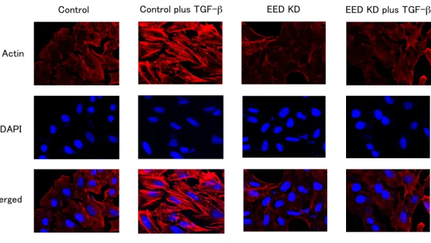
Fig. S5. Knockdown of EED antagonized TGF-β-induced morphological changes of HT29 cells.
2
(A) Cell morphological changes of HT29 cells after TGF-β treatment. Control and EED
3
knockdown HT29 cells were treated without or with TGF-β for 72 hours, and were stained with
4
crystal violet. (B) Immunofluorescence images of cells showing the localization of E-cadherin. The
5
panels of HT29 cells with the same arrangement with (A) were stained with anti-E-cadherin
6
antibody and DAPI. (C) Fluorescence images of cells showing reorganization of actin cytoskeleton.
7
The cells were stained with TRITC-phalloidin (Actin) and DAPI.
8
C
E-cadherin
DAPI
Merged
A
Actin
DAPI
Merged
Control Control plus TGF-!" EED KD EED KD plus TGF-!"
B
Control Control plus TGF-!" EED KD EED KD plus TGF-!"Control Control plus TGF-!" EED KD EED KD plus TGF-!"
1 2 3 4 5 6 7 8
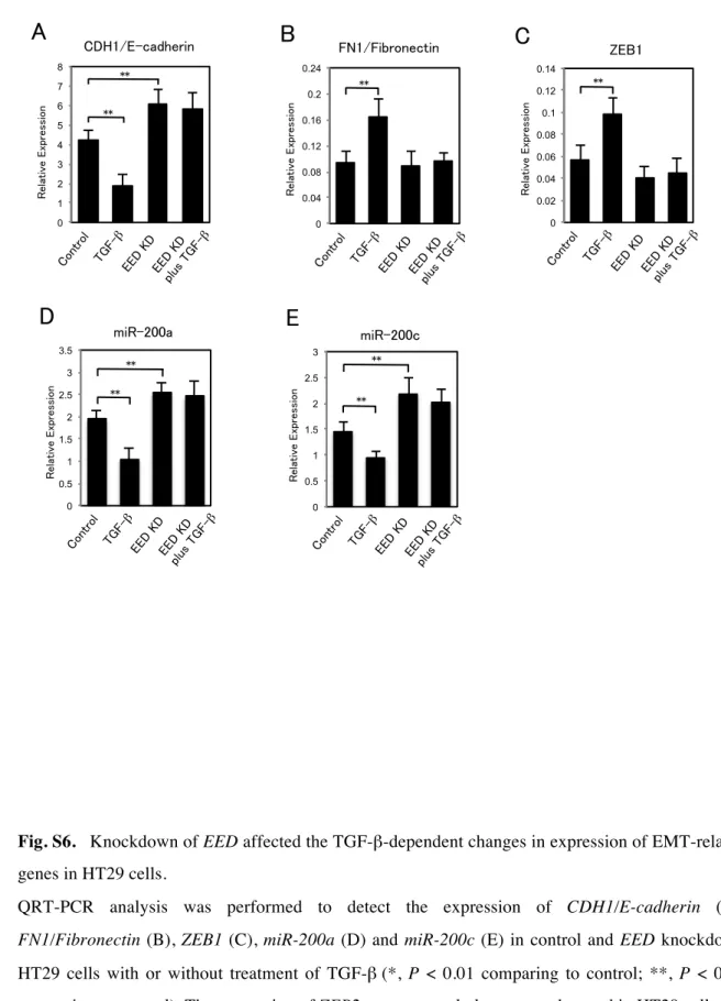
Fig. S6. Knockdown of EED affected the TGF-β-dependent changes in expression of EMT-related
9
genes in HT29 cells.
10
QRT-PCR analysis was performed to detect the expression of CDH1/E-cadherin (A),
11
FN1/Fibronectin (B), ZEB1 (C), miR-200a (D) and miR-200c (E) in control and EED knockdown
12
HT29 cells with or without treatment of TGF-β (*, P < 0.01 comparing to control; **, P < 0.05
13
comparing to control). The expression of ZEB2 was extremely low or not detected in HT29 cells.
14 15
Relative Expression
CDH1/E-cadherin
Relative Expression
FN1/Fibronectin
Relative Expression
A
ZEB1D
B C
E
Relative Expression
miR-200a
Relative Expression
miR-200c
**
**
Control EED
KD EED
KD
plus TGF-! TGF-
!"
**
Control EED
KD EED
KD
plus TGF-! TGF-
!"
**
Cont rol
EED KD
EED KD
plus TGF-! TGF-
!"
0 1 2 3 4 5 6 7 8
0 0.04 0.08 0.12 0.16 0.2 0.24
0 0.02 0.04 0.06 0.08 0.1 0.12 0.14
** **
**
Control EED
KD EED
KD
plus TGF-! TGF-
!"
Control EED
KD EED
KD
plus TGF-! TGF-
!"
0 0.5 1 1.5 2 2.5 3
**
0 0.5 1 1.5 2 2.5 3 3.5