Nucleotide and Amino Acid Sequences in The PreM Region of A Japanese Encephalitis Virus Strain Isolated from A Pool of Aedes albopictus and Ae. butleri Mosquitoes Captured in Peninsula
Malaysia in 1992
Indra VYTHILINGAM1-2, Kouichi MORITA1, and Akira IGARASHI1
1 Department of Virology, Institute of Tropical Medicine, Nagasaki University, 12-4 Sakamoto-1 -chome, Nagasaki City, Japan 852
2 Division of Entomology, Institute for Medical Research, Jalan Pahang, 50588 Kuala Lumpur, Malaysia
Abstract: One strain of Japanese encephalitis (JE) virus isolated from a pool of Ae. albopic- tus and Ae. butleri in Peninsula Malaysia in 1992 was sequenced for its PreM gene region, by direct sequencing the product from reverse transcriptase polymerase chain reaction (RT- PCR). The sequence in the length of 240 nucleotides (nt) and deduced amino acid (AA) se- quence were compared with the published sequences for the various JE virus strains from different geographical regions. This virus strain, MaSAr39692, showed a close homology to the strains from epidemic areas such as Japan, China, Taiwan, Sri Lanka and India. The homology between MaSAr39692 and JaOArS982 strain, for which entire genome sequence was first determined, was 95.8%, with 10 nt and 3 AA sequence divergence.
Key words: Japanese encephalitis virus, PreM gene, nucleotide sequence, amino acid se- quence
INTRODU CTION
Japanese encephalitis (JE) virus is a member of the genus flavivirus, family Flaviviridae (Westaway et al, 1985). All members of the flaviviruses, represented by its prototype yellow fever virus, possess antigenic cross-reactivity, as well as similar physichochemical or molecular characteristics. The virion is the spherical particles of 40 -50 nm diameter with a lipid envelope. They possess 3 structural proteins (C, M, and E) and a single-stranded and positive-sense RNA genome of approximately ll kb in length.
Genomic analysis by olignucleotide fingerprinting showed significant difference among JE virus isolates in geographically distant areas (Hori et al, 1986; Hori, 1986). Relationship amongJE virus isolates was recently analyzed by uncleotide sequencing of PreM gene region of viral RNA (Chen et al, 1990).
The objective of this paper was to study the nucleotide (nt) and deduced amino acid (AA) sequences of the PreM region of a JE virus strain, MaSAr39692, which was isolated from a pool of mosquitoes which were captured in Peninsula Malaysia in 1992.
Received for Publication, June 22, 1994
Contribution No. 3019 from the Institute of Tropical Medicine, Nagasaki University
MATERIALS AND METHODS
Virus: The JE virus strain, MaSAr39692, was isolated from a pool of Ae. albopictus and Ae.
butleri mosquitoes which were captured in Sabak Bernam, Selangor State, Peninsula Malaysia, in 1992, by inoculation to Ae. albopictus, clone C6/36 cell (Igarashi, 1978). The virus strain was passaged 3 times in C6/36 cells at 28°C for 3 days. The infected culture fluid was kept at -70°C as the stock seed virus,
RNA extraction: RNA was extracted from the stock seed virus by a single-step extraction with guanidium thiocyanate and phenol-choloroform mixture as described by Chomczynski and Sacchi (1987).
Rapid RT-PCR: Forty five microliters of H20, 5 pi of 10x buffer and 10 U of RNase in- hibitor (Takara Co., Kyoto, Japan) were added to the tube containing RNA and was vortex- ed. Ten microliters of this template was added to 90 {A of RT-PCR mix containing 100 pmole of each primer, 200 //mole each of 4 deoxynucleotide triphosphates (dNTPs), 10 mM Tris-HCl (pH8.9), 80 mM KC1, 0.5 mg of bovine serum albumin, 0.01% Triton X-100, 10 U of reverse transcriptase (Life Science Inc., UK), 2 U of thermostable Tth DNA polymerase (Toyobo Co., Osaka, Japan), and 10 U of RNase inhibitor. The reaction mixture was overlaid by 2 drops of mineral oil and incubated for 10 minutes at 53°C for reverse transcription reac- tion. PCR amplification was carried out in 35 cycles which comprised of denaturation at 94°C for 1 minutes, annealing at 53°C for 1 minutes and primer extension at 72°C for 1 minutes, in a Thermal Cycler, Iwaki Co., Tokyo, Japan. The last primer extension was prolonged to 5 minutes in order to ensure the chain elongation.
Analysis of amplified DNA product: Seven microliters of PCR product was loaded on a 3% NuSieve 3:1 agarose gel in TAE buffer (40 mM Tris-acetate, pH 8.0, 1 mM EDTA). Gels were stained with ethidium bromide to visualize amplified DNA bands by transilluminator.
DNA sequencing: Seventy microliters of the PCR products were separated from the primers and dNTPs by filtration through a Quick Spin Columns (Boehringer) as described (Tanaka, 1993). Approximately 50-60 //I of the DNA was recovered in a clean tube. Then, the column was again centrifuged to recover DNA completely. The DNA concentration in the specimen was estimated by comparing the optical density of the DNA bands which were visualized by agarose gel electrophoresis to run 1 fA of the specimens with 5 //I of low molecular weight DNA standard marker.
Sequencing was performed using the Taq Dye Deoxy Terminator Cycle Sequencing Kit (Applied Biosystems Inc.). The reaction mixture of 9.5 fA volume was prepared by mixing 4 /A of 5x TACS (sequencing buffer), 1 pi of dNTP mix, 1 pi of each Dye Deoxy N ter- minator (dideoxynucleotide triphosphate fluorescent terminator) and 0.5 /A of Ampli Taq DNA polymerase. To this mixture was added 1.5 fig DNA templates, 3.2 pmole of primer and H20. The reaction mixture was then overlaid with 1 drop of mineral oil, and placed in a Thermal Cycler (Technes, Ltd.). The 25 thermal cyclings were carried out at 96°C 15 sec, 50°C 1 sec, and 60°C 4 minutes.
Twenty microliters of the sequenced reaction mixture was transferred to 1.5 ml tube
containing 2.5 [A of 5% cetyltrimethyl ammoniumbromide (CTAB) and mixed by pipetting, followed by centrifugation at 15,000 rpm for 5 minutes at room temperature. The superna- tant was discarded, and 50 jA of 1.2 M NaCl was added and vortexed briefly to resuspend the pellet. Then, 125 [A of 95% ethanol was added, vortexed and centrifuged at 15,000 rpm for 15 minutes. The supernatant was removed, and the pellet was rinsed with 500 fjtl of 70%
ethanol by vortexing for 2 minutes, followed by centrifugation at 15,000 rpm for 5 minutes at room temperature. The supernatant was removed and the pellet was dried under vacuum.
The dried pellet was resuspended in 6 fA of loading solution (5:1 mixture of deionized formamide: 50 mM EDTA) and the sample was heated at 90°C for 2 minutes, followed by quick cooling on ice. The sample was then loaded onto an Applied Biosystems DNA Se- quencer, Model 373 A according to the instruction of the manufacturer.
RESULTS
The sequence data on 240 nt in PreM gene (nt No. 456 to 695; Sumiyoshi et al, 1987) of the Malaysian JE virus strain MaSAr39692 were compared with the other strains for
Table 1. Nucleotide and amino acid sequence homology (%) of pre-M protein Malaysian strain MaSAr39692 compared to other published works
S tr a i n C o u n t r y N u c le o t i d e s e q u e n c e
A m m o a c i d s e q u e n c e
J a O A r S 9 8 2 J A P A N 9 5 . 8 9 6 . 3
J a G A r J A P A N 9 7 . 5 9 5 . 0
S a g iy a m a J A P A N 9 3 . 8 9 3 . 8
N a k a y a m a J A P A N 9 5 . 0 9 6 . 3
J E (2 ‑ 8 ) C H I N A 9 7 . 9 9 7 . 5
S A ‑ 1 4 C H I N A 9 7 . 5 9 6 . 3
C C ‑ 2 2 3 T A I W A N 9 6 . 3 9 6 . 3
H K 8 2 5 6 T A I W A N 9 7 . 1 9 6 . 3
C C 2 2 3 T A I W A N 9 7 . 1 9 6 . 3
7 5 5 7 2 3 I N D I A 9 5 . 0 9 7 . 5
7 2 4 0 3 8 I N D I A 9 4 . 6 9 6 . 3
6 3 4 9 8 IN D I A 9 4 . 6 9 6 . 3
V N ‑ 1 1 8 V I E T N A M 9 5 . 4 9 6 . 3
P 3 0 7 S R I L A N K A 9 3 . 0 9 5 . 0
M 8 6 4 C A M B O D I A 8 5 . 0 9 1 . 3
M 8 5 9 C A M B O D I A 8 6 . 0 9 3 . 8
B 2 5 8 2 T H A I L A N D 8 5 . 5 9 3 . 8
K E 8 2 0 0 8 T H A I L A N D 8 6 . 0 9 3 .
J E 8 2 7 S A R A W A K 8 6 . 0 9 2 . 5
J K T 1 7 2 4 I N D O N E S I A 8 1 . 0 9 1 . 3 B 1 0 6 5 /8 3 T H A I L A N D 8 4 . 0 9 3 . 8 B 1 0 3 4 /8 3 T H A I L A N D 8 5 . 0 9 3 . 8 W T P /7 0 /2 2 M A L A Y S I A 8 5 . 0 9 5 . 0
similarity. The sequence informations for other JE virus strains were obtained from publica- tion of Chen et al (1990). The MaSAr39692 strain showed 10 nt sequence difference with the JaOArS982 strain for which entire genomic sequence was first determined among JE virus strains (Fig. 1).
The nt differences between MaSAr39692 and other strains were 5-19 nt with the strains from Japan, Taiwan, India and China; 31-36 nt with the strains from Cambodia or Thailand; and 32 -44 nt with the strains from Indonesia or Southern Thailand, respectively.
Therefore, MaSAr96392 belongs to the genogroup of JE virus strains from epidemic areas (Table 1).
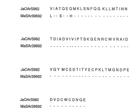
Among 80 AA residues which are encoded by 240 nt sequence in the PreM region of JE virus genome, there were 3 AA divergences between MaSAr39692 and JaOArS982 strains (Fig. 2). The AA sequence divergence between MaSAr39692 and other JE virus strains was 2-7 residues, which is lower than the nt divergence in number (Table 1).
JaOArS982 GUCAUAGCUACGCAGGAGCAAAUGAAGU UGUCGAAUU U
MaSAr39692 C GU C ---C--
JaOArS982 CCAGGGGAAGCUUUUGAUGACCAUCAACAACACGGACA
MaSAr39692 U - -
JaOArS982 UUGCAGACG U UAUCGUGAUUCCCACCUCAAAAGGAGAG MaSAr39692
JaOArS982 AACAGAUGCUGGGUCCGGGCAAUAGACGUCGGCUACAU
MaSAr39692 C - G
JaOArS982 GUGUGAGGACACUAUCACGUACGAAUGUCCUAAGCUCA
MaSAr39692 - -à" '- - U -
JaOArS982 CCAUGGGCAAUGAUCCAGAGGAUGUGGAUUGCUGGUGU MaSAr39692
JaOArS982 GACAACCAAGAA
MaSAr39692 -
Fig. 1. Comparison of the sequences of 240 nucleotides within the pre-M region for JaOArS982 and the Malaysian strain MaSAr39692.
JaOArS982 VIATQEQMKLSNFQG KLLMT1NN
MaSAr39692 L -- S- - H
JaOArS982 TDIADVIVIPTSKGENRCWVRAID MaSAr39692
JaOArS982 VGY MCEDTITYECPKLTMGNDPE
MaSAr39692
JaOArS982 DVDCWCDNQE
MaSAr39692
Fig. 2. Amino acid sequences deduced from the 240 nucleotides used for determining genetic relatedness among JE virus isolates.
DISCUSSION
Burke and Leake (1989) described that epidemics of JE occur in temperate to sub- tropical areas such as Japan, Taiwan, China, Korea, North Vietnam, Northern Thailand, Bur- ma, Nepal, Sir Lanka, and India. While in tropical areas (Malaysia, Indonesia, Southern Thailand, and the Philippines) JE is endemic with relatively samll number of reported cases (Fukunaga et at., 1974). Chen et al (1990) demonstrated that the JE virus strains from the endemic areas, including those from Peninsula Malaysia (JE-827 isolated in 1970) as well as Sarawak (WTP/70/22 isolated in 1968), were genetically different from those of the epidemic areas. However, in our preliminary study, the JE virus strain MaSAr39692, which was isolated from mosquitoes captured in 1992 in Peninsula Malaysia, appears to belong to the genogroup from epidemic areas in Northern or Southern Asia. The result indicates that the JE virus strains circulating in Peninsula Malaysia in recent years are different from those of 20 years ago. Such replacement might have taken place either by introduction of new strains from epidemic areas or by natural mutations throughout 2 decades. The latter possibility ap- pears to be less probable, while the observed difference may be caused by overlooking some epidemic strains which already existed as a minor population 20 years ago.
Vu et al (1993) sequenced 16 JE virus strains isolated in North and South of Vietnam showing that these Vietnamese strains were closely related to the strains from Japan and China. Their result is consistent with ours.
Chen et al (1992) examined 12 JE virus strains from the Indonesian Archipelago show- ing the presence of the 4th genotype of JE virus. Therefore, further studies will be required on JE virus strains isolated from various sources on long-term of observation in order to reveal genomic difference or introduction of new genotypes.
ACKNOWLEDGMENTS
The authors wish to thank Dr. Ismail Mohd Nor, Director of the Institute for Medical Research (IMR), for his permission to publish this paper. The first author was supported by the Japan International Cooperation Agency (JICA) for her travel and stay in Japan under the IMR-JICA Project for the Research on Selected Tropical Diseases in Malaysia, in the fiscal
year 1993.
REFERENCES
1) Burke, D.S. & Leake, CJ. (1988): Japanese encephalitis, pp 63-92. In T. P. Monath (ed.). The Ar- boviruses: Epidemiology and Ecology, Vol. Ill, CRC Press, Boca Raton.
2) Chen, W.-Ru., Tesh, R.B. & Rico-Hesse, R. (1990): Genetic variation of Japanese encephalitis virus in nature. J. Gen. ViroL, 71, 2915-1922.
3) Chen, W.-Ru., Rico-Hesse, R. & Tesh, R.B. (1992): A new genotype of Japanese encephalitis virus from Indonesia. Am. J. Trop. Med. Hyg., 47, 61-69.
4) Chomczynski, P. & Sacchi, N. (1987): Single step method of RNA isolation by guamdium thiocyanate phenol chloroform extraction. Anal. Biochem., 162, 156-159.
5) Fukunaga, T., Rojanasuphot, S., Pisuthipornkul, S., Wungkorbkiat, S., Thammanichanon, A., Chan- tripenkul, P. & Tuchinda, P. (1974): Seroepidemiologic study of arbovirus infection in the northeast and south of Thailand. Biken J., 17, 169-182.
6) Hori, H. (1986): Oligonucleotide fingerprint analysis on Japanese encephalitis (JE) virus strains of dif- ferent geographic origins. Trop. Med., 28, 179-190.
7) Hori, H., Morita, K. & Igarashi, A. (1986): Oligonucleotide fingerprint analysis on Japanese encephalitis virus strains isolated in Japan and Thailand. Acta Virol., 30, 353-359.
8) Igarashi, A. (1978): Isolation of a Singh's Aedes albopictus cell clone sensitive to dengue and chikungunya viruses. J. Gen. Virol., 40, 531-544.
9) Sumiyoshi, H., Mori, C., Fuke, L, Morita, K., Kuhara, S., Kondou, J., Kikuchi, Y., Nagamatsu, H. &
Igarashi, A. (1987): Complete nucleotide sequence of the Japanese encephalitis virus genome RNA.
Virology, 161, 497-510.
10) Tanaka, M. (1993): Rapid identification of flavivirus using the polymerase chain reaction. J. Virol.
Methods, 41, 311-322.
ll) Vu, T.Q.H., Do, Q.H. & Deubel, V. (1993): Genetic studies of Japanese encephalitis viruses from Vietnam. Am. J. Trop. Med. Hyg., 49, 538-544.
12) Westaway, E.G., Brinton, M.A., Gaidamovich, S.Y., Horzinek, M.C., Igarashi, A., Kaariainen, L., Lvov, D.K., Porterfield, J.S., Russell, P.K., & Trent, D.W. (1985): Flaviviridae. Intervirology, 24, 183-192.