Fukushima Medical University
福島県立医科大学 学術機関リポジトリ
This document is downloaded at: 2021-11-07T23:16:46Z
Title Autoantibodies associated with neuropsychiatric systemic lupus erythematosus: the quest for symptom-specific biomarkers
Author(s) Sato, Shuzo; Temmoku, Jumpei; Fujita, Yuya; Yashiro-Furuya, Makiko; Matsuoka, Naoki; Asano, Tomoyuki; Kobayashi, Hiroko; Watanabe, Hiroshi; Migita, Kiyoshi
Citation Fukushima Journal of Medical Science. 66(1): 1-9
Issue Date 2020
URL http://ir.fmu.ac.jp/dspace/handle/123456789/1309
Rights © 2020 The Fukushima Society of Medical Science. This article is licensed under a Creative Commons [Attribution- NonCommercial-ShareAlike 4.0 International] license.
DOI 10.5387/fms.2020-02
Text Version publisher
Vol. 66, No. 1, 2020
[Review]
Autoantibodies associated with neuropsychiatric systemic lupus erythematosus : the quest for symptom
-specific biomarkers
Shuzo Sato, Jumpei Temmoku, Yuya Fujita, Makiko Yashiro
-Furuya, Naoki Matsuoka, Tomoyuki Asano, Hiroko Kobayashi,
Hiroshi Watanabe and Kiyoshi Migita
Department of Rheumatology, Fukushima Medical University School of Medicine, Fukushima, Japan (Received January 14, 2020, accepted January 29, 2020)
Abstract
Systemic lupus erythematosus (SLE) is a chronic autoimmune disease that affects multiple organs, including the central nervous system. Neuropsychiatric SLE (NPSLE) is a severe and potentially fatal condition. Several factors including autoantibodies have been implicated in the pathogenesis of NPSLE. However, definitive biomarkers of NPSLE are yet to be identified owing to the com- plexity of this disease. This is a major barrier to accurate and timely diagnosis of NPSLE. Studies have identified several autoantibodies associated with NPSLE ; some of these autoantibodies are well investigated and regarded as symptom-specific. In this review, we discuss recent advances in our understanding of the manifestations and pathogenesis of NPSLE. In addition, we describe rep- resentative symptom-specific autoantibodies that are considered to be closely associated with the pathogenesis of NPSLE.
Key words: autoantibody, biomarker, neuropsychiatric systemic lupus erythematosus, pathogene- sis, symptom-specific
1. Introduction
Systemic lupus erythematosus (SLE) is a chronic autoimmune disease that affects multiple or- gans, including the central nervous system (CNS)
1). SLE is characterized by the loss of immune toler- ance to nuclear antigens due to development of au- toantibodies and immune complex formation ; the resultant complement activation results in cell de- struction and tissue injury
1,2). Patients with SLE show considerable variability with respect to symp- toms. In particular, neuropsychiatric (NP) SLE is a severe and potentially fatal condition that is charac- terized by CNS manifestations
1,2). In 1999, the American College of Rheumatology Research Com- mittee developed standard nomenclature and case definitions for 19 manifestations of NPSLE (Table 1)
3). NP manifestations usually occur in the early
stage of SLE and 39%
-50% of patients exhibit SLE symptoms. The reported prevalence of NPSLE varies widely from 4%
-91% ; this may be attribut- able to variability in clinical presentations, different selection criteria, and heterogeneity among study populations
4-6). In a recent 3
-year prospective study of 370 SLE patients with no history of CNS in- volvement (excluding non
-specific minor CNS com- plaints and peripheral nervous system symptoms), the prevalence of major CNS events was 4.3% with an estimated incidence of 7.8 events/100 person
-years
7). In spite of recent advances, the diagnosis of NPSLE is typically challenging, due in part to the absence of specific and reliable laboratory or imaging biomarkers
2,8). The diagnosis of NPSLE requires exclusion of other causes such as infection, concur- rent disease, metabolic abnormalities, or drug ad- verse events
1,2,5,6,8). Therefore, investigation of dis-
Corresponding author : Shuzo Sato E-mail : [email protected]©2020 The Fukushima Society of Medical Science. This is an open-access article distributed under the terms of the Creative Commons Attribution-NonCommercial-ShareAlike 4.0 International License (CC-BY-NC-SA 4.0).
https://creativecommons.org/licenses/by-nc-sa/4.0/
1
2 S. Sato et al.
ease
-specific or symptom
-specific autoantibodies observed in NPSLE is a key imperative to facilitate timely and accurate diagnosis. Here, we review re- cent NPSLE studies that focused on the pathogene- sis of NPSLE and the autoantibodies associated with NPSLE manifestations, especially symptom
-specific autoantibodies.
2. Pathogenesis of NPSLE
The pathogenesis of NPSLE is highly complex, with detailed mechanisms yet to be elucidated
1,2,6). The pathogenic pathways suggested thus far include blood
-brain barrier (BBB) dysfunction, vascular oc- clusion, neuroendocrine
-immune imbalance, and tis- sue and neuronal damage caused by autoantibodies and proinflammatory cytokines [interleukin (IL)
-1, IL
-6, IL
-8, IL
-10, IL
-17, type 1 interferons, tumor necrosis factor, colony
-stimulating and macrophage
-stimulating factors]
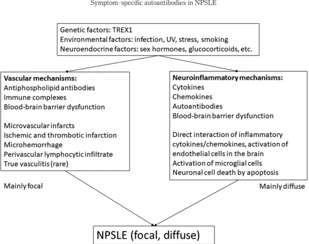
1,2,6,8,9). Predisposing genetic factors, neuroendocrine factors, and environmental factors are also thought to be important. Figure 1 illustrates the currently proposed pathogenetic mechanisms of NPSLE
1,2,6).
2.1 Predisposing factors
Predisposing genetic factors are believed to play an important role in SLE pathogenesis
2,6). In NPSLE patients, mutations in TREX1, which en- codes three
-prime repair exonuclease 1 (DNAse III), were shown to be associated with neurological involvement. Polymorphisms in TREX1 were
found to be associated with neurological involve- ment in European patients with SLE
10). Loss
-of function mutations in TREX1 augment the produc- tion of type 1 interferons in mice and lead to early
-onset cerebral NPSLE
11,12). In addition, HLA
-DRB1*04 and rs10181656(G) alleles were shown to be associated with ischemic cardiovascular disease (CVD) in Caucasian patients with SLE
13,14). More recently, Rullo et al. reviewed recent advances in our understanding of the genetic basis of SLE, including genetic variants in recently identified SLE
-associat- ed loci, the immunological pathways affected by these gene products, and the disease manifestations linked to these loci
15). In addition, environmental factors and neuroendocrine factors play an important role in the development of SLE. In previous stud- ies, silica exposure, smoking, oral contraceptives, postmenopausal hormone therapy, and endometrio- sis were found to be risk factors for SLE
16). Indeed, sex steroid hormones (17β
-estradiol, testosterone, prolactin, progesterone, dehydroepiandrosterone) reportedly impact the immune response and the se- verity of disease in SLE ; this also explains the sex disparity in SLE. Other environmental factors, such as ultraviolet radiation, vitamin D, infection (e.g., Epstein
-Barr virus), vaccination, heavy met- als, solvents, and pesticides are considered as risk factors for SLE
6,16).
2.2 Mechanisms of NPSLE
Recent reports have identified two major mech- anisms for the development of NPSLE, i.e., vascular mechanisms and neuroinflammatory mechanisms (Figure 1)
1,2,6). Among the vascular mechanisms, vasculopathy is implicated in CNS damage in pa- tients with NPSLE ; autopsy studies have shown pathological findings of multi
-focal microinfarcts, small
-vessel noninflammatory vasculopathy and oc- clusion, embolism, cortical atrophy, and microhem- orrhages
1,17,18). Anti
-phospholipid antibodies (aPL) and deposition of immune complexes are likely to be associated with these conditions
1,18-20). Injury to large and small blood vessels mediated by aPL initi- ates vascular damage, finally resulting in focal, and in part, diffuse neuropsychiatric events (seizures, cognitive dysfunction, etc.). The second mecha- nism involves autoimmune inflammation mediated by autoantibodies, resulting in increased permeabili- ty of the BBB, intrathecal formation of immune com- plexes, and production of inflammatory mediators (IFN
-α, IL-6, IL
-8, IP
-10, MCP
-1, etc.)
20,21). Direct CNS tissue injury caused by excitatory amino acid toxicity, oxidative stress, plasminogen activator in-
Table 1. Neuropsychiatric syndromes according tothe criteria proposed by the American College of Rheumatology (Reference 3)
Central NPSLE Peripheral NPSLE Aseptic meningitis1 Guillain-Barre syndrome Cerebrovascular disease1 Autonomic neuropathy Demyelinating syndrome1 Mononeuropathy Headache1 Myasthenia gravis Movement disorder1 Cranial neuropathy
Myelopathy1 Plexopathy
Seizure disorders1 Polyneuropathy Acute confusional state2
Anxiety disorder2 Cognitive dysfunction2 Mood disorder2 Psychosis2
1. Focal NPSLE, 2. Diffuse NPSLE
NPSLE : neuropsychiatric systemic lupus erythema- tosus
hibitor 1 (PAI
-1), and matrix metalloproteinase 9 (MMP9) activity have also been suggested
20,22,23). These processes can cause CNS damage by activa- tion of microglial cells and induction of neuronal cell death by apoptosis
1,6), leading to mainly diffuse NSPLE symptoms, such as acute confusional state and psychosis
1,20). As previously described, BBB dysfunction plays an important role in the pathogen- esis of NSPLE. Normally, the brain is immunologi- cally privileged and is sheltered from foreign sub- stances in the circulation. The BBB limits the entry of soluble molecules and cells into brain pa- renchyma and regulates both uptake into and efflux out of the brain
1,20,24). Although the precise mecha- nism of BBB dysfunction is still unclear, permeabili- ty of the BBB can be affected by both SLE factors (immune complex deposition, cytokine/chemokines) and non
-SLE factors (smoking or hypertension) that induce endothelial dysfunction in brain vascula- ture
1,8,20,25). In this regard, autoantibodies reacting with neuronal cells or those that have been reported as specific for each NPSLE symptom (from the cir- culation or intrathecal production) might be associ- ated with BBB dysfunction. Here, we review the representative autoantibodies that are potentially as- sociated with the pathogenesis of NPSLE.
3. Autoantibodies potentially associated with specific NPSLE symptoms
Table 2 shows the representative autoantibod- ies that have been recently described as potentially associated with NPSLE pathogenesis. More than 100 autoantibodies have been described in patients with SLE or NPSLE
26); however, none of these have been definitively implicated in the complex process of NPSLE pathogenesis. Therefore, exten- sive research is ongoing to establish distinct patho- genic roles for each autoantibody.
3.1 Anti
-phospholipid antibodies (aPL)
The aPL antibody family targets proteins asso- ciated with anionic phospholipids in the plasma membrane that regulate the blood clotting cas- cade ; subsequent activation of procoagulants pro- motes thrombosis and cerebral infarction
27). Anti
-cardiolipin (aCL), anti
-β2GP1 antibodies, and lupusanticoagulant (LAC) are the most widely investigat- ed autoantibodies targeting phospholipids. These have been recognized as major risk factors for NPSLE and are believed to contribute to the devel- opment of thrombosis and other NPSLE symptoms, such as seizures, stroke, chorea, movement disor-
Fig. 1. Pathogenic mechanisms of neuropsychiatric systemic lupus erythematosus (NPSLE)Abbreviations : UV, ultraviolet
4 S. Sato et al.
ders, cognitive dysfunction, and myelopathy
28-32). In vitro studies have demonstrated direct binding of aPL with CNS cells ; in addition, intrathecal passive transfer of IgG isolated from aPL
-positive patients was shown to induce cognitive dysfunction in mice
33). At least, exacerbation of procoagulant state by aPL is believed to be associated with focal NPSLE causing intravascular thrombosis and cere- bral ischemia
20).
3.2 Anti
-ribosomal P protein antibodies (anti
-ribo P) Anti
-ribo P are specific autoantibodies occur- ring in up to 46% of patients ; target epitopes are located in the C
-terminal end of three highly con- served phosphorylated proteins, P0, P1, and P2, which are present in the 60S subunit of ribo- somes
6,34). Many retrospective studies have sug- gested an association between elevated serum or CSF levels of anti
-ribo P and NPSLE manifesta- tions ; however, the results have been contest- ed
35-37). Recent longitudinal studies and prospec- tive studies have shown their association with lupus psychosis
38-41). In a study by Hanly et al., anti
-ribo P was found to be a predictor of psychosis
42). In a mouse model, anti
-ribo P recognized neurons in the hippocampus, cingulate, and primary olfactory piri- form cortex, and induced long
-term depressive
-like behavior when introduced into cerebral ventri- cles
43). Matus et al. reported that anti
-ribo P from psychiatric lupus induced a rapid and sustained in-
crease in calcium reflux and apoptosis in rat neurons expressing cell
-surface P
-antigen protein P331.
The death of these neurons in specific brain regions (such as hippocampus) was found to affect the mem- ory and emotional behavior of rats
44). Direct evi- dence of a pathogenic role for these antibodies in humans is still lacking ; nevertheless, experimental data and prospective studies support the role of anti
-ribo P in the causation of diffuse NPSLE.
3.3 Anti
-N
-methyl
-D
-aspartate receptor antibodies (anti
-NMDA)
Anti
-NMDA antibodies occur in 30%
-40% of patients with SLE ; these have been demonstrated as a subset of double
-stranded DNA (dsDNA) anti- bodies that cross
-react with NMDA receptors, spe- cifically with the NMDA receptor subunit 2 (NR2)
45). NMDA receptors are widely distributed in the brain and localized within glutamatergic synapses ; a par- ticularly high density is observed in the amygdala and hippocampus, which modulate cognitive func- tion, emotional processes, and memory
46,47). Acti- vation of NMDA receptors is critical in learning and memory ; however, prolonged stimulation can cause apoptotic death of neuronal cells. The potential pathogenic role of anti
-NMDA detected in CSF has been demonstrated in both in vitro and in vivo stud- ies
48,49). Interestingly, no neuronal damage was ob- served if the BBB remained intact ; however, sever- al pathological changes were detected when the
Table 2. Representative autoantibodies associated with NPSLETarget of autoantibodies
(autoantibodies) Serum/CSF Prevalence in
SLE patients Associated NPSLE symptoms Phospholipid :
β2-glycoprotein 1 and cardiolipin (aCL-Ab)
Serum, CSF Up to 45% Focal NPSLE (CVD, seizures, chorea)
Diffuse NPSLE (cognitive dysfunction, psychosis, depression, headache)
Ribosomal P protein Serum, CSF 6%-46% Elevated titers in active SLE
(anti-ribosmal P Ab) Diffuse NPSLE (psychosis, depression)
NMDA receptor subtype 2
(anti-NMDA/NR2 Ab) Serum, CSF 30%-40% Diffuse NPSLE (depression cognitive dysfunction) MAP-2 (anti-MAP-2 Ab) Serum, CSF 17%, 33.3% (CSF) Focal NPSLE (seizures, chorea, sensory neuropathy)
Diffuse NPSLE (psychosis, headache) U1 ribonucleoprotein
(Anti-U1RNP Ab)
Structural endothelial proteins (AECA)
Serum, CSF Serum
18% (CSF) 17-75%
NPSLE in general Psychosis, depression
TPI (anti-TPI Ab) Serum, CSF 30%-40% Focal NPSLE (aseptic meningitis) Less frequent in acute confusional state
GAPDH (anti-GAPDH Ab) Serum 47% Increased intracranial pressure, cognitive dysfunc- tion
AECA : anti-endothelial cell antibody ; aCL : anti-cardiolipin ; CVD : cerebrovascular disease ; CSF : cerebrospinal fluid ; GAPDH : glyceraldehyde-3-phosphate dehydrogenase ; MAP-2 : microtubule-associated protein 2 ; NMDA/
NR2 : N-methyl-D-aspartate receptor 2 ; NPSLE : neuropsychiatric systemic lupus erythematosus ; U1-RNP : U1 ri bonucleoprotein ; TPI : triosephosphate isomerase
BBB was disrupted
46-48). Correlation between CSF anti
-NMDA and diffuse NPSLE manifestations has also been reported
50). Furthermore, it has been suggested that anti
-NR2/dsDNA antibodies may also help distinguish SLE patients with central diffuse NPSLE manifestations from patients with peripheral manifestations ; the pathogenic factors and mecha- nisms underlying these manifestations are probably different
51). Then, how do these autoantibodies gain access to the brain in SLE? Yoshio et al. have reported that anti
-NR2/dsDNA antibodies from SLE patients activate endothelial cells and induce expres- sion of surface molecules [intracellular adhesion molecule 1 (ICAM
-1) and vascular cell adhesion molecule 1 (VCAM
-1)] as well as the production of IL
-6 and IL
-8
52). Hirohata et al. also reported that the severity of BBB damage plays a critical role in the development of diffuse NPSLE (acute confusion- al state) through the accelerated entry of larger amounts of anti
-NR2 antibodies into the CNS
53). These results indicate that activation of BBB endo- thelial cells by anti
-NMDA may cause inflammation, disrupt the BBB, and promote entry of autoantibod- ies into the CSF in patients with SLE.
3.4 Anti
-microtubule
-associated protein 2 antibodies (anti
-MAP
-2)
MAP
-2 is one of the abundant groups of cyto- skeletal components predominantly expressed in neurons
54). MAP
-2 regulates the nucleation and stabilization of microtubules, organelle transport protein kinases that are involved in signal transduc- tion
55). In a study by Yamada et al., 33.3% of NPSLE patients tested positive for anti
-MAP
-2 in the CSF
54). Williams also reported that 17% of SLE pa- tients had anti
-MAP
-2 in contrast to 4% of neuro- logic injury/disease control patients
56). Patients who tested positive for anti
-MAP
-2 exhibited neu- ropsychiatric symptoms (psychosis, seizures, neu- ropathy, and cerebritis). Moreover, both anti
-ribo P titers and IL
-6 levels in CSF were significantly higher in NPSLE patients having anti
-MAP
-2, indi- cating some association between them
54).
3.5 Anti
-U1 ribonucleoprotein antibodies (anti
-U1RNP)
Anti
-U1RNP reacts with proteins that are asso- ciated with U1 RNA and form U1 small nuclear ribo- nucleoprotein (snRNP) ; these are detectable in 25%
-47% of SLE patients
57). The snRNP are RNA
-protein complexes that are abundant in the nucleus ; these are involved in the nuclear process- ing of pre
-mRNA along with other proteins compris-
ing the spliceosome
58). In a study by Sato et al., an- ti
-U1RNP in CSF, but not that in serum, was associated with NPSLE and mixed connective tissue disease
59). Recent studies have shown that anti
-RNP as well as anti
-Sm antibodies were less likely to be produced within the CNS ; this was assessed by calculating anti
-RNP and anti
-Sm indices in the CSF (indices of each reflect the intrathecal produc- tion of these antibodies)
60). Nevertheless, the de- tailed role of anti
-U1RNP should be further investi- gated.
3.6 Anti
-endothelial cell antibodies (AECA)
AECAs target a heterogenous group of antigens including structural endothelial proteins (ranging from 10 to 200 kDa) as well as adhesion molecules to endothelial cells ; these are found in a variety of diseases that are characterized by vessel wall dam- age
61). The reported prevalence of AECA in SLE patients ranges from 17% to 75%. Conti et al.
found an association of serum AECA with psychosis and depression in NPSLE
62). AECA can activate endothelial cells by inducing the expression of adhe- sion molecules (ICAM
-1 and VCAM
-1) and stimu- lating the production of cytokines (IL
-1, IL
-8) and chemokines [such as monocyte
-chemotactic protein 1 (MCP
-1)]. AECA can also enhance the produc- tion of tissue factor and von Willebrand factor, pro- moting thrombosis. However, low specificity due to the lack of a standardized detection method and possible presence of natural AECAs (seen in a small percentage of healthy individuals and showing low affinity for their target antigens) limit the use of AE- CAs as diagnostic and prognostic markers
61,62). 3.7 Anti
-triosephosphate isomerase antibodies (anti
-TPI)
Triosephosphate isomerase (TPI) is an impor-
tant glycolytic enzyme that catalyzes the intercon-
version of dihydroxyacetone phosphate and D
-glyc-
eraldehyde
-3
-phosphate
63). TPI is mediated solely
by glycolysis in red blood cells and in brain cells, and
its deficiency is associated with hemolytic anemia
and neurological disorders. TPI is involved in the
stability of neuronal microtubules
63-66). Watanabe et
al. have suggested some association with the patho-
genesis of NPSLE
64-66). Furthermore, in our previ-
ous study, anti
-TPI
-TPI immune complex was de-
tected in the CSF of anti
-TPI
-positive NPSLE
patients
65). In a lupus model mouse, anti
-TPI was
detected in MRL/lpr mice ; in addition, anti
-TPI
was shown to bind to brain tissue in the meninges,
choroid plexus, hippocampus, and periventricular le-
6 S. Sato et al.
sions using anti
-TPI
-producing hybridoma inocula- tion into the brain hemisphere
66). Recently, we re- ported a higher frequency of aseptic meningitis in anti
-TPI
-positive NPSLE patients ; in addition, se- rum anti
-TPI index showed a positive correlation with serum IgG levels
8). These results indicate that anti
-TPI may be associated with NPSLE, main- ly focal NPSLE (aseptic meningitis) ; the underlying mechanism may involve disruption of the BBB via formation of immune complexes in the CNS. In- deed, the underlying mechanism of aseptic meningi- tis in SLE patients is still unclear. A possible mechanism can be considered along with meningeal inflammation similar to that proposed for multiple sclerosis
67): immune cells first pass through the meninges via the bloodstream to the choroid plexus, as a part of immune surveillance. The interaction of autoimmune myelin
-specific T cells with myelin
-loaded antigen
-presenting cells can induce T cell re- activation and production of inflammatory cytokines/
chemokines in the meninges. The additional in- flammatory cells compromise local BBB integrity, which finally triggers immune cell infiltration into the CNS
8,67). In any case, further investigation is needed to clarify the pathogenic role of anti
-TPI in NPSLE.
3.8 Anti
-glyceraldehyde
-3
-phosphate dehydrogenase antibodies (anti
-GAPDH)
GAPDH is a glycolytic enzyme ; however, more recent evidence indicates that mammalian GAPDH performs a number of other functions
68). Its activity contributes to membrane fusion, micro- tubule bundling, phosphotransferase activity, nuclear RNA export, DNA replication and repair, and further, to neuronal cell death
68,69). Takasaki et al. first re- ported that anti
-GAPDH is one of the elements of proliferating cell nuclear antigens, specifically those reactive with serum from SLE patients
69). Further- more, Delunardo et al. demonstrated the reaction of anti
-GAPDH with neuronal cells and its association with cognitive dysfunction in patients with SLE
70). Recently, Sun et al. also reported a positive correla- tion of serum anti
-GAPDH with SLEDAI
-2K, ESR, IgG, and IgM ; in addition, anti
-GAPDH showed an association with increased intracranial pressure, in- dicating its potential role in the induction of brain tissue damage
71). Although the direct effect of anti
-GAPDH is yet to be elucidated, it may serve as a useful biomarker for cerebrovascular damage in NPSLE patients.
4. Conclusion
Neuropsychiatric sequelae are among the main causes of morbidity and mortality in patients with SLE, but they are the least well
-understood aspect of the disease. Appropriate evaluation and accurate classification of NP manifestations is an important focus of NPSLE treatment and research. However, its complex pathogenesis and polymorphic pheno- type hampers the identification of pertinent, robust biomarkers. Further investigation of biomarkers (such as disease
-specific or symptom
-specific auto- antibodies and cytokine/chemokine expression pro- files) is required to increase our knowledge and im- prove the management of NPSLE.
Conflict of interest
The authors declare there is no conflict of inter- est.
References
1. Jeltsch-David H, Muller S. Neuropsychiatric sys- temic lupus erythematosus : pathogenesis and biomarkers. Nat Rev Neurol, 10: 579-596, 2014. doi : 10.1038/nrneurol.2014.148.
2. Magro-Checa C, Zirkzee EJ, Huizinga TW, Steup- Beekman GM. Management of Neuropsychiatric Systemic Lupus Erythematosus : Current Ap- proaches and Future Perspectives. Drugs, 76: 459-483, 2016. doi : 10.1007/s40265-015- 0534-3.
3. [No authors listed]. The American College of Rheumatology nomenclature and case definitions for neuropsychiatric lupus syndromes. Arthritis Rheum, 42: 599-608, 1999.
4. Hanly JG, Urowitz MB, Sanchez-Guerrero J, et al. Neuropsychiatric events at the time of diagno- sis of systemic lupus erythematosus : an interna- tional inception cohort study. Arthritis Rheum, 56: 265-273, 2007.
5. Bertsias GK, Boumpas DT. Pathogenesis, diag- nosis and management of neuropsychiatric SLE manifestations. Nat Rev Rheumatol, 6: 358-367, 2010. doi : 10.1038/nrrheum.2010.62.
6. Govoni M, Bortoluzzi A, Padovan M, et al. The di- agnosis and clinical management of the neuropsy- chiatric manifestations of lupus. J Autoimmun, 74: 41-72, 2016. doi : 10.1016/j.jaut.2016.06.013.
7. Kampylafka EI, Alexopoulos H, Kosmidis ML, et al. Incidence and prevalence of major central ner- vous system involvement in systemic lupus erythematosus : a 3-year prospective study of 370
p a t i e n t s . P L o S O n e , 8: e 5 5 8 4 3 , 2 0 1 3 . doi : 10.1371/journal.pone.0055843.
8. Sato S, Yashiro M, Asano T, Kobayashi H, Wata- nabe H, Migita K. Association of anti-triosephos- phate isomerase antibodies with aseptic meningitis in patients with neuropsychiatric systemic lupus erythematosus. Clin Rheumatol, 36: 1655-1659, 2017. doi : 10.1007/s10067-017-3653-2.
9. McGlasson S, Wiseman S, Wardlaw J, Dhaun N, Hunt DPJ. Neurological Disease in Lupus : To- ward a Personalized Medicine Approach. Front Immunol, 9: 1146, 2018. doi : 10.3389/fimmu.
2018.01146. eCollection 2018.
10. Namjou B, Kothari PH, Kelly JA, et al. Evaluation of the TREX1 gene in a large multi-ancestral lupus cohort. Genes Immun, 12: 270-279, 2011.
doi : 10.1038/gene.2010.73.
11. Ellyard JI, Jerjen R, Martin JL, et al. Identification of a pathogenic variant in TREX1 in early-onset cerebral systemic lupus erythematosus by Whole- exome sequencing. Arthritis Rheumatol, 66: 3382-3386, 2014.
12. Stetson DB, Ko JS, Heidmann T, Medzhitov R. Trex1 prevents cell-intrinsic initiation of auto- i m m u n i t y. C e l l , 1 3 4 : 5 8 7-5 9 8 , 2 0 0 8 . doi : 10.1016/j.cell.2008.06.032.
13. Lundström E, Gustafsson JT, Jönsen A, et al.
HLA-DRB1*04/*13 alleles are associated with vascular disease and antiphospholipid antibodies in systemic lupus erythematosus. Ann Rheum Dis, 72: 1018-1025, 2013. doi : 10.1136/annrheum- dis-2012-201760.
14. Svenungsson E, Gustafsson J, Leonard D, et al. A STAT4 risk allele is associated with ischaemic cerebrovascular events and anti-phospholipid anti- bodies in systemic lupus erythematosus. Ann Rheum Dis, 69: 834-840, 2010. doi : 10.1136/
ard.2009.115535.
15. Rullo OJ, Tsao BP. Recent insights into the ge- netic basis of systemic lupus erythematosus. Ann R h e u m D i s , 7 2 S u p p l 2 : i i 5 6-6 1 , 2 0 1 3 . doi : 10.1136/annrheumdis-2012-202351.
16. Barbhaiya M, Costenbader KH. Environmental exposures and the development of systemic lupus erythematosus. Curr Opin Rheumatol, 28: 497- 505, 2016. doi : 10.1097/BOR.0000000000000318.
17. Rhiannon JJ. Systemic lupus erythematosus in- volving the nervous system : presentation, patho- genesis, and management. Clin Rev Allergy Im- munol, 34: 356-360, 2008. doi : 10.1007/
s12016-007-8052-z.
18. Hanly JG, Walsh NM, Sangalang V. Brain patholo- gy in systemic lupus erythematosus. J Rheuma- tol, 19: 732-741, 1992.
19. Mehta N, Uchino K, Fakhran S, et al. Platelet C4d is associated with acute ischemic stroke and
stroke severity. Stroke, 39: 3236-3241, 2008.
doi : 10.1161/STROKEAHA.108.514687.
20. Hanly JG. Diagnosis and management of neuro- psychiatric SLE. Nat Rev Rheumatol, 10: 338- 347, 2014. doi : 10.1038/nrrheum.2014.15.
21. Yoshio T, Okamoto H, Kurasawa K, Dei Y, Hirohata S, Minota S. IL-6, IL-8, IP-10, MCP-1 and G- CSF are significantly increased in cerebrospinal fluid but not in sera of patients with central neuro- psychiatric lupus erythematosus. Lupus, 25: 997-1003, 2016. doi : 10.1177/09612033166 29556.
22. Ainiala H, Hietaharju A, Dastidar P, et al. In- creased serum matrix metalloproteinase 9 levels in systemic lupus erythematosus patients with neu- ropsychiatric manifestations and brain magnetic resonance imaging abnormalities. Arthritis Rheum, 50: 858-865, 2004.
23. Kwiecin´ski J, Kłak M, Trysberg E, Blennow K, Tarkowski A, Jin T. Relationship between elevat- ed cerebrospinal fluid levels of plasminogen activa- tor inhibitor 1 and neuronal destruction in patients with neuropsychiatric systemic lupus erythemato- sus. Arthritis Rheum, 60: 2094-2101, 2009.
doi : 10.1002/art.24603.
24. Abbott NJ, Patabendige AA, Dolman DE, Yusof SR, Begley DJ. Structure and function of the blood- brain barrier. Neurobiol Dis, 37: 13-25, 2010.
doi : 10.1016/j.nbd.2009.07.030.
25. Gelb S, Stock AD, Anzi S, Putterman C, Ben-Zvi A. Mechanisms of neuropsychiatric lupus : The relative roles of the blood-cerebrospinal fluid barrier versus blood-brain barrier. J Autoimmun, 91: 34- 44, 2018. doi : 10.1016/j.jaut.2018.03.001.
26. Sherer Y, Gorstein A, Fritzler MJ, Shoenfeld Y. Autoantibody explosion in systemic lupus erythematosus : more than 100 different antibod- ies found in SLE patients. Semin Arthritis Rheum, 34: 501-537, 2004.
27. Harris EN, Pierangeli S. Antiphospholipid anti- bodies and cerebral lupus. Ann N Y Acad Sci, 823: 270-278, 1997.
28. Sanna G, Bertolaccini ML, Cuadrado MJ. Neuro- psychiatric manifestations in systemic lupus erythematosus : prevalence and association with antiphospholipid antibodies. J Rheumatol, 30: 985-992, 2003.
29. Hanly JG. Attribution in the assessment of ner- vous system disease in SLE. Rheumatology (Ox- ford), 54: 755-756, 2015. doi : 10.1093/rheuma- tology/keu458.
30. Andrade RM, Alarcón GS, González LA, et al. Sei- zures in patients with systemic lupus erythemato- sus : data from LUMINA, a multiethnic cohort (LUMINA LIV). Ann Rheum Dis, 67: 829-834, 2008.
8 S. Sato et al.
31. Mok MY, Chan EY, Fong DY, Leung KF, Wong WS, Lau CS. Antiphospholipid antibody profiles and their clinical associations in Chinese patients with systemic lupus erythematosus. J Rheumatol, 32: 622-628, 2005.
32. McLaurin EY, Holliday SL, Williams P, Brey RL. Predictors of cognitive dysfunction in pa- tients with systemic lupus erythematosus. Neu- rology, 64: 297-303, 2005.
33. Katzav A, Ben-Ziv T, Blank M, Pick CG, Shoenfeld Y, Chapman J. Antibody-specific behavioral effects : in- tracerebroventricular injection of antiphospholipid anti- bodies induces hyperactive behavior while anti-ribo- somal-P antibodies induces depression and smell deficits in mice. J Neuroimmunol, 272: 10-15, 2014. doi : 10.1016/j.jneuroim.2014.04.003.
34. Mahler M, Kessenbrock K, Raats J, Williams R, Fritzler MJ, Blüthner M. Characterization of the human autoimmune response to the major C-ter- minal epitope of the ribosomal P proteins. J Mol Med (Berl), 81: 194-204, 2003.
35. Nojima Y, Minota S, Yamada A, Takaku F, Aotsuka S, Yokohari R. Correlation of antibodies to ribosom- al P protein with psychosis in patients with sys- temic lupus erythematosus. Ann Rheum Dis, 51: 1053-1055, 1992.
36. Haddouk S, Marzouk S, Jallouli M, et al. Clinical and diagnostic value of ribosomal P autoantibodies in systemic lupus erythematosus. Rheumatology (Oxford), 48: 953-957, 2009. doi : 10.1093/
rheumatology/kep142.
37. Karassa FB, Afeltra A, Ambrozic A, et al. Accura- cy of anti-ribosomal P protein antibody testing for the diagnosis of neuropsychiatric systemic lupus erythematosus : an international meta-analy- sis. Arthritis Rheum, 54: 312-324, 2006.
38. Bonfa E, Golombek SJ, Kaufman LD, et al. Asso- ciation between lupus psychosis and anti-ribosomal P protein antibodies. N Engl J Med, 317: 265- 271, 1987.
39. West SG, Emlen W, Wener MH, Kotzin BL. Neu- ropsychiatric lupus erythematosus : a 10-year prospective study on the value of diagnostic tests. Am J Med, 99: 153-163, 1995.
40. Watanabe T, Sato T, Uchiumi T, Arakawa M. Neu- ropsychiatric manifestations in patients with sys- temic lupus erythematosus : diagnostic and pre- dictive value of longitudinal examination of anti- ribosomal P antibody. Lupus, 5: 178-183, 1996.
41. Briani C, Lucchetta M, Ghirardello A, et al. Neurolu- pus is associated with anti-ribosomal P protein antibodies : an inception cohort study. J Autoimmun, 32: 79-84, 2009. doi : 10.1016/j.jaut.2008.12.002.
42. Hanly JG, Urowitz MB, Siannis F, et al. Autoanti- bodies and neuropsychiatric events at the time of systemic lupus erythematosus diagnosis : results
from an international inception cohort study. Ar- thritis Rheum, 58: 843-853, 2008. doi : 10.1002/
art.23218.
43. Katzav A, Solodeev I, Brodsky O, et al. Induction of autoimmune depression in mice by anti-ribo- somal P antibodies via the limbic system. Arthri- tis Rheum, 56: 938-948, 2007.
44. Matus S, Burgos PV, Bravo-Zehnder M, et al. An- tiribosomal-P autoantibodies from psychiatric lu- pus target a novel neuronal surface protein causing calcium influx and apoptosis. J Exp Med, 204: 3221-3234, 2007.
45. Lauvsnes MB, Omdal R. Systemic lupus erythe- matosus, the brain, and anti-NR2 antibodies. J Neurol, 259: 622-629, 2012. doi : 10.1007/
s00415-011-6232-5.
46. Kowal C, DeGiorgio LA, Nakaoka T, et al. Cogni- tion and immunity ; antibody impairs memo- ry. Immunity, 21: 179-188, 2004.
47. Huerta PT, Kowal C, DeGiorgio LA, Volpe BT, Dia- mond B. Immunity and behavior : antibodies al- ter emotion. Proc Natl Acad Sci U S A, 103: 678-683, 2006.
48. DeGiorgio LA, Konstantinov KN, Lee SC, Hardin JA, Volpe BT, Diamond B. A subset of lupus anti- DNA antibodies cross-reacts with the NR2 gluta- mate receptor in systemic lupus erythemato- sus. Nat Med, 7: 1189-1193, 2001.
49. Husebye ES, Sthoeger ZM, Dayan M, et al. Auto- antibodies to a NR2A peptide of the glutamate/
NMDA receptor in sera of patients with systemic lupus erythematosus. Ann Rheum Dis, 64: 1210-1213, 2005.
50. Arinuma Y, Yanagida T, Hirohata S. Association of cerebrospinal fluid anti-NR2 glutamate receptor antibodies with diffuse neuropsychiatric systemic lupus erythematosus. Arthritis Rheum, 58: 1130-1115, 2008. doi : 10.1002/art.23399.
51. Fragoso-Loyo H, Cabiedes J, Orozco-Narváez A, et al. Serum and cerebrospinal fluid autoantibodies in patients with neuropsychiatric lupus erythema- tosus. Implications for diagnosis and pathogene- sis. PLoS One, 3: e3347, 2008. doi : 10.1371/
journal.pone.0003347.
52. Yoshio T, Okamoto H, Hirohata S, Minota S. IgG anti-NR2 glutamate receptor autoantibodies from patients with systemic lupus erythematosus acti- vate endothelial cells. Arthritis Rheum, 65: 457-463, 2013. doi : 10.1002/art.37745.
53. Hirohata S, Arinuma Y, Yanagida T, Yoshio T.
Blood-brain barrier damages and intrathecal syn- thesis of anti-N-methyl-D-aspartate receptor NR2 antibodies in diffuse psychiatric/neuropsychological syndromes in systemic lupus erythematosus. Ar- thritis Res Ther, 16: R77, 2014. doi : 10.1186/
ar4518.
54. Yamada Y, Nozawa K, Nakano S, et al. Antibodies to microtubule-associated protein-2 in the cerebro- spinal fluid are a useful diagnostic biomarker for neuropsychiatric systemic lupus erythemato- sus. Mod Rheumatol, 26: 562-568, 2016. doi : 10.3109/14397595.2015.1123345.
55. Sánchez C, Díaz-Nido J, Avila J. Phosphorylation of microtubule-associated protein 2 (MAP2) and its relevance for the regulation of the neuronal cyto- skeleton function. Prog Neurobiol, 61: 133-168, 2000.
56. Williams RC Jr, Sugiura K, Tan EM. Antibodies to microtubule-associated protein 2 in patients with neuropsychiatric systemic lupus erythemato- sus. Arthritis Rheum, 50: 1239-1247, 2004.
57. Migliorini P, Pratesi F, Tommasi C, Anzilotti C. The immune response to citrullinated anti- gens in autoimmune diseases. Autoimmun Rev, 4: 561-564, 2005.
58. D e m a B , C h a r l e s N . Au t o a n t i b o d i e s i n SLE : Specificities, Isotypes and Receptors. Anti- bodies (Basel), 5. pii : E2, 2016. doi : 10.3390/
antib5010002.
59. Sato T, Fujii T, Yokoyama T, et al. Anti-U1 RNP antibodies in cerebrospinal fluid are associated with central neuropsychiatric manifestations in systemic lupus erythematosus and mixed connective tissue disease. Arthritis Rheum, 62: 3730-3740, 2010.
doi : 10.1002/art.27700.
60. Hirohata S, Sakuma Y, Yanagida T, Yoshio T. As- sociation of cerebrospinal fluid anti-Sm antibodies with acute confusional state in systemic lupus ery- thematosus. Arthritis Res Ther, 16: 450, 2014.
doi : 10.1186/s13075-014-0450-z.
61. Perricone C, Pendolino M, Olivieri M, Conti F, Valesini G, Alessandri C. Neuropsychiatric mani- festations associated with anti-endothelial cell an- tibodies in systemic lupus erythematosus. Isr Med Assoc J, 17: 171-178, 2015.
62. Conti F, Alessandri C, Bompane D, et al. Autoan- tibody profile in systemic lupus erythematosus with psychiatric manifestations : a role for anti-en- dothelial-cell antibodies. Arthritis Res Ther, 6: R366-372, 2004.
63. Orosz F, Wágner G, Liliom K, et al. Enhanced as- sociation of mutant triosephosphate isomerase to red cell membranes and to brain microtu- bules. Proc Natl Acad Sci U S A, 97: 1026-1031, 2000.
64. Watanabe H, Seino T, Sato Y. Antibodies to trios- ephosphate isomerase in patients with neuropsy- chiatric lupus. Biochem Biophys Res Commun, 321: 949-953, 2004.
65. Sasajima T, Watanabe H, Sato S, Sato Y, Ohira H. Anti-triosephosphate isomerase antibodies in cerebrospinal fluid are associated with neuropsy- chiatric lupus. J Neuroimmunol, 181: 150-156, 2006.
66. Sato S, Watanabe H, Shio K, Kobayashi H, Ohira H. Association of anti-triosephosphate isomerase antibody and MRL/MpJ-Faslpr mouse. J Neuro- immunol, 226: 110-115, 2010. doi : 10.1016/
j.jneuroim.2010.05.041.
67. Russi AE, Brown MA. The meninges : new ther- apeutic targets for multiple sclerosis. Transl Res, 165: 255-269, 2015. doi : 10.1016/j.trsl.2014.08.005 68. Sirover MA. Role of the glycolytic protein, glyc- eraldehyde-3-phosphate dehydrogenase, in normal cell function and in cell pathology. J Cell Bio- chem, 66: 133-140, 1997.
69. Takasaki Y, Kaneda K, Matsushita M, et al. Glyc- eraldehyde 3-phosphate dehydrogenase is a novel autoantigen leading autoimmune responses to pro- liferating cell nuclear antigen multiprotein com- plexes in lupus patients. Int Immunol, 16: 1295- 1304, 2004.
70. Delunardo F, Soldati D, Bellisario V, et al. Anti- GAPDH Autoantibodies as a Pathogenic Determi- nant and Potential Biomarker of Neuropsychiatric Diseases. Arthritis Rheumatol, 68: 2708-2716, 2016. doi : 10.1002/art.39750.
71. Sun J, Li X, Zhou H, et al. Anti-GAPDH Autoanti- body Is Associated with Increased Disease Activity and Intracranial Pressure in Systemic Lupus Ery- thematosus. J Immunol Res, 2019: 7430780, 2019. doi : 10.1155/2019/7430780. eCollection 2019.