Postoperative Changes in Splenic Volume after Spleen-preserving Distal Pancreatectomy with Conservation of the Splenic Artery and
Vein, and after Digestive Tract and Breast Surgery
*
Department of Gastroenterological, General Breast and Thyroid Surgery, Yamagata University Faculty of Medicine
**
Department of Surgery, Gyoda General Hospital
( Accepted March 9, 2018)
Koji Tezuka * , Wataru Kimura * , Ichiro Hirai * , Toshihiro Watanabe * , Shuichiro Sugawara * , Tsuyoshi Fukumoto ** , Shinji Okazaki * , Mitsuhiro Yano * , Osamu Hachiya * , Akihiko Suzuki *
Aim: There is a paucity of information about changes in splenic volume after surgery. The aim of this study was to investigate postoperative changes in splenic volume(SV)and the factors influencing SV after spleen-preserving distal pancreatectomy(SPDP)with conservation of the splenic artery and vein
(CSAV), and after surgery of the digestive tract and breast.
Methods: We investigated 113 patients who underwent SPDP with CSAV(n=7), breast surgery
(n=24), colorectal surgery(n=45), distal gastrectomy(n=27)and total gastrectomy(n=10). SV changes were determined for three years after surgery using volumetry based on computed tomographic imaging, and splenic vein diameter changes after SPDP with CSAV were also determined.
Results: Splenic vein diameter after SPDP with CSAV did not change during 3 years. SV did not change significantly during 3 years after SPDP with CSAV and distal gastrectomy. After breast and colorectal surgery, and total gastrectomy, SV was decreased.
Conclusions: Postoperative SV changes differed according to the type of surgery. SV did not change significantly during 3 years after SPDP with CSAV.
Key words: splenic volume, spleen-preserving distal pancreatectomy, SPDP, digestive surgery, breast surgery
Introduction
The spleen is the largest peripheral lymphoid organ and plays an important role in immune function
1).Overwhelming post-splenectomy infection (OPSI) occurs in 3.2% of all splenectomized patients
2).A method of spleen-preserving distal pancreatectomy (SPDP) with conservation of the splenic artery and vein (CSAV) that emphasized the importance of splenic vein dissection from the pancreatic body to the tail was first reported by Kimura et al
3).Another way of preserving blood flow to the spleen in SPDP is preservation of the
short gastric and left gastroepiploic vessels with excision of the splenic vessels,which is called Warshaw’s procedure
4).Warshaw’ s procedure is technically easier but recently the importance of preserving the splenic vessels in SPDP has been widely recognized
5).
Recently,it has been reported that a small spleen
(<1.1 cm
3/kg) in patients who do not undergo splenectomy is a risk factor for severe infection
6). Therefore,preservation of splenic function is very important in pancreatic surgery.No standard method for assessment of splenic function has yet been established,although some authors have proposed that the appearance of Howell-Jolly bodies ABSTRACT
DOI 10.15022/00004454
Tezuka, Kimura, Hirai, Watanabe, Sugawara, Fukumoto, Okazaki, Yano, Hachiya, Suzuki
in peripheral blood,or measurement of splenic volume (SV),could be useful in this respect
7).
To date,few reports have documented changes in SV after pancreatic surgery,including SPDP with CSAV,and other forms of surgery.
The aim of the present study was to determine the changes in SV after SPDP with CSAV,and after digestive tract and breast surgery,using an image-processing workstation based on computed tomography (CT) imaging data,and to reveal the possible factors that influence postoperative changes in SV.
Materials and Methods
The present study was based on 129 patients who underwent surgery at Yamagata University Hospital between June 2006 and May 2008.Seven patients underwent SPDP with CSAV,29 underwent breast surgery,54 underwent colorectal surgery,29 underwent distal gastrectomy and 10 underwent total gastrectomy.Patients who had conditions possibly influencing SV,such as liver cirrhosis
(n=3),infections at the time of CT scan (n=2),
postoperative hematological disorder (n=1),
portal vein tumor thrombosis due to postoperative recurrence (n=1),and postoperative oxaliplatin chemotherapy (n=2) within 3 years after surgery were excluded.Patients who underwent surgery involving other intra-abdominal organs concomitantly or within 3 years after surgery were also excluded
(n=7).Accordingly,we investigated a final total of 113 patients who underwent SPDP with CSAV
(n=7),breast surgery (n=24),colorectal surgery
(n=45),distal gastrectomy (n=27) and total gastrectomy (n=10).
We verified the preoperative and follow-up
(1,2 and 3 years after surgery) postcontrast CT images of all patients.Data pertaining to pre- and postoperative clinical characteristics [age at surgery,sex,preoperative body weight,BMI,
SV and SV/BMI ratio,pre- and postoperative laboratory data such as white blood cell count,
platelet count,red blood cell count,albumin,and C-reactive protein (CRP) level,surgical procedure,
pathology,stage,adjuvant chemotherapy and
hormonal therapy,and postoperative body weight change] were collected retrospectively from the hospital charts.The staging of each cancer was defined in accordance with the 7th edition of the American Joint Committee on Cancer (AJCC)
staging criteria
8).
SPDP with CSAV was performed for low-grade malignant lesions in the body or tail of the pancreas without obvious stromal invasion or lymph node metastasis,as judged based on imaging modalities and operative findings.For the other operations,
the surgical procedures were selected based on tumor location,size,depth of invasion and lymph node metastasis,and regional lymph node dissections were performed for malignancy.
In the colorectal surgery,distal gastrectomy and total gastrectomy groups,patients with lymph node metastasis received adjuvant chemotherapy with oral fluoropyrimidine drugs for 1–2 years,depending on the situation.
Breast surgery patients received adjuvant chemotherapy for about 6 months after surgery depending on the situation.Female patients with estrogen receptor positive cancer received hormonal therapy with tamoxifen for 5 years and/or LH- RH agonist for 2 years if premenopausal,or with
a Figure 1.
b
c
Fig 1.Splenic volume measurements P.21
The spleen was outlined by manual contouring on a Zio
workstation (a, b). 3-Dimensional computed tomography
images of the spleen were constructed by volume
rendering (c), and the value of SV was calculated
automatically.
aromatase inhibitor for 5 years if postmenopausal.
After adjuvant chemotherapy,patients with estrogen receptor-positive cancer sequentially received hormonal therapy.Postoperative radiotherapy (approximately 50 Gy to the whole breast and an additional 10 Gy for some patients)
was performed for the remnant breast after breast- conserving surgery.
The study was approved by the hospital ethics committee.
Summary of surgical procedures
SPDP with CSAV was performed using Kimura’ s procedure
3).An important aspect of this method is dissection of the splenic vein from the pancreatic body to the tail after mobilization of the spleen and distal pancreas from the retroperitoneum
3).The left gastroepiploic and short gastric vessels were dissected in this cohort.Hand-assisted laparoscopic- SPDP with CSAV was performed using the method reported previously
9),10).For distal gastrectomy,
the left gastroepiploic vessels were dissected.
Splenic volume and splenic vein measurements All CT imaging examinations were performed using multi-detector CT (MDCT) scanners (an Aquilion 64 [Toshiba Medical Systems,Tokyo,
Japan] or a SOMATOM Sensation 32 [Siemens Medical Solutions,Forchheim,Germany]) with a 1-mm slice thickness in the equilibrium phase (120 seconds after initiation of IV contrast injection).SV values were calculated automatically on a Ziostation version 1.17 (Ziosoft Inc.,Tokyo,Japan) using
manual contouring of the spleen.
Splenic vein diameters were measured on axial images on the workstation at the following four points (Fig.2): junction of the splenic vein and portal vein (SV
j); 2 cm distal to the SV
j(SV
j-2);
junction of the splenic vein branches at the splenic hilum (SV
t); 2 cm proximal to the SV
t(SV
t-2).
Statistical analysis
Numerical data are expressed as mean ± standard deviation.Statistical analysis for paired data was done using the Wilcoxon signed rank test.
Categorical variables were analyzed using Fisher’s exact test.Continuous variables were analyzed by Kruskal-Wallis test.Statistical analysis was performed using SAS version 9.4 (SAS Institute Inc.,Cary,NC).Differences at p<0.05 were considered statistically significant.
Results
Pre-and postoperative clinical characteristics of each surgical procedure
Table 1 summarizes the clinicopathological characteristics of the patients.The mean age of the patients overall at the time of surgery was 66.0 ± 11.8 years (range: 35-87).The preoperative mean SV for the patients overall was 117 ± 58 cm
3(range:
27-378 cm
3).
Among the 113 patients,107 (94.7%) had malignant disease.The pathological diagnoses in the patients who underwent SPDP with CSAV included intraductal papillary mucinous neoplasm (IPMN)
with low- or intermediate-grade dysplasia (n=5),
IPMN with an associated invasive carcinoma (n=1)
and pancreatic neuroendocrine neoplasm (n=1).
One patient who underwent SPDP with CSAV received adjuvant chemotherapy with gemcitabine.
Patients in the colorectal surgery group received oral fluoropyrimidine (n=17),and those in the distal gastrectomy (n=4) and total gastrectomy (n=2)
groups received oral fluoropyrimidine.Patients in the breast surgery group received adjuvant hormonal therapy with LH-RH agonist (n=1),LH- RH agonist and tamoxifen (n=2),tamoxifen (n=6),
and aromatase inhibitor (n=7).Recurrence within
SV
jSV
j-22 cm
SV
t-2SV
t 2 cm*
P.22
Fig 2.Splenic vein diameter measurements
Splenic vein diameters were measured on axial images on
the workstation at the following four points: junction of
the splenic vein and portal vein (SV
j); 2 cm distal to the
SV
j(SV
j-2); junction of the splenic vein branches at the
splenic hilum (SV
t); 2 cm proximal to the SV
t(SV
t-2).
Tezuka, Kimura, Hirai, Watanabe, Sugawara, Fukumoto, Okazaki, Yano, Hachiya, Suzuki
3 years was observed in 1 of the 10 patients in the total gastrectomy group.
Postoperative changes in splenic vein diameter of SPDP with CSAV
Postoperative changes in splenic vein diameter after SPDP with CSAV are shown in Table 2.
Splenic vein diameter after SPDP with CSAV was not change significantly during 3 years after surgery.
Postoperative changes in splenic volume
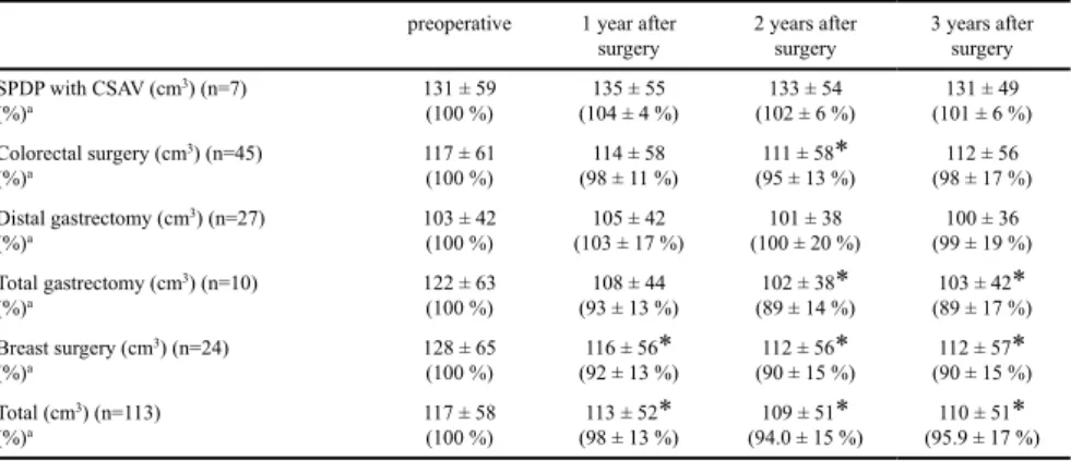
Postoperative changes in SV after each type of surgery are shown in Table 3 and Figure 3.
SV after SPDP with CSAV and distal gastrectomy did not change significantly until 3 years after
surgery.SV after breast surgery was decreased significantly from 1 year after surgery in comparison with the preoperative SV (128±65 vs. 116±56 cm
3,p<0.001).SV after total gastrectomy was decreased significantly from 2 years after surgery in comparison with the preoperative SV (122±63 vs.
102±38 cm
3,p=0.020).In both the breast surgery and total gastrectomy groups,SV reached a plateau from 2 years after surgery.In the colorectal surgery group,SV was decreased significantly at 2 years after surgery in comparison with the preoperative SV (117±61 vs.111±58 cm
3,p=0.006).In the patients overall,SV was decreased significantly from 1 year after surgery (117±58 vs.113±52 cm
3, p=0.006),and reached a plateau from 2 years after surgery.
Table 1. Clinicopathological characteristics of patients
Variable Total (n=113) SPDP with
CSAV (n=7)
Colorectal surgery (n=45)
Distal gastrectomy
(n=27)
Total gastrectomy
(n=10)
Breast surgery
(n=24) P value
Age at surgery 66.0 ± 11.8 67.1 ± 10.8 69.3 ± 9.6 68.1 ± 9.0 67.4 ± 15.0 57.3 ± 13.8 0.017
Sex, male/ female 65 (57.5 %)/
48 (42.5 %) 7 (100 %)/
0 29 (64.4 %)/
16 (35.6 %) 21 (77.8 %)/
6(22.2 %) 8 (80 %)/
2 (20 %) 0/
24 (100 %) <0.001 Preoperative body weight (kg) 58.9 ± 10.6 58.3 ± 6.7 59.2 ± 11.2 58.9 ± 10.6 63.4 ± 13.4 56.5 ± 9.3 0.545 Preoperative BMI (kg/m
2) 23.2 ± 3.4 21.7 ± 2.2 23.2 ± 3.4 22.9 ± 3.4 23.9 ± 3.7 23.8 ± 3.7 0.522
Preoperative SV (cm
3) 117 ± 58 131 ± 59 117 ± 61 103 ± 43 122 ± 63 128 ± 65 0.655
Preoperative SV/BMI ratio 5.0 ± 2.2 6.0 ± 2.4 5.0 ± 2.3 4.5 ± 1.7 5.2 ± 2.9 5.4 ± 2.4 0.514 Preoperative laboratory data
White blood cell count (/μl) Red blood cell count (×10
4/μl) Platelet count (×10
4/μl) Albumin (g/dl) CRP (mg/dl)
5702 ± 1525 424±54 25.4 ± 7.3
4.1 ± 0.5 0.4 ± 0.8
4634 ± 902 424±49 19.6 ± 4.8
4.1 ± 0.2 0.3 ± 0.5
5764 ± 1275 424±56 27.1 ± 7.9
3.9 ± 0.4 0.5 ± 1.0
5727 ± 1650 428±48 25.2 ± 6.4
4.1 ± 0.5 0.4 ± 0.9
5476 ± 1338 438±62 23.5 ± 7.5
4.3 ± 0.6 0.3 ± 0.4
5964 ± 1943 417±56 25.2 ± 6.9
4.2 ± 0.6 0.3 ± 0.5
0.280 0.913 0.079 0.207 0.442 Pathology and Stage
Benign or stage 0/ Ⅰ
Stage Ⅱ/Ⅲ 60 (53.1 %)
53 (46.9 %) 7 (100 %)
0 15 (33.3 %)
30 (66.7 %) 21 (77.8 %)
6 (22.2 %) 6 (60 %)
4 (40 %) 11 (45.9 %) 13 (54.1 %)
<0.001
Adjuvant chemotherapy 35 (30.9%) 1 (14.3 %) 17 (37.8 %) 4 (14.8 %) 2 (20 %) 11 (45.9 %) 0.085
Adjuvant hormonal therapy 16 (14.2 %) 0 0 0 0 16 (66.7 %) <0.001
Radiation therapy 9 (8 %) 0 0 0 0 9 (37.5 %) <0.001
Values are mean ± standard deviation or n (%)
SV splenic volume, SPDP spleen-preserving distal pancreatectomy, CSAV conservation of the splenic artery and vein
a
(Preoperative value - mean BW from 1 to 3 years after surgery)/preoperative value×100 (%)
P.18
Table 1.Clinicopathological characteristics of patients
Table 2. Splenic vein diameter changes after spleen-preserving distal pancreatectomy with conservation of the splenic artery and vein
Table 2. Splenic vein diameter changes after spleen-preserving distal pancreatectomy with conservation of the splenic artery and vein Preoperative
value 1 year after
surgery p value* 2 years after
surgery p value* 3 years after
surgery p value*
SV
j(mm) 6.9 ± 1.7 7.8 ± 1.8 0.578 7.6 ± 1.6 0.469 7.8 ± 1.6 0.469
SV
j-2(mm) 8.2 ± 2.0 8.5 ± 3.0 0.563 8.8 ± 2.6 0.375 8.4 ± 2.1 0.344
SV
t-2(mm) 7.0 ± 1.7 7.3 ± 1.9 0.469 7.5 ± 1.4 0.219 7.3 ± 1.5 0.219
SV
t(mm) 7.0 ± 1.0 7.4 ± 1.1 0.563 7.6 ± 0.8 0.156 7.4 ± 0.8 0.203
Values are mean ± standard deviation
*Comparison between the preoperative splenic vein diameter and splenic vein diameter obtained after surgery by the use of Wilcoxon signed-rank test
P.19
Discussion
Studies from Japanese institutions have reported the mean SV ranging from 112–127 cm
3 11),12).The preoperative mean SV in the present study population was 116 cm
3,and thus within the reported range.
The mean SV in studies from western countries is larger,ranging from 209–219 cm
3 13)-15).Several recent reports have documented changes in splenic volume (SV) in various conditions
11),16)-18).SV is affected by various factors; for example,in adults it is correlated with height
13),weight
13),12),body mass index (BMI) and body surface area
12),and inversely related to age
12),19).It is reported that SV is increased in the presence of higher portal venous
pressure
20),infection
21),inflammatory,hematologic and metabolic disorders
22),cytotoxic chemotherapy including oxaliplatin
16)and certain drugs
23),whereas it is reduced by drugs
23),decreased arterial flow to the spleen
24),and radiation therapy
18).
In this study,SV did not change significantly after SPDP with CSAV.This may be because both blood flow from the splenic artery and venous return through the splenic vein may have been sufficiently preserved.Splenic volume reduction may be a risk factor for severe infection.Therefore,Kimura’ s procedure is excellent in terms of preservation of splenic function.It is reported that 32.4% (12/37)
of patients who underwent Warshaw’ s procedure,
which does not preserve the splenic vessels,had splenic enlargement of more than 20% compared to the preoperative value,at a follow-up CT 6 months after surgery
25).In the present study,splenic enlargement exceeding 20% was not observed after SPDP with CSAV.However,in patients undergoing laparoscopic distal pancreatectomy,
splenic enlargement of more than 20% compared to the preoperative SV was observed in 59% (10/17) of patients treated using Warshaw’s method and 43%
(3/7) of patients in whom the splenic vessels were preserved
17).Splenic venous occlusion after SPDP with CSAV has been described in some reports
26), but in the present study no obvious splenic vein occlusion or stenosis was observed.Splenic vein patency may influence the postoperative SV.Careful
Table 3. Splenic volume change after surgery
preoperative 1 year after
surgery 2 years after
surgery 3 years after surgery SPDP with CSAV (cm
3) (n=7)
(%)
a131 ± 59
(100 %) 135 ± 55
(104 ± 4 %) 133 ± 54
(102 ± 6 %) 131 ± 49 (101 ± 6 %) Colorectal surgery (cm
3) (n=45)
(%)
a117 ± 61
(100 %) 114 ± 58
(98 ± 11 %) 111 ± 58 *
(95 ± 13 %) 112 ± 56 (98 ± 17 %) Distal gastrectomy (cm
3) (n=27)
(%)
a103 ± 42
(100 %) 105 ± 42
(103 ± 17 %) 101 ± 38
(100 ± 20 %) 100 ± 36 (99 ± 19 %) Total gastrectomy (cm
3) (n=10)
(%)
a122 ± 63
(100 %) 108 ± 44
(93 ± 13 %) 102 ± 38 *
(89 ± 14 %) 103 ± 42 *
(89 ± 17 %) Breast surgery (cm
3) (n=24)
(%)
a128 ± 65
(100 %) 116 ± 56 *
(92 ± 13 %) 112 ± 56 *
(90 ± 15 %) 112 ± 57 *
(90 ± 15 %) Total (cm
3) (n=113)
(%)
a117 ± 58
(100 %) 113 ± 52 *
(98 ± 13 %) 109 ± 51 *
(94.0 ± 15 %) 110 ± 51 *
(95.9 ± 17 %) Values are mean ± standard deviation
SPDP spleen-preserving distal pancreatectomy, CSAV conservation of the splenic artery and vein,
a
(splenic volume at each point/preoperative splenic volume)×100 (%)
*P<0.05; Comparison between the preoperative splenic volume and splenic volume obtained after surgery by the use of Wilcoxon signed-rank test
P.20
Table 3.Splenic volume change after surgery
95 100 105 110 115 120 125 130 135 140
1 2 3 4
140
130
120
110
100
pre-ope. 1 years 2 years 3 years
SPDP with CSAV Breast surgery Colorectal surgery Distal gastrectomy Total gastrectomy
0