-8-
Examination of catheter movement
Changes in the position of the internal jugular catheter tip due to head movement : a retrospective study
Department of Anesthesiology, Yamagata University Faculty of Medicine
(Accepted July 16, 2019)
Misato Kurota, Kaneyuki Kawamae
Background: Endotracheal tube displacement due to head and neck movement has been well described. However, no similar study has been conducted with regard to the effect of head movement on internal jugular catheters. We hypothesized that head movement would also affect the internal jugular catheter tip position. This study aimed to determine whether head movement affects the internal jugular catheter tip position in pediatric patients less than 6 years old.
Method: Twenty-four pediatric patients(aged 0-6 y)undergoing elective cardiac surgery were enrolled. Radiography was performed in both neutral and left head rotation positions. Using radiographs, we measured the change in distance between the catheter tip and the level of the upper edge of the 12th vertebra. Changes in catheter tip position between the neutral and left head rotation position were analyzed using a paired t-test.
Results: There was no statistically significant displacement(P = 0.3). The maximum displacement of the catheter tip position was 2.42 cm.
Conclusion: Our data showed that left head rotation did not significantly displace the catheter tip position in most cases. However, in some cases, head movement caused catheter advancement or withdrawal, which could lead to severe complications.
Keywords: pediatrics, jugular vein, displacement, catheter tip
Introduction
Central venous catheter(CVC)placement provides reliable venous access for medications as well as measurement of the hemodynamic status. Several complications are associated with CVC.1)-3)Among them, catheter tip displacement can sometimes cause lethal outcomes. Catheter tip displacement can occur in either direction.
Advancement of the catheter causes mechanical complications such as vascular perforation and cardiac tamponade,4)whereas withdrawal of the catheter increases the risk of thrombosis and infection and can result in insufficient administration of medication.
Increased intrathoracic pressure due to coughing and sneezing, decreased intrathoracic pressure due to tracheal extubation, changing the body position, or physical movement is thought to be the cause of catheter tip displacement.4),5)However, the exact mechanism of displacement remains unclear.
However, endotracheal tube displacement due to head movement has been well described.6)Based on that, we hypothesized that head movement would also affect the internal jugular catheter tip position.
The risk is particularly high in pediatric patients as they are more likely to move and be uncooperative.
The present study aimed to determine whether head movement affects the internal jugular catheter tip position in pediatric patients.
ABSTRACT Yamagata Med J (ISSN 0288-030X)2020;38(1):8-11
DOI 10.15022/00004783
-9-
Kurota, Kawamae
Materials and Methods
This retrospective study was performed at a single center. All pediatric patients(aged 0-6 y)
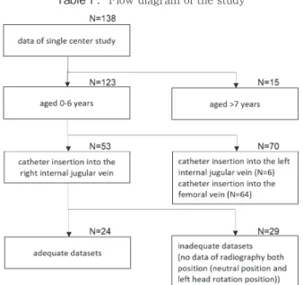
undergoing elective cardiac surgery at Yamagata University Hospital(Yamagata, Japan)between January 2014 and October 2017 were included in this study. Table 1 shows a flow diagram of the study. Patients who underwent catheter insertion into the right internal jugular vein and who had undergone radiographic examination in both neutral and left head rotation positions were enrolled. Using radiographs, one physician measured the change
in distance between the catheter tip and upper edge level of the 12th vertebra(Figure 1). Studies have measured the distance between tracheal bifurcation and catheter tip position. In this study, tracheal bifurcation could not be identified due to the influence of thymus or mediastinal shadow depending on the case. Therefore, the upper edge of the 12th vertebral, which is easy to identify, was used as a reference without shifting the position due to breathing or head position conversion.
Data are expressed as mean±SD(range). A clinically significant displacement was defined as a displacement of >1 cm. This minimal clinically important displacement was selected on the basis
2
Figure.1
Figure.2 2
Figure.1
Figure.2
Figure 1. Method of measuring the distance from the 12 th vertebral upper edge to the tip of the catheter tip.
Figure 3. The number of cases by catheter movement distance.
Figure 2. Individual displacements of the catheter tip from the neutral position. In these box-and- whisker plots, the box boundaries display the first and third quartiles, and the whiskers represent the minimum and maximum values.
The horizontal line within the box represents the median value.
Figure 4. Scatter diagrams between the distance of the catheter movement and the patient’s height.
-10-
Examination of catheter movement
that the length of catheter retention is shorter in children than in adults.7)Changes in the catheter tip position from the neutral position were analyzed using the paired t test. P value < .05 was considered as statistically significant.
Because of the anonymous nature of data, the requirement for informed consent was waived. Study approval was obtained from the institutional review board of the Yamagata University Hospital(No.2018- 145).
Results
Twenty-four pediatric patients were enrolled in this study(Table 2). All patients underwent insertion of a 17-G double lumen catheter under ultrasound guidance. The depth of CVC insertion was determined using the surface landmark method.
Radiographs were obtained in the operating room and intensive care unit. The extent of individual displacement of the catheter tip from the neutral position is shown in Figure 2. The catheter tip moved toward the upper edge of the 12th vertebra by 0.18±0.89(–1.58–2.42)cm(P = 0.3). However, there was no statistically significant catheter tip displacement. The number of cases with catheter movement displacement and distance of displacement are shown in Figure 3. Clinically significant catheter
tip displacement(>1 cm)occurred in 25%(6/24)
of patients. The maximum displacement of the catheter tip position was 2.42 cm. The displacement occurred in an unpredictable manner (advancement in 4 patients and withdrawal in 2). There were no complications associated with catheter displacement.
Scatter diagrams between the distance of catheter displacement and patient’s height is shown in Figure 4. There was no correlation between the height and distance of catheter displacement(r=0.03).
Discussion
This retrospective study investigated whether head movement was related to internal jugular catheter tip migration in pediatric patients(aged 0-6 y). The results showed that left head rotation did not significantly affect the internal jugular catheter tip position in pediatric patients. However, in some cases, left head rotation displaced the catheter tip to over 1 cm.
The mechanism of catheter tip displacement remains unclear. Most existing studies on CVC displacement are case reports.3),4)A previous prospective randomized study has shown that decreased intrathoracic pressure due to tracheal extubation was not associated with a significant postoperative CVC tip displacement in adult patients.8)Our results showed that left head rotation did not significantly affect the internal jugular catheter tip position. However, the result that cannot be missed is that catheter tip displacement of >1 cm due to left head rotation was observed in 25%
of patients. Pediatric patients, especially infants and young children, have difficulty in keeping the
Table.1 Flow diagram of the study
Table 2. Patients characteristics
Age (year) 1.0 ± 1.1 (0-5.3) Weight (kg) 7.8 ± 3.9 (4.2-20.2) Height (cm) 69.3 ± 13.8 (53-110.8) Sex (M/F) 14 / 10
Data are presented as mean ± SD (range) and number
Table.1 Flow diagram of the study
Table 2. Patients characteristics
Age (year) 1.0 ± 1.1 (0-5.3) Weight (kg) 7.8 ± 3.9 (4.2-20.2) Height (cm) 69.3 ± 13.8 (53-110.8) Sex (M/F) 14 / 10
Data are presented as mean ± SD (range) and number Table1.Flow diagram of the study Table2.Patients characteristics
-11-
Kurota, Kawamae
head at rest, and the length of catheter retention is shorter in these patients than in adult patients.7)
The catheter indwelling length for patients aged
<0–6 y is approximately 4–8 cm, and considering the subcutaneous indwelling length, the distance actually indwelled in the blood vessel is considered short. For the reasons stated above, we speculate that even >1 cm of catheter tip displacement may be important from a clinical point of view for pediatric patients.
The relationship between height and catheter displacement was not shown in this study. Previous studies have reported that head rotation >45°
increases the overlap between the internal jugular vein and common carotid artery.9)This anatomical shift due to head rotation may have influenced catheter tip displacement in some cases. In contrast, we cannot deny the possibility that differences in the puncture position and fixation method of the catheter, which we did not investigate in this study, could have influenced catheter tip displacement.
This study has several limitations. Data collection was performed by a single unblinded observer, which may have introduced bias. In addition, we collected data regarding CVC insertions only into the right internal jugular vein, which is the usual site of insertion. We noted a few cases wherein the CVC was inserted into the left internal jugular vein, but we could not investigate those cases. Finally, the left head rotation angle was not uniform, head flexion and extension were likely present in some cases, and puncture position was not constant. In clinical practice, it is impossible for the head movement to be limited to a certain angle or to puncture at the same place at all times. Therefore, it can be said that our result is based on actual clinical practice.
Conclusion
Our data showed that left head rotation did not significantly displace the catheter tip position in pediatric patients. However, in some cases, left head rotation caused catheter advancement or withdrawal, which could lead to severe complications. Physicians and nurses who perform perioperative catheter management should keep in mind that changing the head position may sometimes causes the catheter
to move. Detailed analysis of factors that can affect catheter displacement should be performed in a further study.
This research was presented at the European Anesthesiology Congress in 2018.
Acknowledgement
We would like to thank Editage(www.editage.jp)
for English language editing.
References
1. Malbezin S, Gauss T, Smith I, Bruneau B, Mangalsuren N, Diallo T, et al. : A review of 5434 percutaneous pediatric central venous catheters inserted by anesthesiologists. Paediatr Anaesth 2013;
23: 974-979
2. Miller JA, Singireddy S, Maldjian P, Baker SR:
A reevaluation of the radiographically detectable complications of percutaneous venous access lines inserted by four subcutaneous approaches. Am Surg 1999; 65: 125-130
3. Prabaharan B, Thomas S: Spontaneous migration of central venous catheter tip following extubation. Saudi J Anaesth 2014; 8: 131-133
4. Kim YJ, Kim WY: Sudden hypotension occurring after 4 days of left-sided central catheter placement. J Thorac Dis 2017; 9: E771-E773
5. Rasuli P, Hammond DI, Peterkin IR: Spontaneous intrajugular migration of long-term central venous access catheters. Radiology 1992; 182: 822-824
6. Kim JT, Kim HJ, Ahn W, Kim HS, Bahk JH, Lee SC, et al. : Head rotation, flexion, and extension alter endotracheal tube position in adults and children. Can J Anaesth 2009; 56: 751-756
7. Andropoulos DB, Bent ST, Skjonsby B, Stayer SA:
The optimal length of insertion of central venous catheters for pediatric patients. Anesth Analg 2001; 93:
883-886
8. Struck MF, Jünemann T, Reinhart K, Schummer W:
Central venous catheter tip migration due to tracheal extubation: a prospective randomized study. J Clin Monit Comput 2017; 31: 951-959
9. Ozcelik M, Guclu C, Meco B, Oztuna D, Kucuk A, Yalcin S, et al. : The airway device preference may affect the overlapping of the common carotid artery by the internal jugular vein. Paediatr Anaesth 2016; 26:
1148-1156