DIRECT EFFECT OF PERCUTANEOUS TRANSLUMINAL ANGIOPLASTY ON THE HEART IN PATIENT WITH
PERIPHERAL ARTERY DISEASE
Yasunori Oguma1, Kenji Iino1, Hiroyuki Watanabe1, Toshimitsu Kosaka1, Hitoshi Hasegawa2 and Hiroshi Ito1
(Received 14 January 2009, Accepted 21 January 2009)
1Division of Cardiovascular and Respiratory Medicine, Department of Internal Medicine, Akita University School of Medicine, Akita 010-8543, Japan
2Department of Community Medicine and Primary Care Development, Akita University School of Medicine, Akita 010-8543, Japan
Abstract
Background and Purpose : The patient with peripheral artery disease (PAD) is known to have a poor prognosis for life. Recently, it has been reported that percutaneous transluminal angioplas- ty (PTA) for PAD improves the prognosis. However, the detailed mechanisms remain unclear.
Augmentation index (AIx) or central aortic pressure (CAP) is shown to increase myocardial me- chanical stress and facilitate myocardial hypertrophy. The aim of this study is to clarify the direct cardioprotective effects of PTA mediated by decreasing CAP and AIx.
Methods : 28 patients with PAD were enrolled. They were divided into two groups ; control group (n=12) and PTA group (n=16). Both radial AIx and CAP were measured by using pulse waveform analysis of arterial waveform recorded non-invasively by applanation tonometry in radial artery. We used a plasma B-type natriuretic peptide (BNP) level as a marker of myocardial mechanical stress. Plasma BNP levels, radial AIx and CAP which were measured after PTA or angiography, were compared with the data on admission.
Result : Although plasma BNP levels in the control group were not changed, those in the PTA group were significantly decreased from 68.44 to 42.08 pg/ml (p<0.05). Pulse waveform analysis demonstrated that radial AIx and CAP in the control group remain unchanged. On the other hand, radial AIx and CAP were significantly reduced in the PTA group. The reduction rates of radial AIx, CAP and plasma BNP levels were −11.3%, −8.9% and −34% respectively.
Conclusion : These results suggest that PTA can decrease myocardial mechanical stress in pa- tients with PAD. The mechanisms may be mediated in part by reduction of radial AIx and CAP.
Key words : peripheral artery disease, percutaneous transluminal angioplasty, B-type natri- uretic peptide, augmentation index, central aortic pressure
Introduction
It has been known that peripheral artery disease (PAD) is a predictor of high mortality1). Patients with PAD in lower extremities have a three- to six-fold increased rate of cardiovascular mortality compared with those without Correspondence : Hiroshi Ito, M.D., Ph.D.
Department of Cardiovascular and Respiratory Medicine, Akita University Graduate School of Medicine, 1-1-1 Hondo, Akita 010-8543, Japan
Tel : 81-18-884-6110 Fax : 81-18-836-2612
E-mail : hito@doc.med.akita-u.ac.jp
PAD . As a great advance in recent years in the technique of catheter intervention, percutaneous transluminal angioplasty (PTA) emerged as a novel therapeutic option for PAD. PTA for PAD is shown to improve quality of life of the patients with PAD3). In addition, successful PTA enables PAD patients to reduce oxidative stress caused by repeated muscle ischemia or increase shear stress due to improved ambulatory activi- ty. Although these effects are contributive factors for the improvement of their mortality4), the direct cardioprotec- tive effects of PTA remains unclear.
Recently, a lot of attention has given to augmentation index (AIx) which is defined as an increase in pressure from the first systolic shoulder to the peak pressure of the aortic pressure waveform expressed as a percentage of peak pressure. AIx is not only the index of wave reflectionsbut also a predictor of cardiovascular complications5). The central aortic pressure wave is composed of a forward traveling wave generated by left ventricular ejection and a later-arriving reflected wave from the periphery. As arterial stiffness and stenosis progress, the transmission velocity of both forward and reflected waves increases, causing the reflected wave to arrive earlier in the central aorta and to augment the central blood pressure in late systole6). Augmentation of the central aortic pressure wave is thus a manifestation of wave reflection. Recently, similar information to central pressure wave reflection can be obtained from radial artery pressure wave form as radial AIx7).
In this study, we hypothesized that PTA decreases CAP or AIx and results in reduction of cardiac mechanical stress.
Methods Patient population
Among the patients referred to our Akita University Hospital between February 2007 and October 2008, we included in this study those admitted with PAD consecu- tively. Patients who did not undergo angiography or had renal dysfunction (serum creatinine >2.0 mg/dl) were excluded. Plasma B-type natriuretic peptide (BNP) level were measured before and after PTA. In 28 individuals (3 women and 25 men) who had symptomatic PAD in the
stages Fontaine class II or III and low Ankle-Brachial Index (ABI ; unilateral or bilateral ABI <0.9), 16 of patients underwent PTA. The remaining 12 patients underwent only cardiac catheterization. The objectives and protocols of the study were fully explained, and informed consent was obtained from all the patients.
AIx, CAP and ABI measurements
AIx, CAP, and brachial BP were assessed non-invasively by using the commercially available applanation tonometry device HEM-9000AI (Omron Healthcare Co., Ltd., Kyoto, Japan). Peripheral pressure waveform was recorded over 30 seconds from the radial artery at the wrist with the subject after more than 5 min of rest in a sitting position.
Simultaneously, brachial BP was measured at the opposite arm. The system software allowed on-line recording of the peripheral waveforms, which was assessed visually to ensure that the best possible recordings were obtained and that artifacts from movement were minimized. The peak point of the forward wave and reflected wave were automatically identified using fourth derivatives for each radial arterial waveform and averaged as previously described in order to measure the radial AIx8). As AIx is influenced by the heart rate, an index normalized for a heart rate of 75 bpm was calculated in accordance with Wilkinson et al.9). The reproducibility and reliability of pulse wave analysis by HEM-9000AI have been established by previous reports7). ABI was measured in a supine position using vascular testing device (form PWV/ABI BP-203PRE II ; Omron Health care Co., Ltd., Kyoto, Japan) which simultaneously measured electrocardiograms, bilateral brachial and ankle BPs. We measured brachial BP, radial AIx, CAP, ABI, and plasma BNP levels before and after PTA or angiography of lower limb artery.
Angiography and PTA
The left side of the heart was catheterized from the radial artery in all individuals to evaluate coronary artery and bilateral lower limb artery. PTA was performed for 16 patients by an antegrade or an over-the-bifurcation approach with the use of 6- or 8-French sheaths. After angiography was performed to evaluate the structure of the lesion and runoff, patients were undergone angioplasty with stent implantation. All Stents were
placed from the common iliac artery to the superficial femoral artery.
Statistics
The data were expressed as the mean±standard error of the mean. Percent change of radial AIx, CAP and BNP in the control and the PTA group were compared statisti- cally using the t-test. The paired t-test was used to analyze the difference in the percent change of radial AIx, CAP and BNP between before and after PTA. Values of p<0.05 were considered to be statistically significant.
Result Patient characteristics
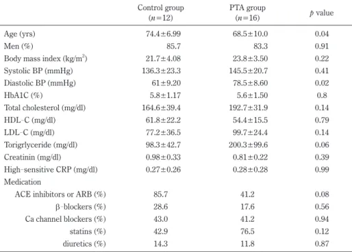
The control group included 12 individuals and the PTA group consisted of 16 individuals. As shown in table 1, the control and the PTA group did not differ significantly in gender, BMI, systolic brachial BP, HbA1c, cholesterol,
triglyceride, serum creatinine levels, high sensitive C-reactive protein, but diastolic BP was significantly higher in the PTA group. The use of angiotensin-con- verting enzyme inhibitors, angiotensin receptor blockers, β-blockers, calcium channel blockers, statins and diuret-
ics were similar between the two groups.
PTA decreases cardiac mechanical stress
BNP plays a regulatory role in the homeostasis of body fluids and blood pressure10). The stretch of cardiomyo- cytes is the most important stimulus of BNP secretion.
Many studies have shown that BNP levels increase in response to hemodynamic parameters, such as left ven- tricular end-diastolic pressure, pulmonary capillary wedge pressure, ejection fraction, and LV volume11). Therefore, BNP is a well recognized marker for cardiac mechanical stress. To investigate the direct effects of PTA on cardiac function, we analyzed the plasma BNP level before and after treatment.
Table 1. Patient characteristics Control group
(n=12)
PTA group
(n=16) p value
Age (yrs) 74.4±6.99 68.5±10.0 0.04
Men (%) 85.7 83.3 0.91
Body mass index (kg/m2) 21.7±4.08 23.8±3.50 0.22
Systolic BP (mmHg) 136.3±23.3 145.5±20.7 0.41
Diastolic BP (mmHg) 61±9.20 78.5±8.60 0.02
HbA1C (%) 5.8±1.17 5.6±1.50 0.8
Total cholesterol (mg/dl) 164.6±39.4 192.7±31.9 0.14
HDL-C (mg/dl) 61.8±22.2 54.4±15.5 0.79
LDL-C (mg/dl) 77.2±36.5 99.7±24.4 0.14
Torigrlyceride (mg/dl) 98.3±42.7 200.3±99.6 0.06
Creatinin (mg/dl) 0.98±0.33 0.81±0.22 0.39
High-sensitive CRP (mg/dl) 0.27±0.26 0.28±0.28 0.99
Medication
ACE inhibitors or ARB (%) 85.7 41.2 0.08
β-blockers (%) 28.6 17.6 0.56
Ca channel blockers (%) 43.0 41.2 0.94
statins (%) 42.9 76.5 0.12
diuretics (%) 14.3 11.8 0.87
Values are mean±SE. HDL-C=high density lipoprotein cholesterol ; LDL-C=low density lipoprotein cholesterol ; hs-CRP=high sensitive C-reactive protein ; ACE=angiotensin converting enzyme ; ARBs=angiotensin receptor blockers
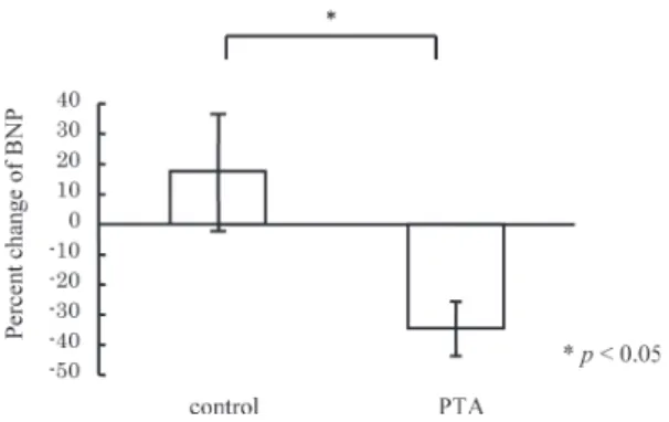
Figure 1 exhibits representative data from one patient in the PTA group. In the patient, angiography showed complete occlusion of the right common iliac artery, his plasma BNP level was 12.7 pg/ml. After PTA treatment, revascularization was obtained and stenosis rate de- creased to 25%. Together with revascularization, his plasma BNP level improved to 4.7 pg/ml. Likewise, we analyzed plasma BNP levels in the PTA group of which ABI was improved from 0.66±0.25 to 0.92±0.04 (p<0.05). The mean plasma BNP levels after PTA was decreased from 68.44 pg/ml to 42.08 pg/ml. Percent change of plasma BNP levels was −34±9.3% (Figure 2).
In contrast, percent change of the plasma BNP levels be- fore and after angiography in the control group was 17±
19.5%. There were significant differences in percent change of the plasma BNP levels between the control group and the PTA group. These results imply that PTA can reduce the cardiac hemodynamic stress and lead to decrease in plasma BNP levels.
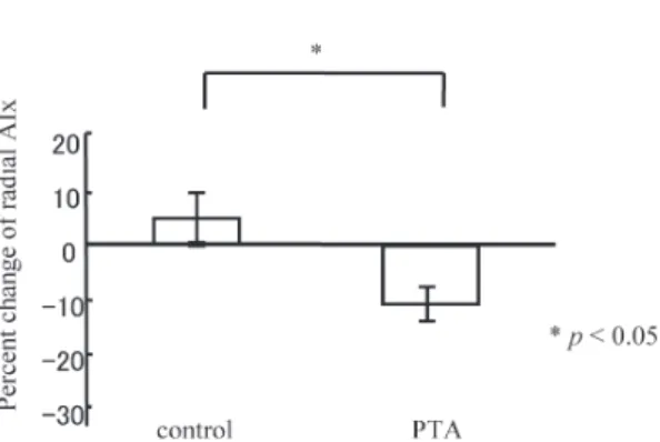
PTA attenuates arterial wave reflections
To explore the detailed mechanism for the cardiopro- tective effects of PTA, we focus on the relief of afterload after PTA. We examined the change in radial AIx before and after PTA. A representative data (Figure 1) indicates Fig. 1. Representative diagram from one PTA patient for data analysis. In the patient, angiography showed complete occlusion of the right common iliac artery. After PTA treatment, stenosis rate decreased to 25%.
Together with revascularization, his plasma BNP level improved from 12.7 pg/ml to 4.7 pg/ml. ABI of his right side improved from 0.48 to 0.91. Furthermore, his radial AIx improved from 79% to 62%. AI, heart rate-corrected AIx, and CAP improved from 80%, 135 mmHg to 65%, 122 mmHg.
Fig. 2. Comparison of the percent change of BNP between the control group and the PTA group.
that radial AIx was decreased from 80% to 65% together with the improvement of ABI (from 0.48 to 0.91). In the PTA group, mean radial AIx before PTA was 86.3±
18.8%. After PTA, radial AIx decreased to 66.4±11.2%.
The percent change of radial AIx in the PTA group was
−11.3±2.4%. On the other hand, the percent change of radial AIx in the control group was only 5.1±5.4% (Fig- ure 3).
Effects of PTA on CAP
In a separate study, we investigated the change in CAP after PTA. In the PTA group, mean CAP before PTA was 147.9±18.7 mmHg. After PTA, mean CAP was decreased to 135.8±23.1 mmHg. The percent change of CAP in the PTA group was −8.9±2.1%. On the other hand, percent change of CAP in the control group was only 0.42±3.4% (Figure 4). Although systemic blood
pressure is known to influence CAP or AIx, these beneficial effects of PTA were obtained independently of BP, because brachial BPs measured before and after treatment were not different (144.1±19.7 mmHg vs.
142.8±20.2 mmHg).
Discussion
In comparison with normal ABI patients, patients with low ABI have high mortality risk in cardiovascular diseases12,13). Low ABI patients were complicated by ischemic heart disease, cerebrovascular disease and renal failure. Therefore, recent study has proposed that ABI is a novel risk factor independent of traditional cardio- vascular risk factors14). Conversely, PTA for PAD is known to those risk factors.
In the past decade, PTA was established as a major therapeutic option for PAD. As a result of successful PTA, patients with PAD can increase exercise tolerance and step up the exercise, resulting in the reduction of the cardiovascular risk. These secondary effects are major reasons for good outcome of successful PTA. However, the direct effect of PTA on the heart remains unresolved.
A lot of study has shown that BNP reflects cardiac mechanical stress. BNP is a well recognized prognostic marker in heart failure. In the present study, we revealed that the plasma BNP levels were significantly decreased after PTA (Figure 2). These results suggest that successful PTA can bring about the reduction of cardiac mechanical stress as well as improvement exercise tolerance.
To explore the detailed mechanism for the cardio- protective effects of PTA, we focused on the relief of cardiac afterload by successful PTA. It has been known that wave reflection during systole increases left ventric- ular pressure, myocardial oxygen requirement leading to left ventricular hypertrophy. It is also reported that AIx significantly increases with increasing cardiovascular risk factors5). Moreover, Sakuragi et al. reported that both left ventricular mass index and plasma BNP levels were correlated with radial AIx in patients with hyper- tension15). In the present study, despite percent change of radial AIx was increased in the control group, percent change of radial AIx in the PTA group was significantly Fig. 3. Comparison of the percent change of radial AIx
between the control group and the PTA group.
Fig. 4. Comparison of the percent change of CAP between the control group and the PTA group.
decreased (5.1±5.4% vs. −11.3±2.4%, p<0.05), indicat- ing that PTA can attenuate arterial wave reflections (Figure 3).
Roman et al. reported that CAP measured by using radial applanation tonometry was more strongly related to vascular hypertrophy, extent of atherosclerosis, and cardiovascular events compared with brachial blood pressure16). In our data, PTA decreased CAP coupled with radial AIx (Figures 3 and 4), suggesting that PTA relieves cardiac afterload and leads to reduction of cardiac mechanical stress and prevention of hypertrophy or heart failure.
Both AIx and CAP have received a great deal of atten- tion after ASCOT-CAFE study17). In the study, despite insignificant differences in brachial blood pressure, CAP of the amlodipine based therapy group was lower than that of the atenolol based therapy group, and central aor- tic systolic pressure wave augmentation was much higher with the atenolol based therapy group than that of the amlodipine based therapy group.
Furthermore augmentation and/or peripheral pulse pressure were significantly associated with clinical out- come. These findings support our idea that the treat- ments to reduce CAP and AIx might improve prognosis of the patients with cardiovascular diseases.
In the present study, we did not observe the change in procollagen III peptide concentrations which reflects car- diac fibrosis after PTA. A possible reason is that procol- lagen III peptide concentrations unchange in the acute phase even after PTA. Long term follow-up might be necessary for evaluating the change in cardiac fibrosis.
In conclusion, PTA is able to decrease myocardial me- chanical stress in patients with PAD. The mechanisms are mediated in part by reduction of radial AIx and CAP.
Acknowledgement
We thank Yuichi Ono and Masaru Ishida for their technical supports in this study.
References
1) McDermott, M.M., Tian, L., Liu, K., Guralnik, J.M., Ferrucci, L., Tan, J., Pearce, W.H., Schneider, J.R.
and Criqui, M.H. (2008) Prognostic value of func- tional performance for mortality in patients with pe- ripheral artery disease. J. Am. Coll. Cardiol., 51, 1482-1489.
2) Howell, M.A., Colgan, M.P., Seeger, R.W., Ramsey, D.E. and Sumner, D.S. (1989) Relationship of sever- ity of lower limb peripheral vascular disease to mor- tality and morbidity : a six-year follow-up study. J.
Vasc. Surg., 9, 691-696. discussion 696-697.
3) Kugler, C.F. and Rudofsky, G. (2005) Do age and co- morbidity affect quality of life or PTA-induced quali- ty-of-life improvements in patients with symptomat- ic pad ? J. Endovasc. Ther., 12, 387-393.
4) Milani, R.V. and Lavie, C.J. (2007) The role of exer- cise training in peripheral arterial disease. Vasc.
Med., 12, 351-358.
5) Nurnberger, J., Keflioglu-Scheiber, A., Opazo Saez, A.M., Wenzel, R.R., Philipp, T. and Schafers, R.F.
(2002) Augmentation index is associated with car- diovascular risk. J. Hypertens., 20, 2407-2414.
6) Shimizu, M. and Kario, K. (2008) Role of the aug- mentation index in hypertension. Ther. Adv. Cardio- vasc. Dis., 2, 25-35.
7) Takazawa, K., Kobayashi, H., Shindo, N., Tanaka, N.
and Yamashina, A. (2007) Relationship between ra- dial and central arterial pulse wave and evaluation of central aortic pressure using the radial arterial pulse wave. Hypertens. Res., 30, 219-228.
8) O'Rourke, M.F. and Gallagher, D.E. (1996) Pulse wave analysis. J. Hypertens. Suppl., 14, S147-157.
9) Wilkinson, I.B., MacCallum, H., Flint, L., Cockcroft, J.R., Newby, D.E. and Webb, D.J. (2000) The influ- ence of heart rate on augmentation index and central arterial pressure in humans. J. Physiol., 525 Pt 1, 263-270.
10) Stein, B.C. and Levin, R.I. (1998) Natriuretic pep- tides : physiology, therapeutic potential, and risk stratification in ischemic heart disease. Am. Heart J., 135, 914-923.
11) Troughton, R.W., Prior, D.L., Pereira, J.J. et al. (2004) Plasma B-type natriuretic peptide levels in systolic heart failure : importance of left ventricular diastolic function and right ventricular systolic function. J.
Am. Coll. Cardiol., 43, 416-422.
12) McKenna, M., Wolfson, S. and Kuller, L. (1991) The ratio of ankle and arm arterial pressure as an inde-
pendent predictor of mortality. Atherosclerosis, 87, 119-128.
13) Kornitzer, M., Dramaix, M., Sobolski, J., Degre, S.
and De Backer, G. (1995) Ankle/arm pressure index in asymptomatic middle-aged males : an indepen- dent predictor of ten-year coronary heart disease mortality. Angiology, 46, 211-219.
14) Criqui, M.H., Ninomiya, J.K., Wingard, D.L., Ji, M.
and Fronek, A. (2008) Progression of peripheral ar- terial disease predicts cardiovascular disease morbid- ity and mortality. J. Am. Coll. Cardiol., 52, 1736- 1742.
15) Sakuragi, S., Maruo, T., Taniguchi, M., Nagase, S., Nakamura, K., Kusano, K.F. and Ohe, T. (2008) Ra- dial augmentation index associated with increase in
B-type natriuretic peptide in patients with hyperten- sion. Int. J. Cardiol., 130, 414-419.
16) Roman, M.J., Devereux, R.B., Kizer, J.R., Lee, E.T., Galloway, J.M., Ali, T., Umans, J.G. and Howard, B.V.
(2007) Central pressure more strongly relates to vascular disease and outcome than does brachial pressure : the Strong Heart Study. Hypertension, 50, 197-203.
17) Williams, B., Lacy, P.S., Thom, S.M., Cruickshank, K., Stanton, A., Collier, D., Hughes, A.D., Thurston, H.
and O'Rourke, M. (2006) Differential impact of blood pressure-lowering drugs on central aortic pressure and clinical outcomes : principal results of the Con- duit Artery Function Evaluation (CAFE) study. Cir- culation, 113, 1213-1225.