Mechanisms of Metabolic Disorders and Memory/Learning Dysfunction Induced by Vitamin B 12 Deficiency and Production of the Vitamin-Enriched Food for Preventing the Deficiency
(ビタミン B
12欠乏症による代謝異常症と記憶・学習障害の発症メカニズムの 解明および欠乏症予防のためのビタミン B
12強化食品の開発)
Tomohiro Bito
2015
Contents
Chapter I Introduction ... 1
Chapter II Preparation of vitamin B12-deficient Caenorhabditis elegans as a model animal ... 8 Section 1 Vitamin B12-deficient Caenorhabditis elegans prepared by the
feeding of vitamin B12-limited Esherichia coli OP-50 as a diet ... 10 Section 2 Vitamin B12-deficient Caenorhabditis elegans prepared by the
treatment of vitamin B12 dodecylamine derivative ... 22 Section 3 Effects of hydroxocobalamin dodecylamine derivatives on the
activities of vitamin B12-dependent enzymes in cultured COS-7 cells ... 42
Chapter III Mechanisms of metabolic disorders and memory/learning dysfunction induced by vitamin B12 deficiency in Caenorhabditis elegans ... 52 Section 1 Vitamin B12 deficiency in Caenorhabditis elegans results in sever
oxidative stress, leading to memory/learning dysfunction ... 54 Section 2 Vitamin B12 deficiency in Caenorhabditis elegans results in
metabolic disorders of polyamines, modulators of synaptic
transmission ... 75
Chapter IV Production of vitamin B12-enriched food for preventing the
deficiency among vegetarians and elderly subjects ... 91
Section 1 Miniaturized high performance thin-layer chromatography of vitamin B12 compounds in foods ... 93
Section 2 Characterization of vitamin B12 compounds in the fruiting bodies of shiitake mushroom (Lentinula edodes) ... 101
Section 3 Characterization of vitamin B12 compounds in organic fertilizers containing purple photosynthetic bacteria ... 116
Section 4 Production and characterization of vitamin B12-enriched lettuce (Lactuca sativa L.) grown using hydroponics ... 123
Chapter V Conclusion ... 139
References……… ... 157
Acknowledgements ... 175
Abbreviations
AdoB12; 5'-Deoxyadenosylcobalamin B12; Vitamin B12 (cobalamin) Cat; Catalase
CBS; Cystathionine-β-synthase CH3-B12; Methylcobalamin C.I.; Chemotaxis index CN-B12; Cyanocobalamin
DNPH; 2,4-Dinitrophenylhydrazine
ESI-MS/MS; Electro spray ionization tandem mass spectrometer GPx, Glutathione peroxidase
GSH, Glutathione or reduced glutathione
GSSH; Glutathione disulfide or oxidized glutathione GSR; Glutathione reductase
GST; Glutathione S-transferase Hcy; Homocysteine
HPLC; High performance liquid chromatography IF; Intrinsic factor
KCN; Potassium cysnide
KPB; Potassium phosphate buffer MCM; Methylmalonyl-CoA mutase MMA; Methylmalonic acid
MS; Methionine synthase
NADPH; Nicotinamide adenine dinucleotide phosphate NGM; Nematode growth medium
NMDA; N-methyl-D-aspartate NOS; Nitric oxide synthase OH-B12; Hydroxycobalamin
qPCR; Quantitative polymerase chain reaction RNS; Reactive nitrogen species
ROS; Reactive oxygen species SAH; S-Adenosylhomocysteine SAM; S-Adenosylmethionine SOD; Superoxide dismutase TBA; Thiobarbituric acid TCA; Trichloroacetic acid
TLC; Thin-layer chromatography RDA; Recommended dietary allowance
Chapter I
Introduction
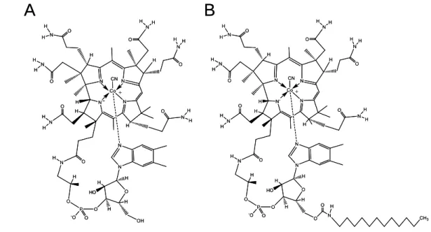
Vitamin B12 or cobalamin (B12) is exclusively synthesized by some anaerobic bacteria and then concentrated in the bodies of higher predatory organisms in the natural food chain [1]. B12 has the highest molecular weight (1355.4) among vitamins and a complicated structure with a cobalt complex ion (Fig. 1). B12 compounds contain a corrin ring with a cobalt atom coordinated by the lower axial ligand (base-on conformation), which is an unusual ribonucleoside with dimethylbenzimidazole as a base moiety. Although B12 is officially defined as cyanocobalamin, the upper axial ligand of B12 can be substituted by several functional groups (hydroxo, methyl, or 5’-deoxyadenosyl). The coordinated dimetnylbenzimidazole of the lower ligand can be dissociated from cobalt atom (base-off conformation). The base-on and base-off conformations appear to play a role in the stability of different forms of B12 as well as their ability to bind to proteins [2].
Fig.1. Structures of vitamin B12. (A) CN-B12, cyanocobalamin; (B) OH-B12, hydroxocobalamin; (C) CH3-B12, methylcobalamin; (D) AdoB12, adenosylcobalamin.
R= OH- (B) CH3- (C) CN- (A)
(D)
B12 has a complex process for gastrointestinal absorption of dietary B12, which is normally bound to proteins in food [3, 4] (Fig. 2). B12 is released from the proteins by the action of HClpresent in the stomach and the formed free B12 is immediately bound by haptocorrin (HC, a B12-binding protein produced by the salivary grands) [5].
Subsequently, B12 is released from HC by the action of pancreatic proteases (e.g.
trypsin) and the formed free B12 is bound by intrinsic factor (IF, a B12-binding protein produced by the stomach cells). The IF-B12 complex is absorbed by enterocytes of the ileum; the IF-B12 complex is specifically bound by the receptor (such as cubilin and megalin) involved in gastrointestinal absorption of B12 and then internalized into the enterocytes by the receptor-mediated endocytosis [6, 7]. The IF-B12 complex liberated from the receptor is transferred to lysosomes of enterocytes. In lysosomes, proteases are responsible for digestion of the protein component of the complex, and subsequent lysosomal exit of B12 involves LMBD1 transporter [8, 9]. Free B12 is transported cellularly and/or processed as coenzyme forms and then exported into the portal circulation. The exported B12 is normally bound to transcobalamin-II (TC-II, a B12-binding protein).
Fig.2. Gastrointestinal absorption mechanism of vitamin B12 Salivary
Stomach Gastric
Pancreas Cholecyst
Ileum
×
HC
B12 Food
IF
gland
acid
HC
HC B12
HCB12
HCB12
HC B12 ×
HC
B12
B12
B12
B12
IF IF IF
B12
B12 IF IF
B12 IF
Salivary
Stomach Gastric
Pancreas Cholecyst
Ileum
×
HCHC
B12
B12 Food
IF IF
gland
acid
HCHC
HC B12
HC HC BB1212
HCB12HCHCB12B12
HCB12HCHCB12B12
HC B12 ×
HC HC BB1212
HCHC
B12 B12
B12 B12
B12 B12
B12 B12 IF
IF IFIF IFIF
B12 B12
B12 IF BB1212
IF IF IF IF
B12 IF BB1212
IF IF
On the target cell surface, the TC-II-B12 complex is specifically recognized by TC receptor CD 320 [10]. The TC-II-B12 complex is transported inside the cells by the receptor-mediated endocytosis. Fig. 3 shows the summary of transport and processing of B12 in the mammalian cells. The endocytic vesicle containing the TC-II-B12 complex is transferred to lysosomes, in which proteases are responsible for digestion of the protein component of the complex [11]. Subsequently, the export of B12 from lysosomes to cytosol involves LMBD1 (cblF) [8, 9]. Cytosolic B12 [cob(III)alamin] is bound to the tracking protein methylmalonic aciduria CblC type (MMACHC) and then reduced to cob(II)alamin (one electron-reduced form of cobalt atom) by the action of methylmalonic aciduria CblD type (MMADHC) protein. Cob(II)alamin is further processed and then converted into two coenzyme forms, 5'-deoxyadenosylcobalamin (AdoB12) and methylcobalamin (CH3-B12) (Fig. 1), which function as the coenzymes for mitochondrial methylmalonyl-CoA mutase (MCM; EC 5.4.99.2) [12] and cytosolic methionine synthase (MS; EC 2.1.1.13) [13], respectively. MS catalyzes the methylation of homocysteine (Hcy) to form methionine (Met) using 5-methyltetrahydrofolate as methyl donor. The prosthetic B12 functions as transient acceptor of the methyl group, with cycling of B12 between cob(I)alamin (two electron-reduced cobalt atom) and CH3-B12. Whereas, cytosolic cob(II)almamin is transported into the mitochondria by the mediation of an unknown system and then reduced to cob(I)alamin and synthesized to AdoB12 by cob(I)alamin adenosyltransferase (MMAB; EC 2.5.1.17) [14, 15]. Methylmalonic aciduria CblA type (MMAA) protein functions as an escort protein that transfers the synthesized AdoB12 directly to MCM [16, 17].
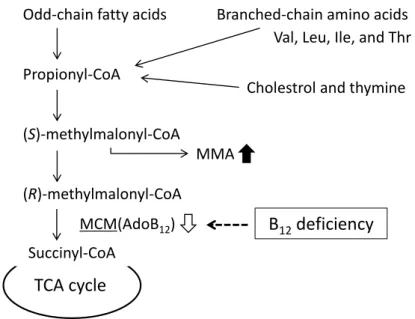
MCM catalyzes the coversion of (R)-methylmalonyl-CoA to succinyl-CoA, an intermediate of TCA cycle, and plays important roles in catabolic pathways of branched chain-amino acids (valine, leucine, isoleucine, and threonine), odd-numbered fatty acids, cholesterol, and thymine (Fig. 4) [18].
Fig. 3. Transport and cellular processing of vitamin B12. Abbreviation: LMBD1; the limb region 1, MCM; methylmalonyl-CoA mutase, MMAA; methylmalonic aciduria CblA type, MMAB; methylmalonic aciduria CblB type, MMACHC; methylmalonic aciduria CblC type, MMADHC; methylmalonic aciduria CblD type, MS; methionine synthase, TC- II; transcobalamin II.
Fig. 4. Vitamin B12-dependent methylmalonyl-CoA metabolic pathway.
MS catalyzes the synthesis of Met from Hcy with 5'-methyltetrahydrofolate (CH3-THF) (Fig. 5) and plays important roles in sulfur amino acid, folate, polyamine, and
Mitochondria Cytosol
Cob(I) MMAB MCM MMAA
AdoB12
Cob(II)
OH-B12 TC-II
OH-B12 TC-II (III) Lysosome
LMBD1 Cob(III) Cob(III)?
MMACHC MMADHC
Hcy Met MS CH3-B12
Mitochondria Cytosol
Cob(I) MMAB MCM MMAA
AdoB12 AdoB12
Cob(II)
OH-B12 TC-II OH-BOH-B1212
TC-II OH-B12 TC-II OH-BOH-B1212
TC-II (III) Lysosome
LMBD1 Cob(III) Cob(III)?
MMACHC MMADHC
Hcy Met MS CH3-B12 CH3-B12
Odd-chain fatty acids
Propionyl-CoA
(S)-methylmalonyl-CoA
(R)-methylmalonyl-CoA
Succinyl-CoA
TCA cycle
Branched-chain amino acids Val, Leu, Ile, and Thr
MMA
Cholestrol and thymine
MCM(AdoB12) B12deficiency
S-adenosylmethionine (SAM) metabolisms; in particular SAM involves various cellular methylation reactions such as DNA, RNA, proteins, and lipids [19, 20]. Urinary excretion of methylmalonic acid (MMA) and/or increased serum (or plasma) Hcy are clinically used as indexes of B12 deficiency [21, 22].
Fig. 5. Vitamin B12-dependent methionine metabolic pathway.
Several studies have suggested that B12 deficiency is linked to serious health complications such as growth retardation, megaloblastic anemia, metabolic disorders, and neuropathy [23], although the underlying disease mechanisms are poorly understood [24]; in particular, further research is required to elucidate the precise mechanisms of B12-deficient neuropathy in elderly people and vegetarians, high risk populations. As described above, B12 deficiency causes peripheral neuropathy, spinal cord disorders, and cognitive dysfunction. Hyperhomocysteinemia (HHcy) is reportedly associated with vascular dementia and Alzheimer Disease (AD) [25-27]. Moreover, significant accumulation of Hcy potentiates oxidative injury in vascular disease and AD [28, 29]. McCaddon et al. [30] proposed that biological consequence of oxidative stress caused by HHcy induced AD. However, the precise mechanism of B12-deficient neuropathy is not clear.
Ebara et al. [22] demonstrated that B12 deficiency in rats caused serine and threonine metabolic disorders, which are attributable to impairment of the adenylyl
Met
Hcy MS THF
CH
3- THF
SAM SAH DNA synthesis
(CH3-B12)
B12deficiency
cyclase system. The N-methyl-D-aspartate (NMDA) receptor involving in the long-term memory preservation is significantly modulated by Hcy and/or polyamines [31].
However, there is no information on whether B12 deficiency induces the metabolic disorders of polyamines. In addition, the precise mechanism of the cognitive dysfunction caused B12 deficiency in humans [32] is poorly understood.
Developing animal models of B12 deficiency is essential for investigating the molecular mechanisms that are defective in this metabolic disorder. However, such animal models have proven difficult to generate because animals must be fed with a B12-deficient diet for long periods to achieve B12 deficiency [33]. The lack of robust B12-deficient animal models has limited investigations to the biochemical mechanisms induced by B12 deficiency.
Caenorhabditis elegans offers several advantages for genetic and biochemical studies, including a short lifespan, a 3 day life cycle, a completely sequenced genome,
<1000 somatic cells, and the ability to change reproductive rates, life cycle, and locomotive behavior [34]. In addition, many molecular and cellular processes are conserved between nematodes and mammals. Most human disease genes and pathways are present in the worm [35]. Thus, this animal has been widely used as a model organism for studying a variety of biological processes. However, whether B12 is an absolute requirement for normal growth and physiological function in C. elegans is unknown. If a method for creating viable B12-deficient worms can be found, C. elegans could serve as a suitable model organism for studying the effects of B12 deficiency.
In the present thesis, I describe the preparation and characterization of B12-deficient C. elegans grown under B12-deficient conditions (Chapter II, Section 1). Furthermore, I characterized various B12 dodecylamine derivatives as potent inhibitors of B12-dependent MS and MCM in worms and mammalian cells (Chapter II, Section 2 and 3), indicating that B12-deficient C. elegans is readily prepared using the dodecylamine derivatives. In Chapter III, I describe the molecular mechanisms of metabolic disorders and memory/learning dysfunction induced by B12 deficiency in C.
elegans.
Strict vegetarians and/or elderly people are at a high risk of developing B12
deficiency [36]. A large number of people have low serum B12 levels that result more commonly from malabsorption of protein-bound B12 (food-bound B12 malabsorption) rather than pernicious anemia [37]. Food-bound B12 malabsorption is found in people with certain gastric dysfunctions, particularly atrophic gastritis with low stomach acid secretion, which prevails in elderly people [38, 39]. It is therefore necessary to identify plant foods containing high free B12 to prevent vegetarians or elderly people from developing B12 deficiency. Thus, I developed a simple method for analyzing B12
compounds in various food samples using miniaturized high-performance TLC (Chapter IV, Section 1). In Chapter IV, Section 2, I characterized B12 compounds in the fruiting bodies of Shiitake mushroom as a plant-derived food. Finally, I describe production and characterization of a B12-enriched vegetable to prevent vegetarians or elderly people from developing B12 deficiency. (Chapter IV, Section 3 and 4).
Chapter II
Preparation of Vitamin B
12-deficient Caenorhabditis elegans as a model animal
The major signs of B12 deficiency are megaloblastic anemia and neuropathy [23].
The underlying cause(s) of various symptoms (growth retardation, metabolic disorders, and neuropathy) caused by B12 deficiency are not still understood [24, 40]. Developing animal models of B12 deficiency is essential for investigating the molecular mechanisms that are defective in the metabolic disorders linked to such diseases. However, they cannot be readily prepared because animals (e.g. rats) must be fed with a B12-deficient diet for long periods toachieve B12 deficiency [33]. C. elegans has been widely used as a model organism for studying a variety of biological processes [34]. In addition, the enzymes involved in human methylmalonic aciduria caused by B12 deficiency have been studied in C. elegans [41, 42]. However, whether B12 is an absolute requirement for normal growth and physiological function in C. elegans is unknown. If a method for creating viable B12-deficient worms can be found, C. elegans could serve as a suitable model organism for studying the effects of B12 deficiency.
The B12 analogue with a modification of side chain of C-ring, B12[c-lactam], has been reported to antagonize B12 in the cultured HL 60 cells [43]. Subcutaneous administration of OH-B12[c-lactam] to rats has indicated that the analogue has the ability to act as a potent inhibitor of the mammalian B12-dependent enzymes to make the rats B12 deficiency [44]. However, considerably high concentration of OH-B12[c-lactam] must be administered for several weeks with osmotic mini pumps in order to prepare B12-deficient rats because IF hardly binds OH-B12[c-lactam] [44].
Although McEwan et al. [45] demonstrated high affinity binding of IF to a B12
dodecylamine derivative, our preliminary experiments indicated that this derivative was inactive in B12-dependent microorganisms typically employed in B12 the bioassay. If the B12 alkylamine derivatives act as potent inhibitors of mammalian B12-dependent enzymes in vivo, B12-deficient animals would be readily prepared by oral administration
of the derivative.
In Chapter II, I describe the preparation and characterization of B12-deficient C.
elegans grown under B12-deficient conditions (Section 1). Furthermore, I characterized various B12 dodecylamine derivatives as potent inhibitors of B12-dependent MS and MCM in worms and mammalian cells (Section 2 and 3), indicating that B12-deficient C.
elegans is readily prepared using the dodecylamine derivatives.
Section 1 Vitamin B
12-deficient Caenorhabditis elegans prepared by the feeding of vitamin B
12-limited Escherichia coli OP-50 as a diet
Introduction
B12 deficiency results in growth retardation, metabolic disorders, and neuropathy [24, 40]. However, the underlying disease mechanisms are poorly understood. To investigate the precise mechanisms of these B12-deficient symptoms, B12-deficient rat have been used as a human model animal. However, they require for long periods to make them B12 deficiency [33]. Therefore, the lack of robust B12-deficient animal models such rats has limited investigations to the biochemical mechanisms induced by B12 deficiency. Recently, the new model animal turning into rats is demanded.
C. elegans has been widely used as a model organism for studying a variety of biological processes [24]. Many molecular and cellular processes are conserved between nematodes and mammals. Most human disease genes and pathways are present in the worm [35]. However, whether B12 is an absolute requirement for normal growth and physiological function in C. elegans is unknown.
In this section, I describe a novel method for inducing B12 deficiency in C. elegans and the effects of B12 deficiency on various biomarkers, and characterize the physiological roles of B12 in this model organism.
Materials and Methods
Organisms and growth conditions
The N2 Bristol wild-type C. elegans strain was maintained at 20°C on nematode growth medium (NGM) plates using the Escherichia coli OP50 strain as the food source [46]. To induce B12 deficiency, worms were grown on 1.7% (w/v) agar plates containing M9 medium (3 g/L KH2PO4, 6 g/L Na2HPO4, 0.5 g/L NaCl, 1 g/L NH4Cl, 1 mmol/L
MgSO4, 50 μmol/L CaCl2, 2 g/L glucose, 4 mg/L thiamine hydrochloride, and 5 mg/L cholesterol) in l L H2O. Plates containing B12-supplemented (100 g/L cyanocobalamin) M9-medium each received one egg obtained from worms grown on NGM plates with B12-deficient OP50 E. coli (described below). Eggs were allowed to hatch and develop into egg-laying adult worms. The adult worms were then removed from each plate, eggs were collected, and each egg was transferred onto a new control plate. After this procedure was repeated at least 10 times, the resultant worms were used as experimental controls.
Preparation of B12-deficient E. coli cells
E. coli OP50 was grown in M9 medium (3 g/L KH2PO4, 6 g/L Na2HPO4, 0.5 g/L NaCl, 1 g/L NH4Cl, 1 mmol/L MgSO4, 50 μmol/L CaCl2, 2 g/L glucose, and 4 mg/L thiamine hydrochloride) at 37°C for three days. Cells were inoculated every three days into fresh M9 medium and used as a food source for C. elegans. The B12 content (0.2
g/g wet weight) of E. coli cells grown in the M9 medium was significantly reduced compared with cells grown in the standard Luria-Bertani medium (11.1 g/g wet weight).
Preparation of B12-deficient C. elegans
C. elegans were grown at 20°C on B12 (100 g/L)-supplemented medium using B12-deficient E. coli OP50 as a food source. An individual worm egg was transferred onto each plate, which contained fresh B12-deficient medium seeded with the B12-deficient E. coli OP50. After the eggs hatched, worms were allowed to grow until they became adults and had laid eggs (yielding the F1 generation). Individual eggs were removed from the plate and each was transferred onto a fresh plate containing B12-deficient medium and grown to maturity under the same conditions (yielding the F2
generation). This process was repeated for five generations. After three days, adult worms of each generation (F1-F5) were used for experiments. Fig. 6 shows the preparative procedure of B12-deficient worms.
Fig. 6. Preparation of B12-deficient C. elegans. Single eggs from C. elegans adults grown on NGM plate were transferred onto individual plates containing B12-supplemented M9 seeded with B12-deficient E. coli OP50 (control) and grown for three days. The eggs hatched and developed into adult worms, which then laid eggs. The adult worm was removed from each plate and individual eggs were transferred individually onto fresh identical plates. This procedure was repeated at least 10 times.
These worms were used as experimental control worms. To prepare B12-deficient worms, single eggs from the control worm were transferred onto individual plates containing B12-deficient M9 medium seeded with B12-deficient E. coli and allowed to reach maturity and lay eggs (F1 generation). F1 worms obtained from these eggs were used to generate F2 worms following the same procedures. This process was repeated to prepare F5 B12-deficient worms.
Vitamin B12 assay
F1-F5 worms grown under B12-supplemented or B12-deficient conditions were harvested and incubated for 1 h at 20°C in fresh M9 medium to remove any residual E.
coli cells. Worms (2 g wet weight) were then disrupted using a hand homogenizer (AS ONE Corp., Osaka, Japan) and sonicated (6 kHz for 60 s) three times. The worm homogenate was resuspended in 100 mL of 57 mmol/L sodium acetate buffer (pH 4.8) containing 0.05% (w/v) KCN and boiled for 30 min. The extract was centrifuged at 15,000 × g for 15 min at 4°C and the supernatant was used for assaying B12
concentrations by standard microbiological methods utilizing Lactobacillus delbrueckii subsp. lactis ATCC 7830, as described previously [40].
B12-related biomarker assays
F1-F5 worms grown under B12-supplemented or B12-deficient conditions were collected and washed in M9 medium as described above. The harvested worms were resuspended in 0.5 mL of 100 mmol/L potassium phosphate buffer (pH 7.0) at 2°C and homogenized using a hand homogenizer. The cell homogenate was centrifuged at 15,000 × g for 15 min at 4°C and the supernatant was used as a crude homogenate for subsequent biomarker assays.
MMA and Hcy, two indices of B12 deficiency, were assayed using the high performance liquid chromatography (HPLC) methods of Al-Dirbashi et al. [47] and Febriani et al. [48], respectively. MCM-[49] and MS-[50, 51] activities were assayed at 37°C as previously described. Total- and holo-enzyme activities were determined in the presence or absence of each B12 coenzyme (Ado-B12 for MCM and CH3-B12 for MS).
Analysis of egg-laying capacity, life cycle, and lifespan
Measurement of egg-laying capacity was based on the method of Byerly et al. [52].
Individual L4-stage worms grown under B12-supplemented or B12-deficient conditions were selected, transferred onto the fresh plates containing the same culture medium, and incubated for one day at 20°C. After laying eggs, each worm was removed from the plate, and the eggs were counted. Egg counting was performed in triplicate.
The life cycle and lifespan of B12-deficient F5 worms were determined at 20°C using the synchronization method of Johnson and Wood [53]. Worms were scored as dead when they no longer responded to prodding with a pick. In each survival experiment, 100 worms were used.
Protein quantitation
Proteins were assayed by the method of Bradford [54] using ovalbumin as a standard.
Statistical analysis
The effects of B12 deficiency on various C. elegans biomarkers were evaluated by one-way ANOVA, and a post-hoc analysis was performed using Tukey’s multiple comparison tests. Analyses were performed with GraphPad Prism 3 for Windows version 2.01 (GraphPad software Inc., La Jolla, CA, USA). All data are presented as the mean ± SD. Differences were considered statistically significant when p < 0.01.
Results and Discussion
Effects of B12-deficient growth conditions on various B12-related biomarkers in C.
elegans
Nematodes grown on B12-supplemented M9 medium with B12-deficient E. coli OP50 as a food source (control) showed identical growth rates that were identical to that of worms grown under normal conditions, indicating that the experimental control conditions were adequate for the normal growth of C. elegans. The control worms were able to ingest both B12-enriched agar medium and B12-deficient E. coli cells. Therefore, they would mainly absorb sufficient amount of free B12 from the agar medium because
of low B12 content in E. coli.
Under B12-deficient conditions, the B12 content of the worms decreased gradually over four generations (Fig. 7). The B12 concentration in F5 generation worms was only 4% compared with that in the control worms. These results indicate that dietary B12
deprivation over five generations leads to a significantly decreased B12 status in C.
elegans.
Fig. 7. B12 content is reduced in worms grown under B12-supplemented and -deficient conditions. The B12 content of F1-F5 worms grown under control (black bar) and B12-deficient (white bars) conditions was assayed using microbiological methods.
Data represent the mean ± SD of ten independent experiments. Different letters (a-e) indicate values that are significantly different (p < 0.01); identical letters indicate values that are not significantly different.
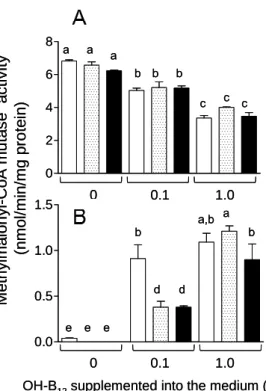
The MMA and Hcy indices of B12 deficiency were assayed in C. elegans grown under both control and B12-deficient conditions. There was a significant increase in the levels of both compounds between F3 and F5 generation worms (Fig. 8A and B). Hcy and MMA levels were approximately four and five times greater, respectively, in F5 worms grown under B12-deficient conditions than in the control worms. Although holo-MCM activity significantly decreased in F4 and F5 worms grown under B12-deficient conditions (Fig. 8E), the total MCM activity (holo- and apo-enzymes) increased (Fig.
8C). The result indicates that apo-MCM activity is significantly increased by B12
deficiency.
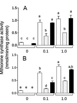
Fig. 8. Vitamin B12 deficiency changes the concentration of various B12-related biomarkers. (A) Methylmalonic acid (MMA) and (B) homocysteine (Hcy) content, (C) total- and (E) holo-methylmalonyl-CoA mutase (MCM) activity, and (D) total- and (F) holo-methionine synthase (MS) activity were measured in extracts of worms grown under B12-supplemented (black bar) and B12-deficient conditions (white bars) for up to 5 generations. For (E) holo-MCM and (F) holo-MS activities, measurements were made in the absence of specific coenzymes. Data represent mean ± SD of ten independent experiments; a-e indicate values that are significantly different, p < 0.01.
Both total- and holo-MS activities significantly decreased with each generation until the F4 generation and were maintained at a constant level thereafter (Fig. 8D and F). These results indicate that F5 generation worms grown under B12-deficient conditions develop severe B12 deficiency. However, one day after transfer of B12-deficient worms onto the B12-supplemented medium, the level of these B12-related biomarkers recovered considerably (unpublished data). These results indicate that B12 functions as a cofactor for both MCM and MS in C. elegans and that B12-dependent changes in both MCM and
MS enzyme activities occur in C. elegans and mammals [55, 56]. However, using the nematode model, the time needed to produce a severely B12-deficient animal is only 15 days (i.e. five generations).
Holo-MS activity was rapidly decreased by F1 generation under B12-deficient conditions (Fig. 8F), but holo-MCM activity was not changed until the F4 generation (Fig. 8E). These results indicated that MS is more sensitive to cellular B12
concentrations than MCM. Yamada et al. have demonstrated that most MS activity is derived from holo-enzyme in B12-sufficient or -deficient mammals because the apo-enzyme is very unstable [55]. In contrast, Nakao et al. have indicated that holo-MCM activity was less than 5% of the total enzyme activity in B12-sufficient rats and that a marked increase in the apo-enzyme activity occurred under B12-deficient conditions [56]. In this C. elegans study, holo-MCM activity was 97% of the total enzyme activity in the control worms and holo-enzyme activity gradually decreased in the F1 (92%), F2 (50%), and F3 (24%) generations, under B12-deficient conditions.
However, the specific activity of holo-MCM did not change until the F4 generation even if the B12 content of the worms significantly decreased along with increased MMA content. The details of the occurrence of this MCM-independent increase in MMA concentration soon after the onset of B12 deficiency remain to be elucidated.
B12 deficiency affects C. elegans egg-laying capacity, life cycle and lifespan
Egg-laying rates significantly decreased in B12-deficient worms (Fig. 9A), which also showed a significantly prolonged life cycle compared with the control worms (Fig.
9B). Similarly, B12-deficient rats have been reported to show severe growth retardation [21] and infertility [57].
The lifespan of B12-deficient F5 worms were significantly decreased by B12
deficiency (Fig. 9C). The maximal lifespan of B12-deficient worms was reduced to 18 days, compared with a lifespan of 24 days in the control worms (Fig. 9C). These data demonstrate for the first time that B12 deficiency significantly reduces the lifespan of
animals.
B12 deficiency causes severe growth retardation and various metabolic disorders in mammals [21]. The B12 coenzyme AdoB12 functions as a coenzyme of MCM, catalyzing the isomerization of (R)-methylmalonyl-CoA to succinyl-CoA in the mitochondria.
Odd-numbered fatty acids, branched chain amino acids, and cholesterols are metabolized by methylmalonyl-CoA to the tricarboxylic acid (TCA) cycle intermediate succinyl-CoA via MCM [12]. When MCM activity was significantly decreased by B12
deficiency, MMA abnormally accumulated in the cells [21].
Fig. 9. Vitamin B12 deficiency reduces egg-laying capacity and lifespan, and increases the length of the life cycle in C. elegans. (A) Total number of eggs per worm, (B) the length of the life cycle (h), and (C) the lifespan were determined in the control (black bar) and B12-deficient (white bars) F5 worms. Data represent mean ± SD of ten independent experiments; a-d indicate values that are significantly different, p < 0.01.
The elevated concentration of MMA mainly inhibits mitochondrial respiration because of competitive inhibition of succinate dehydrogenase (EC 1.3.99.1) by MMA [57]. TCA cycle inhibition by MMA accumulation contributes to various metabolic disorders associated with B12 deficiency [21], including severe growth retardation (prolonged life cycle) in the B12-deficient worms.
Many studies have demonstrated that there is a relationship between B12 deficiency and infertility in males and females [58, 59]. However, the mechanism whereby B12
deficiency causes infertility is poorly elucidated. CH3-B12 functions as a coenzyme of MS, which catalyzes the methyl transfer from CH3-THF to Hcy, resulting in the donation of a methyl group to Hcy, forming Met [13]. MS is important to re-synthesize Met and to metabolize CH3-THF. THF is the precursor for the methylene derivative of folate, which is essential for thymidine supply and normal DNA replication in cells [60].
Furthermore, Met is one of the amino acid building blocks of protein and acts as the universal methyl group donor (SAM) for a large number of methylation reactions.
Yamada et al. [61] reported that reduced testicular MS activity is the primary cause of pathological impairment of spermatogenesis owing to B12 deficiency and that methionine supplementation to the diet can reduce this impairment. Bennet [62] has reported that B12 deficiency may lead to recurrent fetal death owing to the elevated Hcy levels. The epigenetic regulation of gene expression involves remodeling of chromatin by either the addition of methyl group to DNA and/or the post-translational modification of histone amino acid residues. SAM is a critical substrate for histone methyltransferases, whereas S-adenosylhomocysteine (SAH) is a potent inhibitor of the enzymatic reaction [63]. It has been observed that the concentration of SAM is reduced, with a concomitant increase in SAH concentration, in B12-deficient animal models or humans [64]. Our experiments indicated that SAM/SAH ratios significantly decrease in B12-deficient C. elegans relative to control worms (Details see in Chapter III, Section 2). The decreased SAM/SAH ratios may lead to abnormal epigenetic regulation of gene expression, including gene relevant to fertility.
These observations suggest that decreased egg-laying and prolonged life cycle
found in B12-deficient worms are because of various B12-associated metabolic disorders, which results in abnormal epigenetic regulation of the expression of certain genes.
Approximately 1 % of B12-deficient worms showed a specific morphological abnormality (Fig. 10), similar to the short and plump “dumpy” mutant phenotype that is formed because of disordered cuticle collagen biosynthesis [65, 66]. However, there is no information available on the relationship between B12 deficiency and collagen biosynthesis. Hcy, which is significantly increased by B12 deficiency, has been shown to interfere with post-translational modifications of collagen directly by inhibiting lysyl oxidase (EC 1.4.3.13), which is involved in collagen cross-linking [67]. This observation and our data indicate a possible link between B12 deficiency and collagen biosynthesis including post-translational modifications of collagen cross-linking.
Fig. 10. B12-deficient C. elegans show morphological changes. Differential interference microscopy images of the (1) control and (2) B12-deficient worms were obtained using an IX71 microscope (OLYMPUS Corp., Tokyo, Japan). The length of individual worms was measured using Image J software, bar = 200 m.
Above results indicate that B12 is essential for C. elegans growth and that prolonged B12 deficiency induces a number of phenotypes, including decreased egg-laying capacity (infertility), prolonged life cycle (growth retardation), and a reduced lifespan.
Therefore, I propose that C. elegans is an ideal model organism for investigating the mechanisms driving such B12-deficient phenotypes, as B12 deficiency can be induced in this animal in only 15 days. However, there are some limitations to this animal model;
for example, C. elegans does not have any blood corpuscle systems. Moreover, bioinformatic analyses indicate that C. elegans also does not have any orthologs of three
B12-transport proteins (HC, IF, and TC-II) involved in human gastrointestinal absorption and subsequent blood circulation of B12. Thus, C. elegans is not suitable for use as a model organism to study the mechanisms of some human B12-deficient disease phenotypes, such as megaloblastic anemia and dysfunctions of intestinal absorption and transport of B12. However, this animal is widely used as a model organism for studying the mechanisms of fertilization [68] and embryonic cell division [69]. Moreover, C.
elegans is often used for understanding human brain and neuronal disorders [70] in addition to the effects of certain molecules on learning and memory [71]. C. elegans may become a suitable organism and a powerful new tool for the study of B12-deficient human diseases such as infertility [58, 59, 61], fetal death [62, 72], neuropathy [73], and cognitive impairment [74].
Summary
B12 deficiency has been linked to developmental disorders, metabolic abnormalities, and neuropathy; however, the mechanisms involved remain poorly understood. C.
elegans grown under B12-deficient conditions for five generations develop severe B12
deficiency associated with various phenotypes that include decreased egg-laying capacity (infertility), prolonged life cycle (growth retardation), and reduced lifespan.
These phenotypes resemble the consequences of B12 deficiency in mammals, and can be induced in C. elegans in only 15 days. Thus, C. elegans is a suitable animal model for studying the biological processes induced by vitamin deficiency.
Section 2 Vitamin B
12-deficient Caenorhabditis elegans prepared by the treatment of vitamin B
12dodecylamine derivative
Introduction
When C. elegans was grown under B12-deficient conditions for five generations (approximately 15 days), worm developed severe B12 deficiency (Chapter II, Section 1). The B12-deficient worms showed various phenotypes including decreased egg-laying capacity (infertility), prolonged life cycle (growth retardation), and reduced life span.
These phenotypes resemble those of B12-deficient mammals. If development of animal models of B12 deficiency is facilitated by the use of a potent inhibitor of B12-dependent enzymes, B12-deficient animals would be prepared in a short time. McEwan et al. [45]
synthesized ribose-5'-carbamate derivatives of B12 and demonstrated high-affinity binding of IF to certain alkylamine derivatives. My preliminary experiments indicate that these alkylamine derivatives lack detectable biological activity in certain microorganisms that require B12 for growth, such as Escherichia coli 215, L. delbrueckii ATCC 7830, and Euglena gracilis Z. In this section, I show that CN-B12 dodecylamine derivative potently inhibited the B12-dependent enzymes MCM and MS of C. elegans.
Materials and Methods
Chemicals
CN-B12, 1, 1'-carbonyldiimidazole, and dodecylamine were purchased from Wako Pure Chemical Industries (Osaka, Japan). Phenyl-Toyopearl 650M was purchased from Tosoh Corporation (Tokyo, Japan).
Organisms and culture conditions
The maintenance of worms and preparation of control or B12-deficient worms was
performed as described in Chapter II, Section 1.
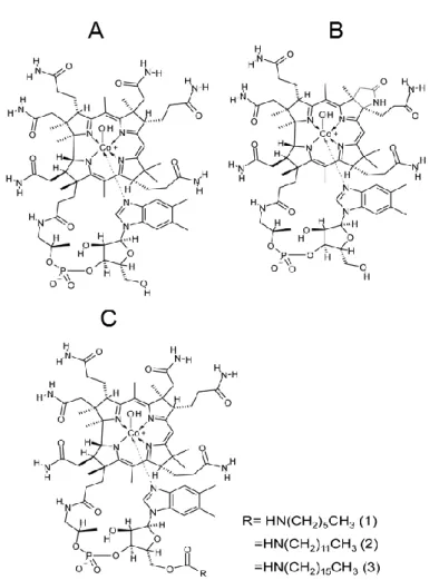
Preparation of the CN-B12 dodecylamine derivative
The CN-B12 dodecylamine derivative (Fig. 11) was prepared according to the method by McEwan et al. [45]. In brief, solid 1, 1'-carbonyldiimidazole (26 mg) was added to CN-B12 (0.1 g) dissolved in dimethyl sulfoxide (1.2 mL) at 30°C, and the mixture was stirred for 25 min. Dodecylamine (0.27 mmol) was added to the mixture and then stirred for 24 h at 25°C. The B12 compound was extracted twice from the mixture with 2 mL of phenol/dichloromethane (1:2, v/v) and then re-extracted twice from the combined phenol fractions with 2 mL of distilled water. The water-soluble fractions were combined and chromatographed using a TSK gel-Toyopearl 650M column (2.4 × 24 cm) equilibrated with 250 mL of 25% (v/v) ethanol. Unreacted and modified CN-B12 compounds were eluted with 250 mL of 25% (v/v) and 60% (v/v) ethanol. The fractions containing products were combined, the solvent was evaporated under reduced pressure, and the residue was dissolved in 5 mL of 80% (v/v) ethanol.
The product was further purified using silica-gel 60 TLC with the solvent 1-butanol:2-propanol:water (10:7:10, v/v/v). After drying the TLC sheet, the CN-B12
derivative was collected, extracted with 80% (v/v) methanol, and the solvent was evaporated under reduced pressure. The residue was dissolved in a small amount of 18% (v/v) acetonitrile and then purified using a reversed-phase HPLC column (Wakosil-II 5C18RS, φ 4.6 × 150 mm) and a Shimadzu HPLC system (SCL-10A VP System controller, DGU-20A 3R Degassing unit, LC-20AB Liquid chromatograph, SPD-20A UV/VIS detector, and CTO-20AC Column oven). The CN-B12 derivative was eluted with a linear gradient of acetonitrile (18%
-
28% for 8.5 min and 28%-
100%for 13 min, 1.0 mL/min) at 40°C and monitored by measuring absorbance at 254 nm.
The peak fraction was collected, the solvent was evaporated under reduced pressure, and the residue was used for the following experiments. The purified dodecylamine
derivative of CN-B12 (approximately 94% purity) was dissolved in distilled water, and its concentration was determined at 361 (ε =13,000) as described previously [45].
Fig. 11. Structures of CN-B12 and its dodecylamine derivative. (A) CN-B12 (B) CN-Cbl dodecylamine derivative.
Preparation of C. elegans treated with CN-B12 dodecylamine derivative
Control worm eggs were transferred to and hatched on an M9 plate containing the CN- B12 derivative (100 g/L) and E. coli OP50 and grown to maturity.
Determination of the levels of CN-B12 and its derivative in C. elegans
Worms grown in the absence or presence of CN-B12 or its dodecylamine derivative were harvested and incubated for 1 h at 20°C in fresh M9 medium to remove any residual E. coli. The washed worms (1.0 g wet weight) were homogenized in 0.5 mL of 100 mmol/L potassium phosphate buffer (pH 7.0) at 4°C using a manual homogenizer (AS ONE Corp., Osaka, Japan) and sonicated (6 kHz for 60 s) three times on ice. The
A B
A B
worm homogenate was resuspended in 25 mL of 57 mmol/L sodium acetate buffer (pH 4.8) containing 0.05% (w/v) KCN, boiled for 30 min, and centrifuged at 15,000 ×g for 15 min at 4°C. The B12 content of the supernatant fraction was determined using a microbiological assay as described previously [75].
CN-B12 and the B12 derivative were extracted from treated worms using the same conditions as described above. The supernatant fraction (20 mL) that was chromatographed using a Sep-Pak Plus C18 cartridge (Waters Corp., Milford, MA, USA) to separate the CN-B12 derivative from CN-B12. After the cartridge was washed with 10 mL of distilled water, CN-B12 and the CN-B12 derivative were eluted sequentially with 10 mL of 20% (v/v) ethanol solution and 60% (v/v) ethanol, respectively. The solvent of each fraction was evaporated under reduced pressure and then dissolved in 100 L of distilled water. CN-B12 was determined using the microbiological B12 assay method described above, and the CN-B12 derivative was assayed using a Shimadzu HPLC system (SCL-10A VP System controller, DGU-20A 3R Degassing unit, LC-20AB Liquid chromatograph, SPD-20A UV/VIS detector, and CTO-20AC column oven) with a CDS ver. 5 chromato-data processing system (LAsoft, Ltd., Chiba, Japan). Samples (20 μL) were chromatographed using a reversed-phase HPLC column (Wakosil-II 5C18RS, φ 4.6 ×150 mm) and eluted (1.0 mL/min) with a linear gradient of methanol (20%-90% for 30 min) containing 1% (v/v) acetic acid at 40°C. The B12 derivative was monitored by its absorbance at 254 nm, and its retention time was 30.3 min.
Determination of coenzyme forms of the B12-dodecylamine derivative
All procedures were performed in the dark. Hydroxo (OH)- and adenosyl (Ado)-forms of the B12-dodecylamine derivative were prepared from the CN-B12
derivative. In brief, the CN-B12 dodecylamine derivative was dissolved in distilled water, bubbled with nitrogen gas for 30 min, reduced with sodium tetrahydroborate until the solution turned dark brown, and neutralized with 1.0 mol/L HCl. Most of the CN-B12
derivative was converted to the OH-B12 dodecylamine derivative. To prepare AdoB12
dodecylamine derivative, a small amount of 5'-iodo-5'-deoxyadenosine was added to the reduced form of the B12 dodecylamine derivative, and this solution was neutralized as described above. The neutralized preparations were desalted using a Sep-pak Vac 20 cc (5 g) C18 cartridge (Waters corp.), and the desalted and concentrated solutions were treated with silica-gel 60 TLC as described above to separate the OH- or Ado-form of the dodecylamine derivative and unmodified CN-B12 dodecylamine derivative. After drying the TLC sheet, the OH- or Ado-form of the dodecylamine derivative was collected, extracted with 80% (v/v) ethanol, and the solvent was evaporated to dryness under reduced pressure. Each residue was dissolved in a small amount of distilled water and used as the authentic OH-B12 dodecylamine or AdoB12 dodecylamine derivatives. I was unable to prepare a CH3-B12 dodecylamine derivative with high purity.
To extract B12 derivatives, including its coenzyme forms from worms, 100 mL of 80% (v/v) ethanol was added to lyophilized samples of worms treated with the CN-B12
dodecylamine derivative. The samples were heated at 98°C for 30 min under reflux and cooled to room temperature [76], centrifuged at 10,000 × g for 10 min, and the supernatant was evaporated under reduced pressure. The residue was dissolved in 5 mL of distilled water and centrifuged at 10,000 × g for 10 min to remove insoluble material.
The supernatant fraction was chromatographed a Sep-Pak Plus C18 cartridge, which was washed with 10 mL of distilled water and eluted with 2 mL of ethanol. The ethanol elute was evaporated under reduced pressure and the residue was dissolved in 100 L of distilled water. The levels of coenzyme forms of the B12 dodecylamine derivative in worms treated with the CN-B12 dodecylamine derivative were determined using HPLC as described above. The retention times of OH-, CN-, and Ado-forms of B12
dodecylamine derivatives were 22.0, 30.6, and 36.2 min, respectively.
Assays for B12-related biomarkers
The worms (1.0 g wet weight) grown each condition were assayed B12-related biomarkers described in Chapter II, Section 1.
Inhibition of MCM and MS activities by the CN-B12 dodecylamine derivative
My previous study showed that the levels of apo-MCM are increased up to approximately 97% of the total MCM (holo- and apo-MCMs) in B12-deficient worms.
Thus, a cell homogenate of B12-deficient worms was used as an apo-MCM preparation in the following experiments (Chapter II, Section 1). Lineweaver-Burk analysis was used to determine the Km value of worm MCM for AdoB12 in reaction mixtures containing 0, 2.5, 5, and 10 mol/L of AdoB12. Dixon plots [77] were used to determine the apparent Ki value of MCM in reaction mixtures containing 0, 1, 3, 5, and 10 mol/L of CN-B12 or its dodecylamine derivative.
A cell homogenate of B12-supplemented (control) worms was used as a holo-MS enzyme preparation, because most MS activity is derived from the holoenzyme in B12-supplemented or -deficient worms. Holo-MS activity was determined in reactions containing 0, 1, 3, 5, and 10 mol/L of CN-B12 or its dodecylamine derivative.
Western blotting
Worms grown in the absence of CN-B12 or in the presence of CN-B12 or the CN-B12
dodecylamine derivative were homogenized in 100 mmol/L potassium phosphate buffer (pH 7.0) at 4°C. Each homogenate was centrifuged at 15,000 ×g for 10 min and the supernatant fraction was analyzed. We used a precast slab gel (PAGEL, type NPG-520L, ATTO Corporation, Tokyo, Japan) for electrophoresis of samples through a 5%
-
20%(w/w) linear gradient of polyacrylamide in the presence of SDS. After electrophoresis, proteins were transferred to a PVDF membrane (Immuno-Blot PVDF, Bio-Rad Laboratories, Inc. Hercules, CA, USA) in a Trans-Blot SD semi-dry electrophoretic
transfer cell (Bio-Rad). The PVDF membrane was probed with an anti-MS antibody (ab66039, abcam®, Cambridge, MA, USA). We performed the immunodetection reactions using an anti-mouse IgG antibody secondary antibody (Promega KK, Tokyo, Japan) coupled to horseradish peroxidase and an immunoblot-staining kit for peroxidase (EzWestBlue, ATTO), according to the manufacturer’s instructions. A Protein Ladder One Triple-color kit (Nacalai Tesque Inc., Kyoto, Japan) was used to determine molecular mass. After the treated PVDF membrane was photographed using a digital camera (Coolpix 4300, Nikon, Japan), the intensities of the protein bands were calculated of Image J software [78].
Quantitative PCR analysis (qPCR)
Total RNA was prepared from worms using Sephasol®-RNA1 (Nacalai Tesque).
Poly(A)+ mRNA prepared from total RNA using the Poly (A)+ Isolation kit from Total RNA (Nippon Gene, Tokyo, Japan) was used to synthesize cDNA using PrimeScriptTM
Ⅱ 1
st Strand cDNA Synthesis kit (Takara Bio, Otsu, Japan). Primer pairs used for qPCR analysis were designed using GENETYX software (GENETYX Corporation, Tokyo, Japan) (Table 1). Gene-specific primers were selected such that the resulting PCR products were approximately 100 bp. A CFX ConnectTM Real-Time System (Bio-Rad) with SYBR Premix Ex Taq (Takara Bio) was used to perform qPCR. The level of the mRNA encoding β-actin was used as an internal standard. The qPCR experiments were repeated at least three times for each cDNA prepared from three preparations of worms.Analysis of egg-laying capacity and life cycle
Measurements of egg-laying capacity and life cycle were performed according to the method described in Chapter II, Section 1. Individual worms grown in the presence of CN-B12, its dodecylamine derivative or in the absence of B12 were transferred to fresh plates containing the respective culture media and incubated for 1 day at 20°C. After
laying eggs, each worm was removed from the plate, and the eggs were counted in triplicate.
Table 1. Primer pairs used for the qPCR analysis
Primer pairs for the qPCR were designed using GENETYX software. Each gene is involved in the metabolisms of B12, including MCM (mmcm-1), MS (metr-1), methylmalonic acidemia cobalamin A complementation group (mmaa-1), cob(I)alamin adenosyltransferase (mmab-1), methylmalonic aciduria B12 C type (cblc-1), and MS reductase (mtrr-1). For normalization,
-actin (act-1) mRNA levels served as the internal standard.
Protein quantitation
Protein concentrations were determined using the Bradford method [54] with ovalbumin as a standard.
mmcm-1
metr-1
mmaa-1
mmab-1
cblc-1
mtrr-1
act-1
Gene name Primer sequences (5' to 3')
ATTAAAGCAGGGCAGCAAGGAC and GGCACGGATATGGTTCAACTTGG AAAGACCACGACAAGCCACTGA and CGCTTGATGATTCGAGCACGTT
AAGACCGCTGATTCGTGTACTC and GCATGTCAGCCAATGACAATCC TCCAAGAGAGGTATCCTTACCC and CTCCATATCATCCCAGTTGGTG TTGGAAAGAAGGCGACAGGA and TTGTTTCTGCAGCCAGACCA GCTCTTCTGATTTGGGAAGAGG and CGACGCTCATTGTTGTACAGAG ATGTTGCTGGAGCAGCTTTC and AGTGTCCACCGTAGATTGGATG
Statistical analysis
The effects of the CN-B12 dodecylamine derivative on C. elegans phenotypes were evaluated using one-way ANOVA with Tukey’s multiple comparison test (GraphPad Prism 3 for Windows version 2.01; GraphPad Software Inc., La Jolla, CA, USA). All data are presented as the mean ± standard deviation (SD). Significant differences were defined as p < 0.05.
Results and Discussion
Effects of the CN-B12 dodecylamine derivative on B12-related biomarkers of C. elegans Although McEwan et al. [45] demonstrated high affinity binding of the gastric B12-binding protein IF to the CN-B12 dodecylamine derivative, my preliminary experiments indicated that this derivative was inactive in B12-dependent microorganisms typically employed in B12 the bioassay. Therefore, I evaluated effects of the CN-B12 dodecylamine derivative on B12-related phenotypes using C. elegans as a model. Table 2 shows the concentrations of B12 and its dodecylamine derivative in homogenate prepared from adult worms grown in the presence of the CN-B12 derivative for 3 days. Remarkably, the B12 concentration of worms grown in the presence of the CN-B12 derivative was only 35% compared with that of control worms and was similar to that of worms grown for two generations (6 days) in the absence of B12 (Chapter II, Section 1). In contrast, the CN-B12 dodecylamine derivative was absorbed and accumulated by worms grown in the presence of the CN-B12 derivative (approximately 110 ng/g wet weight). These results suggest that the CN-B12 dodecylamine derivative did not inhibit the uptake of B12 in the intestine, but it was readily accumulated in worms and significantly decreased their B12 concentrations.
To determine whether the dodecylamine derivative detected in the treated worms was converted into other forms of B12, corrinoid compounds were extracted using 80%
(v/v) ethanol from worms grown in the presence of the dodecylamine derivative and analyzed using reversed-phase HPLC.
Table 2. Contents of CN-B12 and the CN-B12 dodecylamine derivative in worms
Control and treated worms were grown on plates containing CN-B12- and CN-B12 dodecylamine derivative-supplemented (each at 100 μg/L) M9 media for 3 days, respectively. Corrinoids were extracted from the treated worms by boiling with KCN at acidic pH. CN-B12 and the CN-B12
dodecylamine derivative were separated each other using a Sep-Pak Plus C18 cartridge and their levels were determined using the microbiological assay and HPLC, respectively. Data represent the mean ± SD of five independent experiments.
The retention times of authentic OH-B12, CN-B12, AdoB12, CH3-B12, OH-B12
dodecylamine, CN-B12 dodecylamine, and AdoB12 dodecylamine were 3.5, 8.1, 9.5, 12.8, 22.0, 30.6, and 36.2 min, respectively (Fig. 12A-D). The compounds extracted from worms exposed to the derivative eluted with retention times of 3.8-30.3 min (Fig.
12E). A major peak with the retention time of 30.3 min was identical to that of authentic CN-B12 dodecylamine, and peaks were not detected with retention times of CN-B12 (8.1 min), OH-B12 dodecylamine (22.0 min), or AdoB12 dodecylamine (36.2 min). These results indicate that the CN-B12 dodecylamine derivative accumulated by worms was not converted to any other B12-related compound, including its coenzyme forms.
CN-B12 CN-B12 dodecylamin derivative
Control worms 132.2 ± 26.7 -
Treated worms 46.5 ± 7.8 110.0 ± 17.2
(ng/g wet weight)