表
題
MRSA におけるダプトマイシンとバンコマイシン交差耐性メカ
ニズムの解明
(Elucidation of cross-resistance mechanism to daptomycin and
vancomycin in MRSA)
論 文 の 区 分
博士課程
著
者
名
ティティアナンパコーン カネート
担当指導教員氏名
教授 崔 龍洙
所
属
自治医科大学大学院医学研究科
人間生物学系 専攻
生体防御医学 専攻分野
微生物・免疫学 専攻科
2020年1月10日申請の学位論文
CONTENTS
ACKNOWlEDGEMENTS ... i
ABSTRACT ... iii
LIST OF TABLES ... iv
LIST OF FIGURES ... v
LIST OF ABBREVIATIONS ... vi
CHAPTER I INTRODUCTION ... 1
CHAPTER II LITERATURE REVIEW ... 3
1.1. Staphylococcus aureus (S. aureus) ... 3
1.2. S. aureus cell envelop ... 3
1.2.1) Cell wall (CW) ... 3
1.2.2) Plasma membrane ... 11
1.3. The emergence of methicillin-resistant S. aureus (MRSA) ... 16
1.4. Antibiotics for treatment of MRSA infection ... 18
1.4.1) Vancomycin (VCM) ... 18
1.4.2) Daptomycin (DAP) ... 19
1.5. VCM and DAP resistance in MRSA ... 21
1.5.1) VCM resistance ... 21
1.5.2) DAP nonsusceptibility ... 26
1.5.3) Cross-resistance between DAP and VCM ... 29
CHAPTER III OBJECTIVES ... 31
CHAPTER IV MATERIALS AND METHODS ... 34
4.1 Materials ... 34
4.1.1) Chemicals and reagents ... 34
4.1.2) Bacterial strain ... 35
4.1.3) Primers and plasmid ... 39
4.2 Methods ... 43
4.2.1) Bacterial strains and drug susceptibility test ... 43
4.2.3) Population analysis profiling-area under the curve (PAP-AUC) analysis ... 44
4.2.4) DNA extraction and purification ... 44
4.2.5) MLST and single nucleotide polymorphisms (SNPs) determination by whole
genome sequencing ... 45
4.2.6) Gene replacement into the chromosome ... 46
4.2.7) In vitro induction by stepwise DAP exposure ... 46
4.2.8) Transmission electron microscopy (TEM) ... 47
4.2.9) Evaluation of membrane surface charge ... 47
4.2.10) Determination of L-PG production ... 48
4.2.11) RNA extraction and RNA expression analysis ... 49
4.2.12) Statistical analysis ... 49
CHAPTER V RESULTS ... 50
5.1 Reassessment of VCM and DAP susceptibility
... 505.2 Comprehensive mutation identification
... 515.3 Detection of genes reported to be associated with decreased susceptibility to VCM or
DAP in S. aureus
... 595.4 Substitution of mprF with mutated mprF identified in the cross-resistant DNS strain
caused cross-resistance of DS strain to VCM and DAP
... 635.5 Cross-resistance resulting from mprF mutation was found in in vitro selected mutants
... 645.6 Cross-resistance and CW thickness
... 665.7 mprF mutation and membrane surface charge
... 685.8 mprF mutation and L-PG production
... 705.9 Transcriptional analysis on representative DNS strains with both single and
cross-resistance and their DS counterparts
... 72CHAPTER VI DISCUSSION ... 84
ACKNOWLEDGEMENTS
I would like to express my deepest gratitude and appreciation to Professor Longzhu Cui
for his guidance supervision, suggestion, knowledge, thinking skill and encouragement
throughout the study.
I am also deeply thankful to Assistant Professor Yoshifumi Aiba and Postdoctoral
Researcher Xin-Ee Tan for their valuable suggestions on the experiments and comments on
my manuscript and thesis.
I would like to thank all members of Bacteriology Laboratory at Jichi Medical
University for their help on my experiments and manuscript proofreading. My appreciation
also goes to every staff of Graduate School of Medicine, Jichi Medical University for their
management of financial support of Special Student Scholarship and Research Assistance
Scholarship, Jichi Medical University. Many thanks also go to Dr. Tom Kouki (Division of
History and Cell Biology, Jichi Medical University) for technical assistance to operate
transmission electron microscopy.
In addition, we greatly appreciate Dr. Yoshitaka Yamamoto from Dokkyomed
Koshigaya Hospital, Professor Intetsu Kobayashi from Toho University, Professor Harumi
Yano from Tsukuba University, Professor Naohisa Fujita from Kyoto Prefectural University
of Medicine, Dr. Go Matsumoto from Shinshu University, Dr. Jun Ogawa and Dr. Susumu
Kawanishi from Tsuyama Chuo Hospital, and BEI Resources of the National Institute of
Allergy and Infectious Diseases for kindly provided the MRSA isolates in this study.
This work has been supported by, the Japan Agency for Medical Research and
Development J-PRIDE (grant No. JP17fm0208028, JP18fm0208028, and JP19fm0208028 to
LC), JSPS KAKENHI (Grant No. 18K15149 to KK, 15H05654 and 19K08960 to SW,
17K15691 to YS, 19K15740 to MK and 17K19570 to LC), the Takeda Science Foundation
(LC) and Jichi Medical University Young Investigator Award (YA). The funders had no role
in the study design, data collection and analysis, decision to publish, or preparation of the
manuscript.
Finally, I am much indebted and deeply grateful to my family for their love,
understanding, and encouragement throughout my life.
ABSTRACT
We first reported a phenomenon of cross-resistance to vancomycin (VCM) and
daptomycin (DAP) in methicillin-resistant Staphylococcus aureus (MRSA) in 2006, but
mechanisms underlying the cross-resistance remain incompletely understood. Here, we present
a follow-up study aimed at clarifying the genetic mechanism of cross-resistance. Using 12 sets
of paired DAP-susceptible (DS) and DAP-nonsusceptible (DNS) MRSA isolates from 12
patients who had DAP monotherapy, we (i) assessed susceptibility to VCM and DAP, (ii)
compared whole-genome sequences, (iii) investigated the identified mutations of
cross-resistance, and (iv) determined the impact of altered gene expression and metabolic pathway
on the cross-resistance. We found that all 12 DNS strains exhibiting cross-resistance carried
mutations in mprF, while one DNS strain with resistance to only DAP carried a lacF mutation.
On the other hand, among the 32 vancomycin-intermediate S. aureus (VISA) strains isolated
from patients treated with VCM, 5 out of the 18 strains showing cross-resistance to VCM and
DAP carried a mprF mutation, while 14 strains resistant to only VCM had no mprF mutation.
Moreover, substitution of mprF in a DS strain with mutated mprF resulted in cross-resistance
and vice versa. The mprF mutation elevated lysyl-phosphatidylglycerol (L-PG) production,
positive membrane surface charges, and cell wall (CW) synthetic pathways. These results
demonstrated that the mprF mutation contributed to cross-resistance to VCM and DAP in
MRSA.
LIST OF TABLES
Table 1: Genes involved in peptidoglycan synthesis ... 7
Table 2: Genes involved in teichoic acid synthesis ... 11
Table 3: Genes involved in phospholipid synthesis ... 15
Table 4: Genes contributed to VCM-resistance in VCM-resistant Staphylococcus aureus
(VRSA) ... 22
Table 5: Gene mutations contributed to the development of VISA ... 25
Table 6: Clinical isolates from the same patient with DAP treatment ... 35
Table 7: Worldwide VISA collection upon VCM treatment ... 37
Table 8: Bacterial transformation ... 38
Table 9: Primers used in this study ... 39
Table 10: Plasmid used in this study ... 42
Table 11: Summary of MIC, gene mutation, MLST, doubling time ... 53
Table 12: Daptomycin and vffancomycin MICs determined by using broth microdilution
method ... 55
Table 13: Summary of MIC and gene mutation of VISA strains ... 60
Table 14: MICs of DAP and VCM in the mprF substitution of H-set isolates ... 63
Table 15: Summary of MIC, doubling time and mutations in mprF and lacF on in vitro
derivatives of the C-1 and K-1 strains ... 65
Table 16: Representatives of genes differentially expressed between cross-resistant strain
H-5 and susceptible strain H-3 ... 74
Table 17: Representatives of genes differentially expressed between
daptomycin-nonsusceptible strain K-2 and -suscptible strain K-1 ... 80
LIST OF FIGURES
Figure 1: Schematic model of peptidoglycan synthesis. ... 6
Figure 2: Schematic model of teichoic acid production. ... 10
Figure 3: Schematic model of phospholipid production. ... 14
Figure 4: Brief history of antibiotic therapies and resistance in S. aureus. ... 17
Figure 5: Schematic model of inhibition of peptidoglycan synthesis by VCM. ... 19
Figure 6: Schematic model of cell membrane disruption upon DAP exposure. ... 20
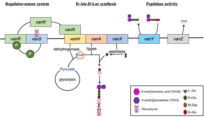
Figure 7: Schematic model of vanA operon-mediated VCM resistance in VRSA. ... 22
Figure 8: Schematic model of reduced DAP susceptibility mediated by mprF mutation. ... 29
Figure 9: Population analysis profiles of clinical MRSA isolates in DAP treatment group. . 56
Figure 10: Sequence alignment for lacF of K-1 and its DAP-resistant mutants. ... 57
Figure 11: The location of mprF mutations in DNS strains and VISA strains. ... 58
Figure 12: Comparison of cell-wall size in clinical MRSA isolates ... 67
Figure 13: Measurement of membrane surface charge ... 69
Figure 14: Evaluation of membrane L-PG production. ... 71
Figure 15: Gene expression in contribution to cross-resistance in DNS strain H-5. ... 78
LIST OF ABBREVIATIONS
Abbreviation
Definition
ABC transporter
ATP-binding cassette transporter
ACC
acetyl-CoA carboxylase
acetyl-CoA
acetyl-coenzyme A
ACP
Acyl carrier protein
acyl-P
acyl-phosphate
agr
accessory gene regulator
Ala
alanine
AMPs
antimicrobial peptides
BHI
brain-heart infusion
Ca
2+calcium ion
CAMPs
cationic antimicrobial peptides
ccr
cassette chromosome recombinase
cDNA
complementary deoxyribonucleic acid
CDP
cytidine diphosphate
CFU
colony forming unit
CL
cardiolipin
cls
cardiolipin synthease
CLSI
clinical and laboratory standards institute
CM
cell membrane
CO
2carbon dioxide
COGs
clusters of orthologous groups
CTP
cystidine triphosphate
CW
cell wall
DAG
diacylglycerol
DAP
daptomycin
dgkB
diacylglycerol kinase
DNS strain
DAP-nonsusceptible strain
DS strain
DAP-susceptible strain
EDTA
ethylenediaminetetraacetic acid
EUCAST
European committee on antimicrobial susceptibility testing
F-6P
fructose-6-phosphate
fabG
β-ketoacyl-ACP reductase
fabI
Enoyl-A ACP reductase
fabZ
β-hydroxyacyl-ACP
FASII
type II fatty acid synthesis
FDA
US Food and Drug Administration
fem
factor essential for methicillin resistance
G3P
glycerol-3-phosphate
Glc1P
glucose-1-phosphate
Glc6P
glucosamine-6-phosphate
Abbreviation
Definition
glmM
phosphoglucosamine-mutase
glmS
glucosamine-F-6P aminotransferase
glmU
UTP-Glc1P uridyltransferase
Glu
glutamic acid
Gly5
pentaglycine
graSR
glycopeptide resistance-associated
GSH
glutathione
gtaB
UTP: α-Glc1P uridyltransferase
GUVs
giant unilamellar vesicles
H
2O
2hydrogen peroxide
HCl
hydrochloric acid
JMUB
Jichi medical university bacterialbank
KCl
potassium chloride
L-PG
lysyl-phosphatidylglycerol
LTA
lipoteichoic acid
ltaA
glycolipid permease
ltaS
LTA synthase enzyme
Lys
lysine
MAN
N-acetylmannosamine
Mg
2+magnesium ion
MH
Muller-Hinton
MIC
minimum inhibitory concentration
MLST
multi-locus sequence typing
MOPS
3-(N-morpholino) propanesulfonic acid
mprF
multiple peptide resistance factor
mraY
phospho-NAM-pentapeptide translocase
MRSA
methicillin-resistant Staphylococcus aureus
murA
UDP-N-acetylglucosamine transferase
murB
UDP-N-acetylmuramate dehydrogenase
murG
undecaprenyl-PP-NAM-pentapeptide-UDP-NAM transferase
N/A
not available
NAG
N-acetylglucosamine
NAM
N-acetylmuramic acid
NaOAc
sodium acetate
NARSA
network on antimicrobial resistance in Staphylococcus aureus
OD
optical density
PAP-AUC
population analysis profiling-area under the curve
PBP
penicillin-binding protein
PEP-PTS
lactose phosphoenolpyruvate phosphotransferase system
PG
phosphatidylglycerol
PG-P
PG-phosphate
Abbreviation
Definition
PL
phospholipid
Poly-GroP
polyglycerolphosphate
PtdOH
phosphatidic acid
rot
repressor of toxins
SCCmec
staphylococcal cassette chromosome mec
SD
standard deviation
SNP
single nucleotide polymorphism
TAs
teichoic acids
tar
teichoic acid ribitol
TCRS
two-component regulatory system
TEM
transmission electron microscopy
TLC
thin-layer chromatography
TMSs
transmembrane segments
TSB
tryptic soy broth
UDP
uridine diphosphate
UDP-Glc
UDP glucose
UDP-GlcNAc
UDP-N-acetyl-glucosamine
UPRT
uracil phosphoribosyltransferase
UTP
uridine triphosphate
VCM
vancomycin
VISA
VCM-intermediate Staphylococcus aureus
vraSR
VCM-resistance-associated sensor/regulator
VRSA
VCM-resistant Staphylococcus aureus
VSSA
VCM-susceptible Staphylococcus aureus
WT
wild type
CHAPTER I
INTRODUCTION
Infections with methicillin-resistant Staphylococcus aureus (MRSA) are serious
clinical problems in all parts of the world causing high morbidity and mortality. MRSA is
resistant not only to the β-lactam antibiotics, but also the other classes of antibiotics such as
aminoglycosides, tetracyclines, or fluoroquinolones, restricting the available antibacterial
agents for MRSA treatment [1-4]. Vancomycin (VCM), a glycopeptide antibiotic exerting
bactericidal activity by binding to D-Ala-D-Ala residues of peptidoglycan to inhibit bacterial
cell wall (CW) synthesis, is the first-line antibiotic against MRSA infections [5]. Emergence
of MRSA with reduced susceptibility to VCM has therefore further limits the scarcely available
treatment options [3, 4, 6-8].
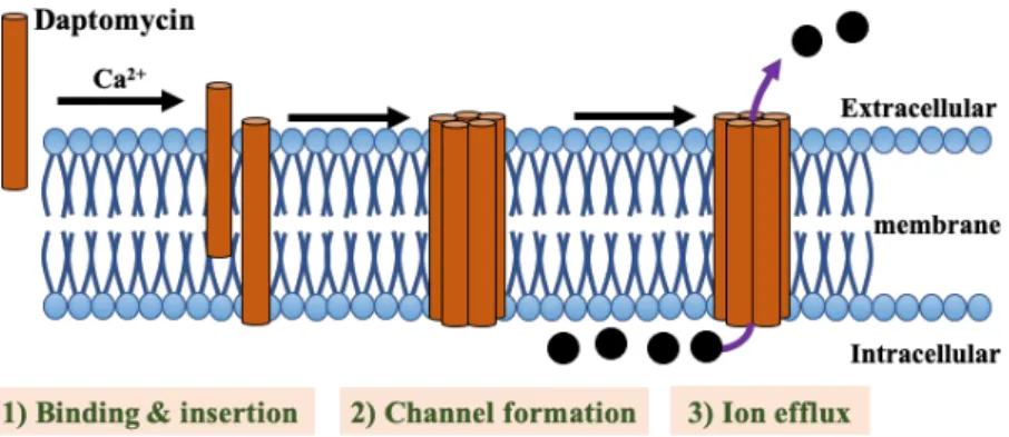
Daptomycin (DAP), a cyclic lipopeptide antibiotic with potent bactericidal activity, is
frequently used as salvage therapy after failure of VCM treatment [9]. In the presence of
calcium, anionic DAP molecule attained its active cationic peptide form which will then insert
its lipophilic tail into the negative-charged cell membrane (CM) [10, 11]. The interaction
between DAP and CM causes potassium leakage and membrane depolarization that ultimately
contribute to cell death [12]. These mean that DAP and VCM differ in not just chemical
structure, but also in their bactericidal mechanisms [7, 13]. Nevertheless, MRSA strains with
cross-resistance to DAP and VCM have been frequently isolated from patients treated with
either DAP or VCM, with the first isolation reported by our group in 2006 [8, 14-16].
Multiple peptide resistance factor (MprF) is known to mediate DAP-nonsusceptibility
in S. aureus by alteration of net surface charges on CM. Mutation of mprF gene causes a
gain-in-function, in which lysinylation of phosphatidylglycerol (PG) will be enhanced and thus
increasing membrane lysyl-PG (L-PG) production [17, 18]. These positively-charged L-PG
will then be translocated from the inner membrane to the outer leaflet of CM by flippase
domain of MprF protein, causing an increased net positive charge on CM [19]. Eventually, the
more positively-charged CM surface will serve as a protective barrier against DAP binding [20,
21]. Besides changes in CM properties, increased thickness of CW is also proposed to cause
ineffective binding of DAP to CM [14, 22]. DAP-nonsusceptibility is accompanied by an
increased expression of genes involved in CW metabolism, such as murAB or pbp2, a response
similar to those induced by VCM and the other CW-targeting agents [23, 24]. As a salient
feature of VCM-intermediate S. aureus (VISA), CW thickening could be a potential factor of
VCM resistance in DAP-nonsusceptible strain. In fact, mutations in either walK, encoded for
the sensor protein kinase of a two-component regulatory system, or vraSR, involved in cell
envelop homeostasis, both of which resulted in CW thickening, is sufficient to cause
DAP/VCM cross-resistance [25, 26]. However, phenotypic change in CW thickness was not
consistently observable in all DAP-nonsusceptible strains [22, 27]. Consequently, the
mechanism(s) conferring resistance of S. aureus to the two different classes of antibacterial
agents remains largely unknown.
This study clarifies the mechanism of cross-resistance between DAP and VCM in
clinically isolated MRSA. A total of 12 sets of DAP-susceptible (DS) and DNS MRSA isolates
collected from different hospitals in Japan were compared for their genotypic and phenotypic
characteristics. Our results suggested that DAP and VCM cross-resistance was regulated by
mprF mutation via increased L-PG production, subsequent alteration of membrane surface
charge, and CW biosynthetic pathways. This proposed mechanism was supported by
transcriptional analysis that revealed an enhanced CW/CM metabolism in cross-resistant strain
and was found to contribute more substantially to DAP and VCM cross-resistance than changes
in CW thickness.
CHAPTER II
LITERATURE REVIEW
1.1. Staphylococcus aureus (S. aureus)
S. aureus are Gram-positive spherical bacteria (round and grape-like shape) that grow
under both aerobic and anaerobic conditions, called facultative anaerobic organism. The
bacteria can grow on rich agar medium forming yellowish-orange colonies, which is caused by
production of staphyloxanthin pigment following carbohydrate fermentation [28]. A positive
catalase test (active bubbling as a result of conversion of hydrogen peroxide (H
2O
2) to water
and CO
2) is used to distinguish staphylococci from the other members of Gram-positive cocci.
While positive-coagulation, activating the transition of fibrinogen to fibrin, differentiated S.
aureus from the other Staphylococcus species [29, 30]. S. aureus normally harmlessly inhabit
mucous membrane and skin (ranged from 20 to 50%) and were found on clothing or medical
equipment [31]. However, S. aureus is also one of the most common pathogenic bacteria
frequently isolated from community- and healthcare-associated infections, causing various
diseases such as pneumonia, osteomyelitis, skin infection or sepsis, as well as
enterotoxin-mediated food poisoning [30, 32]. High prevalence of S. aureus and difficulties in treating
staphylococcal infections due to its rapid adaptation against many types of antibiotics lead to
the increased rate of morbidity and mortality [33]. Thus, understanding the mechanism of drug
resistance are crucial for development of new strategies against resistant bacteria.
1.2. S. aureus cell envelop
1.2.1) Cell wall (CW)
The prokaryotic cells have a simpler structure than eukaryotic cells because they lack
many membrane-bound organelles such as ribosome, mitochondria or nucleus [34]. However,
unlike eukaryotic cells, a complex multilayer structure known as CW peptidoglycan, locates
outside bacterial CM to support cell structure and protects bacteria from environmental stresses
[35]. Different properties of CW between Gram-positive and Gram-negative bacteria have also
been used for bacterial characterization, with a thicker peptidoglycan observed in
Gram-positive bacteria [36]. The peptidoglycan (murein) is consists of three important parts; 1)
disaccharide units composed of alternating N-acetylglucosamine (NAG) and N-acetylmuramic
acid (NAM) connected with β-1,4 linkage, which serves as backbone for peptidoglycan [37],
2) pentapeptide side chains (L-Ala-D-Glu-L-Lys-D-Ala-D-Ala) cross-linked to L-Lys of
another peptide chain at the 4
thamino acid (D-Ala) with 3) pentaglycine cross-link, resulting
in the cleavage of the terminal D-Ala residue [38].
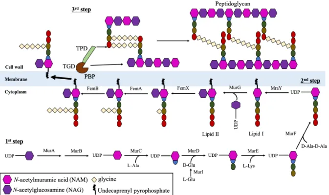
There are three stages of peptidoglycan biosynthesis (Figure 1). In the first stage,
nucleotide sugar-linked precursors (uridine diphosphate-NAM (UDP-NAM) and UDP-NAG)
are synthesized in the cytoplasm. glmS (glucosamine-F-6P aminotransferase) catalyzed the
conversion of fructose-6-phosphate (F-6P), the substrate from glycolysis pathway, to
glucosamine-6-phosphate (Glc6P), which are then used for the generation of uridine
UDP-NAG. The latter reaction of which is catalyzed by glmM (phosphoglucosamine-mutase) and
glmU (UTP-glucose-1-phosphate uridyltransferase) [39]. The UDP-NAM molecules are
generated from UDP-NAG molecules through reactions with two transferase enzymes murA
(UDP-NAG transferase) and murB (UDP-N-acetylenolpyruvoylglucosamine reductase) [40].
Finally, the ligase enzymes (MurC - F) catalyzed the construction of pentapeptide by adding
L-Ala, D-Glu, L-Lys and dipeptide D-Ala (D-Ala-D-Ala) sequentially to UDP-NAM,
generating the Park’s nucleotide. The second stage of peptidoglycan biosynthesis which occurs
in inner membrane, involved mraY (phospho-NAM-pentapeptide translocase)-catalyzed
transferring of Park’s nucleotide to lipid carrier undecaprenyl-diphosphate located at CM,
forming “lipid I” (NAM-(pentapeptide)-pyrophosphoryl-undecaprenol). The UDP-NAG
synthesized in the first stage is then linked to lipid I by murG
(undecaprenyl-PP-NAM-pentapeptide-UDP-NAM transferase), generating “lipid II”
(NAG-β-(1,4)-NAM-(pentapeptide)-pyrophosphoryl-undecaprenol). These lipid II-molecules are the building block
for CW synthesis. Five glycyl-tRNA (glycine residues) were added at the D-Lys position of
pentapeptide side chain by femXAB (factor essential for methicillin resistance) and the D-Glu
position were deaminated by couple enzymes murT/gatD before lipid II molecules can be
cross-linked to form peptidoglycan polymers [41, 42]. The final stage involved translocation
of complete lipid II molecules (peptidoglycan) across cytoplasmic membrane for cross-bridge
formation with the other lipid II, as catalyzed by penicillin binding proteins (PBPs), a family
of proteins which serve as either mono-functional or bi-functional transglycosylase and/or
transpeptidase [43]. Transglycosylase catalyzed the cross-linking between NAM from one
peptidoglycan with NAG of other peptidoglycan, with release of lipid carrier; while
transpeptidase cross-linked the L-Lys of pentapeptide side chain to D-Ala (4
thposition) of
another pentapeptide chain with pentaglycine (Gly5) bridge following cleavage of the terminal
D-Ala residue (5
thposition) [44-46].
Figure 1: Schematic model of peptidoglycan synthesis.
NAG and NAM are the backbone of peptidoglycan which is the building block for generation
of CW. NAG is synthesized from the substrate of glycolysis pathway and converted to NAM
as catalyzed by MurAB. Then, Mur ligases (MurC to F) catalyzed the formation of
pentapeptide by sequentially added amino acids L-Ala, D-Glu, L-Lys and dipeptide D-Ala
(D-Ala-D-Ala) to NAG, generating the Park’s nucleotide. MraY-mediated membrane
translocation subsequently attached Park’s nucleotide to lipid carrier
undecaprenyl-diphosphate at CM, yielding lipid I. The following MurG-catalyzed NAG linkage to Park’s
nucleotide generated lipid II. The five glycine residues are attached to lysine residue of lipid II
in a process catalyzed by FemX/A/B proteins. After translocation of pentaglycine-lipid II to
outer membrane, penicillin binding proteins (PBPs) generated CW from peptidoglycan units
through transglycosylation and transpeptidation. This figure is modified from previous study
[47].
Table 1: Genes involved in peptidoglycan synthesis
Genes
Description
glmS
glucosamine-6-phosphate synthase
fructose-6-phosphate à glucosamine-6-phosphate
glmM
phosphoglucosamine mutase
glucosamine-6-phosphate à glucosamine-1-phosphate
glmU
glucosamine-1-phosphate acetyltransferase
glucosamine-1-phosphate à UDP-N-acetylglucosamine (UDP-NAG)
murA
UDP-N-acetylglucosamine 1-carboxyvinyltransferase
UDP-NAG + phosphoenolpyruvate à UDP-NAG enolpyruvate
murB
UDP-N-acetylenolpyruvoylglucosamine reductase
UDP-NAG enolpyruvate + NADPH à UDP-N-acetylmuramic acid
(UDP-NAM)
murC
UDP-N-acetylmuramate-L-alanine ligase
UDP-NAM + L-Alanine à UDP-NAM-L-Alanine
murI
glutamate racemase
L-Glutamine à D-Glutamine
murD
UDP-N-acetylmuramoylalanine-D-glutamate ligase
UDP-NAM-L-Alanine + D-Glutamine à UDP-NAM dipeptide
murE
UDP-N-acetylmuramoyl-L-alanyl-D-glutamate-L-lysine ligase
UDP-NAM dipeptide + L-lysine à UDP-NAM tripeptide
murF
UDP-N-acetylmuramoyl-tripeptide-D-alanyl-D-alanine ligase
UDP-NAM-tripeptide + D-Ala-D-Ala à UDP-NAM pentapeptide
mraY
phospho-N-acetylmuramoyl-pentapeptide transferase
UDP-NAM pentapeptide + cis-undecaprenyl phosphate à lipid I
murG
UDP-NAG-NAM-pentapeptide pyrophosphoryl-undecaprenol NAG
transferase
lipid I + UDP-NAG à lipid II
femX
lipid II:glycine glycyltransferase
lipid II + glycyl-tRNA
Glyà lipid II-Gly1
femA
aminoacyltransferase
lipid II-Gly1 + 2 (glycyl-tRNA
Gly) à lipid II-Gly3
femB
aminoacyltransferase
lipid II-Gly3
+ 2 (glycyl-tRNA
Gly) à lipid II-Gly5
murT/gatD
lipid II isoglutaminyl synthase
lipid II-Gly5 à lipid II(D-isoglutamine-NH
2)-Gly5-
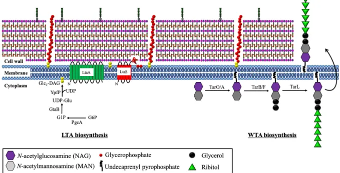
Teichoic acids (TAs), anionic glycosylated poly(alditolphosphates), also located on
CW. TAs served as a surface antigen and are involved in cell division, maintenance of cell
shape, cation homeostasis and protection of cells against extreme conditions (salt or
temperature) as well as antimicrobial peptides (AMPs) [48, 49]. There are two types of TAs:
wall teichoic acids (WTAs) and lipoteichoic acids (LTAs) [48, 50]. The anionic WTAs consist
of glycopolymers of ribitol-phosphate repeats that covalently linked to NAM residues in every
ninth peptidoglycan (Figure 2) by WTA linkage unit [51]. The WTA synthesis occurs in
cytoplasm and is regulated by many tar (teichoic acid ribitol) genes [52]. This process is
initiated by the synthesis of WTA linkage unit. tagO gene first transferred the NAG residue to
undecaprenyl phosphate carrier anchored at inner CM. NAG transferase tagA then catalyzed
the formation of β-1,4-linked N-acetylmannosamine (MAN) and NAG disaccharide complex
by transferring MAN to C4 hydroxyl residue of NAG. Following that, phosphoglycerol is
attached to C4 hydroxyl residue of MAN in MAN-NAG complex by glycerophosphate
transferase tarBF genes. Finally, ribitol-repeating units are attached to the glycerol phosphate
of the disaccharide linkage unit by polymerase enzyme tarL gene [53, 54] and the complete
WTAs polymers are translocated to outer cellular membrane by ABC-dependent transporter
complex (TarGH) before attached to CW peptidoglycan. The enzymes involved in WTAs
linkage to peptidoglycan in S. aureus have not been elucidated.
The ribitol repeat units in WTAs are important for cation binding or phage attachment,
as well as protection from antibiotic actions [55-61]. In addition, the negatively-charged
phosphate group in the ribitol-repeating units of WTAs allow metal cations binding, which is
important for CW rigidity [62]. Previous reports have shown that bacteria lacking WTA have
increased mortality rate due to imbalanced ion homeostasis. On the other hand, increased
concentration of bound Mg
2+in bacteria with enhanced WTA synthesis improved bacterial
survival [63, 64]. Moreover, metal cations binding at WTA increased surface positive charges
which then contributed to bacterial resistance against many antibiotics [58-61].
Unlike WTAs, the anionic LTAs is consisted of polyglycerolphosphate (poly-GroP)
chain, which are attached to diglucosyl-diacylglycerol (Glu
2-DAG) at CM and extended into
the peptidoglycan layer [65]. The process of LTA synthesis takes place in cytoplasm (Figure
2). Firstly, glucose-6-phosphate, a substrate in glycolysis, is converted to glucose-1-phosphate
(Glc1P) by PgcA (α-phosphoglucomutase) enzyme. After that, GtaB (UTP:
α-glucose-1-phosphate uridyltransferase) enzyme produces UDP-Glc (uridine diα-glucose-1-phosphate glucose) from
Glc1P. Two UDP-Glc moieties will be transferred to diacylglycerol (DAG) at CM by YpfP
(glycosyltransferase) enzyme, generating Glc
2-DAG [66], which are then translocated to the
outer leaflet of membrane by LtaA (glycolipid permease) [67]. Finally, LTA synthase (LtaS)
cleaves GroP subunits from head group of CM phospholipids and adds onto Glc
2-DAG to
generate the poly-GroP chains (Figure 2) [68]. LTAs is required for cell division, bacterial
invasion, as well as protection from cationic antimicrobial peptides [69-71].
Figure 2: Schematic model of teichoic acid production.
Wall teichoic acids (WTAs) and lipoteichoic acids (LTAs) are located between peptidoglycan
contributing to CW rigidity. WTAs is comprised of N-acetylmannosamine
(MAN)-N-acetylglucosamine (NAG) disaccharides attached to undecaprenyl pyrophosphate and
anchored with glycerol and polyribitol by a series of enzymes: tarO/A, tarB/F and tarL.
Meanwhile, glucose-6-phosphate (G6P) is the precursor for LTA production, from which Glu
2-DAG will be generated and attached with polyglycerophosphate cleaved from CM by LtaS
membrane protein.
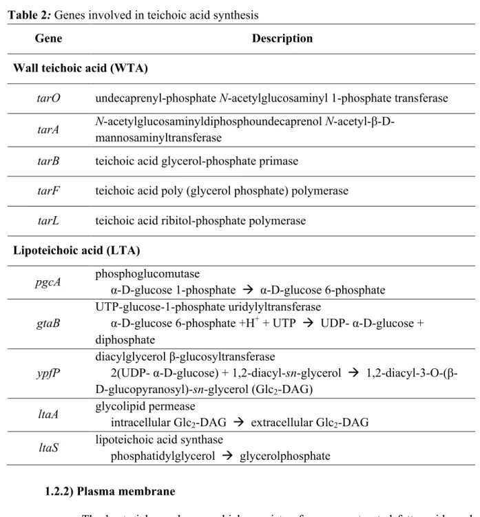
Table 2: Genes involved in teichoic acid synthesis
Gene
Description
Wall teichoic acid (WTA)
tarO
undecaprenyl-phosphate N-acetylglucosaminyl 1-phosphate transferase
tarA
N-acetylglucosaminyldiphosphoundecaprenol
N-acetyl-β-D-mannosaminyltransferase
tarB
teichoic acid glycerol-phosphate primase
tarF
teichoic acid poly (glycerol phosphate) polymerase
tarL
teichoic acid ribitol-phosphate polymerase
Lipoteichoic acid (LTA)
pgcA
phosphoglucomutase
α-D-glucose 1-phosphate à α-D-glucose 6-phosphate
gtaB
UTP-glucose-1-phosphate uridylyltransferase
α-D-glucose 6-phosphate +H
++ UTP à UDP- α-D-glucose +
diphosphate
ypfP
diacylglycerol β-glucosyltransferase
2(UDP- α-D-glucose) + 1,2-diacyl-sn-glycerol à
1,2-diacyl-3-O-(β-D-glucopyranosyl)-sn-glycerol (Glc
2-DAG)
ltaA
glycolipid permease
intracellular Glc
2-DAG à extracellular Glc
2-DAG
ltaS
lipoteichoic acid synthase
phosphatidylglycerol à glycerolphosphate
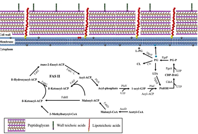
1.2.2) Plasma membrane
The bacterial membrane, which consists of monounsaturated fatty acids and
lacks sterols, is different from eukaryotic cells. It is composed of 40% phospholipids (PLs) and
60% proteins. Most proteins are embedded in the membrane for selective transport, secretion
of molecule or protection from harmful substrate. The amphipathic PLs consist of a polar head
(hydrophilic) that is attached with two non-polar tails (hydrophobic) by ester bond [72]. These
PLs bilayer completely surround a bacterial cell to avoid the leakage of intracellular molecules
such as DNA and ribosome, and protect the bacterial cell from environmental stresses such as
high osmolarity and extreme pH. Besides, PLs bilayer contained many membrane-bound
proteins which are involved in energy production through selective permeation of protons. In
addition, many antibiotics as well as human immunity targeting CM are associated with PLs
properties [73].
There are three types of PLs; PG (phosphatidylglycerol), L-PG (lysyl-PG) and
cardiolipin (CL) [74]. The first step of PL synthesis involved acyl-acyl carrier protein
(acyl-ACP) elongation via type II fatty acid synthesis (FASII) cycle (Figure 3). Initially, acetyl-CoA
is converted into malonyl-CoA by acetyl-CoA carboxylase (ACC). The malonyl-CoA is then
covalently linked with ACP by malonyl-CoA:ACP transacylase enzyme fabD forming
malonyl-ACP. After that, β-ketoacyl-ACP is generated from malonyl-ACP by 3-oxoacyl-ACP
synthase. Both malonyl-ACP and β-ketoacyl-ACP will be used for the production of acyl-ACP,
a multi-step process catalyzed by fabG (β -ketoacyl-ACP reductase), fabZ (β
-hydroxyACP) and fabI (enoyl reductase) [75]. Acyl-ACP is subsequently transformed into
acyl-phosphate (acyl-P) by membrane associated protein PlsX. Other than FASII cycle, acyl-P can
be generated from extracellular fatty acids by either of the two fatty-acid binding proteins
(FakB1 and FakB2) depending on the properties of the fatty acids, with the former specific for
saturated fatty acids while the latter is specific for unsaturated fatty acids [76]. Following that,
membrane-bound acyl transferase PlsY will catalyzed the acylation of glycerol-3-phosphate
(G3P) to 1-acyl-G3P using acyl-P as the substrate [77], and PlsC, another acyltransferase, will
transfer a second acyl-ACP to the carbon-2 position of 1-acyl-G3P to form phosphatidic acid
(PtdOH) [78]. The synthesis of PtdOH is completed when a long chain acyl-ACP generated
from FASII cycle is added to the 2
ndposition of 1-acyl-G3P [79]. Then, phosphatidate
cytidylyltransferase CdsA converts PtdOH and cystidine triphosphate (CTP) to cytidine
diphosphate-DAG (CDP-DAG), which are then changed into phosphatidylglycerolphosphate
(PG-P) when CDP-diacylglycerol-G3P 3-phosphatidyltransferase PgsA catalyzes the
substitution of cytidine monophosphate in CDP-DAG with G3P [80]. The
phosphatidylglycerophosphatase enzyme PgpP dephosphorylates PG-P into PG, that is the key
intermediate for phospholipid production [81]. The generated PG can then be converted to
L-PG and CL by MprF and CL synthases (cls1 & cls2), respectively (as explained below) [82,
83]. The turnover of PLs turnover is regulated by phosphorylation of DAG to PtdOH by
diacylglycerol kinase DgkB, in the process of LTA production [84].
PLs is a selective barrier for ions, proteins and some other molecules, whereby it
regulates the bacterial membrane fluidity and hence the penetration of molecules into
intracellular compartment. The properties of PLs are dependent on three factors: 1) temperature,
2) carotenoid (in replacement of cholesterol in eukaryotic cell membrane) and 3) saturated or
unsaturated fatty acids [85-87]. The structure of PL is more rigid (crystallization) at low
temperature and make it difficult for the membrane molecules to move. This reduced cell
permeability, causing the cells to be easily broken. In contrast, high temperature increased the
distance between PL and ultimately leads to nonselective permeability and loss of membrane
structure. On the other hand, the carotenoid which are randomly inserted between PLs rendered
CM not too tight during low temperature, while preventing the uptake of unwanted substrates
during high temperature [88, 89]. Lastly, fatty acids comprising the tailed part of PLs are made
up of saturated/unsaturated fatty acids depending on the presence of double bonds between
carbon atoms. Increased amount of unsaturated fatty acids in CM enhances fluidity due to
increased distance between PLs. Overall, these factors indicated that the balance of PLs
distance is crucial for maintenance of bacterial structure and membrane permeability [90].
Figure 3: Schematic model of phospholipid production.
Acyl-ACPs (acyl-acyl carrier proteins) generating through FASII pathway by using acetyl CoA
as substrate is an intermediate substrate for phospholipid synthesis. The synthesis of
phosphatidic acid (PtdOH) from Acyl-ACPs or LTA recycle produces phospholipids (PGs)
that generate the other kind of phospholipids such as positive-charge PG by mprF or cardiolipin
(CL) by cls or lipoteichoic acid (LTA) by ltaS.
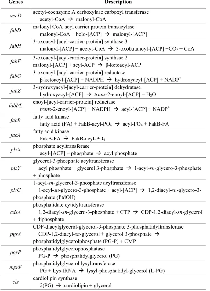
Table 3: Genes involved in phospholipid synthesis
Genes
Description
accD
acetyl-coenzyme A carboxylase carboxyl transferase
acetyl-CoA à malonyl-CoA
fabD
malonyl CoA-acyl carrier protein transacylase
malonyl-CoA + holo-[ACP] à malonyl-[ACP]
fabH
3-oxoacyl-[acyl-carrier-protein] synthase 3
malonyl-[ACP] + acetyl-CoA à 3-oxobutanoyl-[ACP] +CO
2