【症例】Successful Treatment of Severe Ischemic Foot Ulcer with Sterile Maggots: The First Case in This Country
5
0
0
全文
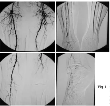
(2) 654. 日血外会誌 14巻 6 号. Fig. 1. Contrast arteriogram shows only small collateral vessels in the left lower extremity and no vessels suitable for use in a distal bypass procedure.. dl; number of platelets 18×104; BUN 48 mg/dl; creatinine 2.4. resistant staphylococcus aureus (MRSA).. mg/dl; sHba1c 15.3%.. Course of the disease after admission: Transplantation of bone. ECG: Left ventricular hypertrophy; sporadic supraventricular. marrow mononuclear cells (1×10 9) to the left lower leg was. premature contractions.. conducted in September, 2003 and transplantation of peripheral. Echo test of the cervical artery: Left, no significant stenosis;. mononuclear cells (1×10 9 ) was conducted in October and. right, 70% stenosis of the internal carotid artery.. November of 2003. Following these procedures, the API of. ABI: 0.15 (left), 0.61 (right).. the left lower leg improved from 0.2 to 0.3. However infection. Ultrasonic echo test of the heart: Good left ventricular function.. of the foot become aggravated and the wounds enlarged.. Aortography (Fig. 1): Left common iliac artery: 50% stenosis.. Finally the infected 1st and 2nd toes had to be surgically. Left superficial femoral artery occluded, left popliteal artery. debrided and amputated on February 6, 2004 (Fig. 2).. occluded. Left peroneal artery: multiple stenosis. Decrease. Although morphine hydrochloride 200 mg/day was adminis-. in arterial blood flow was observed in the anterior tibial. tered for pain at rest, control of the pain was difficult. The. artery and in the lower leg. Blood flow of the left foot was. pain at the time of blood dialysis conducted three times a. extremely deficient, especially at the inner part surrounding. week was so strong that continuous subcutaneous injection. the ulcers.. of narcotic analgesics (morphine hydrochloride) was necessary. Findings in the retina: Diabetic retinopathy was found in. to control it. As the infection was found to extend to the. both eyes (already treated with light coagulation).. whole foot, amputation under the knee was indicated. At this. Thermography: Lowering of the temperature was observed. point the patient and her family were informed of the ineffec-. at the toes of the left foot. The temperature of the left foot. tiveness of the conventional treatment of diabetic gangrenes. was 1.5˚C lower than that of the right foot.. and ulcers and that amputation was the only method remained.. Transcutaneous oxygen pressure of at toes of the left foot: 20. At the same time all the methods reported at that moment in. mmHg (right toe: 75 mmHg).. the whole world as regards the treatment were explained to. Microbial examination of pus that came from ulcer: Methicillin-. the patient herself. As the patient expressed a strong desire to. 28.
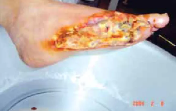
(3) 2005年10月. Mitsui et al.: Maggot Ulcer Therapy. 655. receive the treatment with sterile maggots, application for implementation of the therapy was presented to the Committee of Standards of Official Conduct of Okayama University School of Medicine and approved. Maggots were kindly provided from Dr. R. Russel (Director, Medical Entomology, Institute of Clinical Pathology and Medical Research, Westmead Hospital, Westmead, NSW, Australia). Transport and medical application of maggots to patients in our University are granted by the Bureau of Drug and Medical Safety, Department of Health and Social Welfare, Okayama Prefecture Government Office. Safety to use those imported maggot to. Fig. 2. Before maggot debridement therapy.. Fig. 3. After maggot debridement therapy (2 weeks).. Fig. 4. After maggot debridement therapy (3 months).. patients was assured by Dr. R. Russel. He reported every batch of second instara larvae of Lucilia sericata had been checked “disinfected ” and had passed a microbiological check-up as described by R. A. Sherman et al. and were suitable for use in wound debridement. Second instara larvae of Lucilia sericata (maggot) were placed on the wounds of the patient (3–4 larvae/cm2). The wound was covered with a plastic board with small holes (0.2 mm) to enable the respiration of larvae. The time needed for larvae to become pupae (about 5 days) was set as 1 course and 3 courses were repeated in succession (Fig. 3). The area of the wound before treatment of 38 cm2 decreased markedly to 15 cm2 after 1 week and 6 cm2 after 3 weeks. Granulation tissue at the bottom of the wound exhibited a good scarlet colour and became liable to bleeding. After 3 months the ulcer became completely epithelialized and the patient was discharged from the hospital (Fig. 4). Rehabilitation was started 3 weeks after the treatment and walking with a stick became possible after 5 months.. Discussion The fact that maggots are useful for the treatment of ulcers has been recognized from ancient times.1-3) It is reported that the natives of Australia and doctors of traditional medicine. of the arms and legs with bred maggots, used maggots after. of Burma used maggots to treat ulcers thousands of years. coming home for treatment of patients with chronic infection. ago. In Europe and the United States famous medical scientists. of the bone (osteomyelitis) and reported on the effectiveness. such as Ambroise Pare (1510–1590) in France, and Baron. of the therapy. Thereafter, the therapeutic method has been. Dominique-Jean Larrey, a French army doctor, described in. used continuously in hospitals of Europe and United States. their books the effectiveness of maggots for treatment of. and more than 100 papers were published during the period. ulcers. W. S. Baer, Professor of Orthopaedics of Johns. between 1930 and 1940. However, after the discovery of. Hopkins University, who participated in the World War I as. penicillin by A. Fleming in 1928, the therapeutic method. an army doctor and observed in the field the healing of wounds. using maggots was considered obsolete and gradually lost. 29.
(4) 656. 日血外会誌 14巻 6 号. popularity. This therapeutic method became rapidly forgotten. Drawbacks of the treatment with maggots are:. by people after World War II as a result of the development. 1) Danger that the maggots become settled in the site of. of surgical operations during the war, appearance of various. treatment (myiasis) cannot be denied.15-17). kinds of antibiotics and resultant decreases in various kinds. 2) As the maggots move around the site of ulcers, patients. 4). of infectious diseases. However, ulcers resistant to antibiotics. sometimes feel a sense of discomfort that something is creep-. developed in the 1990’s due to excessive use of the agents. ing.18). and diseases that were prone to lead to diabetes mellitus,. 3) Sometimes ulcers do not improve with the use of sterile. arteriosclerosis, ischemia and so forth increased and the state. maggots.17). of these diseases became more and more serious. Increased. In such cases it is necessary to stop the treatment at once. occurrence of nonhealing ulcers brought about by such. and switch to conventional methods. It seems necessary to. changes put the treatment of ulcers with maggots again on. increase clinical experience in this country. At the same time,. 1, 3). the stage as a therapeutic method.. As the usefulness of the. it is necessary to continue to study the mode of life of maggots/. method was proved in 1995, the treatment established a firm. flies further.. position in the medical treatment of today with stress on. It seems possible that the treatment mentioned above de-. evidence based medicine (EBM).5) Maggots are now used. vised for treatment of nonhealing ulcers may become a new. widely all over the world in around 2000 medical institutes. standard method also in Japan for treatment of serious cases. as maggot debridement therapy (MDT).. 6-8). of ulcers in the four extremities.19) Furthermore, the treatment. The following 4 mechanisms have been reported to explain. may possibly be useful 1) for treatment of ulcers caused by. the effectiveness of maggots. However, precise mechanisms. other diseases such as post-deep venous thrombosis,20) collagen. remain to be clarified.. diseases and autoimmune diseases,21) 2) for ulcers of the body. 1) Necrotic tissues of the wounds are liquefied by a large. other than the extremities such as the trunk, the buttocks,22). amount of digestive fluids maggots secrete and become. the head,23) and so forth, 3) for control of infection with bacteria. absorbed and digested. As a result, the wounds become. resistant to ordinary antibiotics such as MRSA and so forth.. cleaned (debridement action).9). Thus, further investigations and studies are needed.24, 25). 2) As ammonium compounds, calcium carbonate and. While this represents the first case in which this treatment. allantoin secreted by maggots keep the wounds alkaline, the. was carried out in Japan, a system has already been established. proliferation of bacteria is prevented and, as a result, the. in Europe and in the United States in order to make it possible. infection is controlled.. 10). for public institutions to supply sterile maggots.26) The present. 3) Proteolytic enzymes secreted by maggots prevent the proliferation of bacteria.. study was made possible by obtaining sterile maggots from. 11). Australia. It is necessary to make a system in this country in order to make domestic supply of maggot possible.27). 4) Growth of granulation tissues at the site of wounds is promoted due to the strengthening of local immune reac-. Conclusion. tions.12, 13) Treatment with maggots is advantageous in that 1) anaesthesia. The treatment with sterile maggots was conducted for a. is not necessary, 2) there are no apparent contraindications,. patient of severe ischemic ulcers of the lower leg with pro-. 3) the treatment is cheap as compared with conventional. gressed infarction (diabetic gangrene). The ulcers healed. methods of treatment (administration of antibiotics, surgical. completely after 3 months. Although the method is already. treatment and so forth)8) and 4) simultaneous use of other. established for the treatment of severe ischemic ulcers of the. methods of treatment is possible. Furthermore, the treatment. arms and legs (diabetic gangrene) in Europe and in the United. has a long history of use in Europe and in United States and. States, this is the first time that the treatment was performed. is used widely.. 14). It is to be expected that the treatment will. in this country with good results. Thus, it was decided to. become widely used in Japan.. report the present case in some detail.. 30.
(5) 2005年10月. Mitsui et al.: Maggot Ulcer Therapy. 657. We would like to express sincere thanks to Professor R. A.. from the greenbottle larva Lucilia sericata used for the. Sherman (University of California, Irving), Professor R. Russel. clinical debridement of non-healing wounds. Br. J. Dermatol., 148: 14-23, 2003.. and Ms. Merilyn J. Geary (Westmead Hospital, ICPMR, Univer-. 14) Stege, H. and Mang, R.: Wound debridement and treatment. sity of Sydney) for advice in conducting this project.. of ulcus cruris. Conservative and operative therapeutic procedures. Hautarzt, 54: 1053, 2003.. References. 15) Sherman, R. A.: Wound myiasis in urban and suburban. 1) Fleischmann, W., Grassberger, M. and Sherman, R. A.:. United States. Arch. Intern. Med., 160: 2004-2014, 2000.. Maggot Therapy, A Handbook of Maggot-Assisted Wound. 16) Miller, K. B., Hribar, L. J. and Sanders, L. J.: Human myiasis. Healing, Thieme Medical Publisher, 2004.. caused by Phormia regina in Pennsylvania. J. Am. Podiatr.. 2) Sherman, R. A.: Maggot therapy for treating diabetic foot. Med. Assoc., 80: 600-602, 1990.. ulcers unresponsive to conventional therapy. Diabetes Care,. 17) Nuesch, R., Rahm, G., Rudin, W., et al.: Clustering of. 26: 446-451, 2003.. bloodstream infections during maggot debridement therapy. 3) Alderman, C.: New success for old treatment. Nurs Stand.,. using contaminated larvae of Protophormia terraenovae.. 10: 26-27, 1996.. Infection, 30: 306-309, 2002.. 4) Sherman, R. A., Hall, M. J. and Thomas, S.: Medicinal. 18) Kitching, M.: Patients’ perceptions and experiences of. maggots: An ancient remedy for some contemporary. larval therapy. J. Wound Care, 13: 25-29, 2004.. afflictions. Annu. Rev. Entomol., 45: 55-81, 2000.. 19) Stoddard, S. R., Sherman, R. A. and Mason, B. E.: Maggot. 5) Sherman, R. A.: Maggot versus conservative debridement. debridement therapy. An alternative treatment for. therapy for the treatment of pressure ulcers. Wound Repair. nonhealing ulcers. J. Am. Podiatr. Med. Assoc., 85: 218-. Regen., 10: 208-214, 2002.. 221, 1995.. 6) Drisdelle, R.: Maggot debridement therapy: a living cure.. 20) Sherman, R. A., Tran, J. M. and Sullivan, R.: Maggot. Nursing, 6: 17, 2003.. therapy for venous stasis ulcers. Arch. Dermatol., 132: 254-. 7) Summers, J. B. and Kaminski, J. M.: Management of pressure. 256, 1996.. ulcers. JAMA, 17: 2210, 2003.. 21) Mumcuoglu, K. Y., Ingber, A., Gilead, L., et al.: Maggot. 8) Wolff, H. and Hansson, C.: Larval therapy—an effective. therapy for the treatment of intractable wounds. Int. J. Clin.. method of ulcer debridement. Clin. Exp. Dermatol., 28:. Dermatol., 38: 623-627, 1999.. 134-137, 2003.. 22) Semple, L.: Use of larval therapy to treat a diabetic patient’s. 9) Lerch, K., Linde, H. J. and Lehn, N.: Bacteria ingestion by. pressure ulcer. Br. J. Nurs., 12 (15 Suppl): S6-13, 2003.. blowfly larvae: an in vitro study. Dermatology, 207: 362-. 23) Sherman, R. A., Wyle, F. and Vulpe, M.: Maggot therapy. 366, 2003.. for treating pressure ulcers in spinal cord injury patients. J.. 10) Wollina, U., Liebold, K., Schmidt, W. D., et al.: Biosurgery. Spinal Cord Med., 18: 71-74, 1995.. supports granulation and debridement in chronic wounds—. 24) Beasley, W. D. and Hirst, G.: Making a meal of MRSA—. clinical data and remittance spectroscopy measurement.. the role of biosurgery in hospital-acquired infection. J.. Int. J. Dermatol., 41: 635-639, 2002.. Hosp. Infect., 56: 6-9, 2004.. 11) Schmidtchen, A., Wolff, H., Rydengard, V., et al.: Detection. 25) Wolff, H. and Hansson, C.: Larval therapy for a leg ulcer. of serine proteases secreted by Lucilia sericata in vitro and. with methicillin-resistant Staphylococcus aureus. Acta. during treatment of a chronic leg ulcer. Acta Derm.. Derm. Venereol., 79: 320, 1999.. Venereol., 83: 310-311, 2003.. 26) Sherman, R. A. and Wyle, F. A.: Low-cost, low-mainte-. 12) Young, A. R., Meeusen, E. N. and Bowles, V. M.: Charac-. nance rearing of maggots in hospitals, clinics, and schools.. terization of ES products involved in wound initiation by. Am. J. Trop. Med. Hyg., 54: 38-41, 1996.. Lucilia cuprina larvae. Int. J. Parasitol., 26: 245-252, 1996.. 27) Wayman, J., Nirojogi, V., Walker, A., et al.: The cost. 13) Chambers, L., Woodrow, S., Brown, A. P., et al.: Degradation. effectiveness of larval therapy in venous ulcers. J. Tissue. of extracellular matrix components by defined proteinases. Viability, 10: 91-94, 2000.. 31.
(6)
図
関連したドキュメント
In [1, 2, 17], following the same strategy of [12], the authors showed a direct Carleman estimate for the backward adjoint system of the population model (1.1) and deduced its
Eskandani, “Stability of a mixed additive and cubic functional equation in quasi- Banach spaces,” Journal of Mathematical Analysis and Applications, vol.. Eshaghi Gordji, “Stability
Since the augmented Tchebyshev transform of a lower Eulerian poset is lower Eulerian, in the case of lower Eulerian binomial posets we obtain a particularly elegant rule: to invert
Let X be a smooth projective variety defined over an algebraically closed field k of positive characteristic.. By our assumption the image of f contains
It can be shown that cubic graphs with arbitrarily large girth exist (see Theorem 3.2) and so there is a well-defined integer µ 0 (g), the smallest number of vertices for which a
In Section 3 the extended Rapcs´ ak system with curvature condition is considered in the n-dimensional generic case, when the eigenvalues of the Jacobi curvature tensor Φ are
We show that a discrete fixed point theorem of Eilenberg is equivalent to the restriction of the contraction principle to the class of non-Archimedean bounded metric spaces.. We
We present sufficient conditions for the existence of solutions to Neu- mann and periodic boundary-value problems for some class of quasilinear ordinary differential equations.. We
