はじめに
内頸動脈内血栓(internal carotid thrombus; ICT)は内頸動 脈内に存在する血栓を指し,頭蓋内血管へ飛散した場合に広 範な梗塞巣を形成し重篤な後遺症を残しうる.ICT の考えら れる成因としてこれまで様々な考察がなされているが,その 治療法については今のところ定まった見解はない.今回,我々 は抗凝固療法を行い頸動脈エコーで経時的な血栓の縮小を確 認できた ICT の 1 例を経験したため報告する. 症 例 症例:49 歳,男性 主訴:失語,右片麻痺 既往歴:検診で不整脈の指摘歴あり(詳細不明). 家族歴:特記事項なし. 生活歴:煙草 20 本 / 日・30 年間の喫煙歴あり,缶ビール 1~2 本 / 日・30 年間の飲酒歴あり. 職業歴:鉄骨解体業. 現病歴:生来健康であり,ADL は自立していた.2014 年 5月某日の午前 11 時 5 分頃までは普段と変わりはなかった が,11 時 10 分頃,仕事現場で倒れているところを同僚に発 見され,11 時 49 分に当院へ搬送された. 入院時現症:身長 168.0 cm,体重 62.8 kg,血圧 111/85 mmHg, 脈拍 94/ 分・整でその他一般理学所見に明らかな異常を認め なかった.両側の頸部血管雑音も聴取しなかった.意識レベ ルは JCS I-3 であり,左共同偏視,全失語の状態で顔面を含む 高度な右片麻痺(上下肢ともに MMT: 1/5),および右半身の 感覚障害を認めていた.脳卒中スケール(NIHSS)スコアは 21点であった. 検査所見:血液検査では WBC 6,500/μl,Hb 16.1 g/dl,Plt 28.8×10⁴/μl と貧血は認めず血小板数も正常であった.また TG 483 mg/dl,HDL-C 36 mg/dl,LDL-C 133 mg/dl と脂質異常 症を認めたが,HbA1c,BNP,D-dimer は正常範囲内であっ た.CRP は 0.04 mg/dl であった.心電図,胸部単純 X 線では 異常所見を認めなかった.頭部単純 CT で明らかな出血所見 や早期虚血性変化を認めなかったが,頭部単純 MRI で左前 大脳動脈(anterior cerebral artery; ACA)および中大脳動脈 (middle cerebral artery; MCA)領域に新鮮梗塞巣を認め(DWI-ASPECTS: 9点),頭蓋内 MRA では左 A2 部および M2 部以遠 の描出が不良であった(Fig. 1A, B). 経過:搬入後,塞栓性に頭蓋内主幹動脈の分枝が閉塞を来 した超急性期脳梗塞と診断し,最終健常確認時刻より 1 時 間 33 分後に t-PA を投与した.投与終了後明らかな症状の変 化を認めなかった.発症第 2 日目に施行した頸動脈エコーで は左内頸動脈(internal carotid artery; ICA)起始部に 6×7×
17 mmの等輝度な巨大血栓を認め(Fig. 2A),同日に施行した 頭部 3D-CTA でも同部位の造影欠損所見を確認した(Fig. 3). また,同日に頭部単純 MRI を再検したところ,MRA では左 A2部・M2 部の再開通が確認され(Fig. 1C),T2*では明らか な出血性梗塞の所見を認めなかった.再検で新規病巣を認め ないことより,塞栓症の原因と考えられた左 ICA の血栓に対 してまずは保存的加療を行い,症状悪化時には血栓回収術を 行う方針とした.同日より未分画へパリンによる抗凝固療法 を開始し,APTT が元値の 1.5~2.0 倍となるように調節を 行った.発症第 3 日目よりワルファリンの内服を開始した.
坂井 翔建
1)上床 武史
1)*
石束 光司
1)杉森 宏
1) 要旨: 症例は 49 歳の男性.全失語,右片麻痺を主訴に搬送された.頭部 MRI で左前大脳動脈・中大脳動脈領 域に超急性期梗塞を認め,MRA では左 A2 部・M2 部の閉塞が示唆された.t-PA を投与したが症状は改善せず,頸 動脈エコーで左内頸動脈起始部に 6×7×17 mm の巨大血栓を認め,脳梗塞の原因と考えられた.抗凝固療法を開 始後に血栓は徐々に縮小し最終的に消失した.内頸動脈内血栓(internal carotid thrombus; ICT)に対しては抗凝 固療法が有効であり,その効果の確認には頸動脈エコーが有用であった 1 例を報告する.(臨床神経 2017;57:14-20)
Key words: 内頸動脈内血栓,抗凝固療法,頸動脈エコー
*Corresponding author: 佐賀県医療センター好生館脳卒中センター脳血管内科〔〒 840-8571 佐賀県佐賀市嘉瀬町中原 400〕
1)佐賀県医療センター好生館脳卒中センター脳血管内科
(Received August 30, 2016; Accepted December 2, 2016; Published online in J-STAGE on December 23, 2016) doi: 10.5692/clinicalneurol.cn-000951
同日の頸動脈エコーで ICA 内の血栓は 6×7×10 mm まで縮 小していることが確認され,その後徐々にサイズは小さくな り最終的には第 31 病日に消失を確認した(Fig. 2B~F).その
間に行った経頭蓋ドプラで明らかな HITS(high-intensity transient signal)を認めず,定期的に撮像した頭部単純 MRI で も新規梗塞巣を認めなかった.またその際に撮像した頸部 Fig. 1 Brain MRI on admission (A, B) and on the second day in hospital (C).
On admission, DWI (axial, 1.5 T; b = 1,000, TR 3,000 ms, TE 70 ms) shows high-intensity area in the left anterior cerebral artery (ACA) and middle cerebral artery (MCA) territory (A) and MRA (3D-TOF, 1.5 T; b = 0, TR 20 ms, TE 6.91 ms) shows occlusions at the A2 and M2 portion (B, arrow). On the second day after admission to the hospital, MRA (3D-TOF, 1.5 T; b = 0, TR 22 ms, TE 6.91 ms) shows recanalization of the left ACA and MCA (C).
Fig. 2 Temporal changes of thrombus on carotid ultrasonography.
Carotid ultrasonography shows sequential changes in size of thrombus at the origin of the left internal carotid artery (ICA). Panels A through F depict images at the 2nd day (A), 5th day (B), 11th day (C), 23rd day (D), 30th day (E) and 31st day (F), respectively.
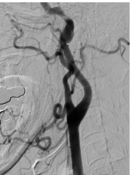
MRA上,血栓が縮小していく様子が確認された(Fig. 4).発症 第 45 日目に血管造影検査を施行し,左 ICA 起始部に明らか な血栓透亮像や内膜のびらん,解離腔を認めなかった(Fig. 5). 今回,その他の塞栓源検索として Holter 心電図や経胸壁心エ コー,凝固因子異常・膠原病・血管炎マーカーなどの検査を 施行したが有意な所見を認めなかった.腫瘍マーカーの検査 は行っていないが,胸部~骨盤部の単純 CT では明らかな腫 瘍性病変や大動脈弓部の石灰化は疑われなかった.また,経 食道心エコー検査はプローベ挿入に際しての怒責による血栓 の飛散を危惧し今回施行しなかった.代替検査として経頭蓋 ドプラ下で気泡を静注し,Valsalva 負荷・解除をさせたとこ ろ,明らかな HITS を認めず右左シャントの存在も示唆され なかった.経過とともに右上肢麻痺・失語は改善し,上腕は 軽度挙上できるようになり簡単な内容の会話も可能となった (NIHSS: 7 点,mRS: 4).一方,高度な右下肢麻痺は後遺した ため,発症第 47 日目に回復期リハビリテーション病院へ転院 した(Fig. 6).
Fig. 3 Neck CT-angiography on the second day in hospital.
Neck CT-angiography shows a filling defect at the origin of the left internal carotid artery (ICA) (arrows).
Fig. 4 Temporal changes of thrombus on neck MRA (upper: time of flight, lower: source image).
Neck MRA shows sequential changes in size of thrombus at the origin of the left internal carotid artery (ICA). Panels A through D depict images at the 2nd day (A), 7th day (B), 17th day (C), 26th day (D), respectively.
本例は頸部の内頸動脈内に発生した血栓が主因と考えられ た塞栓性脳梗塞であるが,その血栓の成因や経過は一般的な 動脈硬化に伴ったものとは異なると考えられる.ICT に関す る報告はこれまでに多くなされており,脳虚血症例において のその有病率は 0.05~0.7%とされている1).動脈硬化に起因 した ICT の場合は当然内頸動脈起始部が好発部位となるが, そ れ 以 外 の 原 因 に よ る 動 脈 内 血 栓 で あ れ ば, 総 頸 動 脈 (common carotid artery; CCA)や siphon 部・床上部などの報
告も少なからずみられる2)~7).これは,いずれの部位であっ ても血管の分岐および屈曲という要素に,炎症や凝固異常な どの要因が加わり動脈内血栓を生じるものと思われ,発症部 位の多様性に寄与しているものと考えられる. ICTの成因についてこれまでの報告では,動脈硬化巣のプ ラーク破綻8)9)や鉄欠乏性貧血・本態性血小板増多症10),外 傷11)12),一酸化炭素中毒13),薬剤(ステロイド14))などの 様々な疾患や背景因子の関与が考察されている.比較的若年 に ICT を認める場合には,単なる動脈硬化性変化以外の因子 が基盤にあることが多いようである.本例では当初,鉄骨解 Fig. 5 Left carotid angiography on the 46th day after admission.
Angiography shows no filling defect at the origin of the left internal carotid artery (ICA).
Fig. 6 The clinical course.
On admission, anticoagulation therapy was initiated and continued. Aphasia, right facial palsy and upper limb palsy were gradually improved. His final NIHSS was 7 and the patient was transferred to a rehabilitation hospital on the 47th day after admission.
体業に従事していた背景から作業中に受けた左頸部の鈍的外 傷で血管内皮障害を起こした可能性が考えられたが,後の病 歴聴取により明らかな外傷機転は働いていないことが判明し ている.また若年ではあるが脂質異常症・喫煙歴があること と頸動脈エコーで所々に軽度の動脈硬化性変化を認めていた ことから,動脈硬化巣が血栓形成の基礎病変となっていた可 能性も考えられたが,血栓消失後にその近辺に明らかな動脈 硬化性変化は確認されなかった.本例では検索した限りで特 殊な基礎疾患を認めておらず,頸動脈内膜剝離術や血栓回収 術なども行っていないためその明確な原因は不明であるが, 頸動脈エコーや CTA,血管造影などの画像所見15)16)からは動
脈硬化以外の何らかの病態が関与した free floating thrombus (FFT)と考えられた.FFT についてもこれまでに報告があり (Table 1),脳卒中患者の中で本症例のような狭窄病変を有し ない FFT の頻度は 0.18%17)と決して高くはないが,FFT 患 者の 90%以上は神経症状を合併する18)とされているため早 急かつ的確な治療が要される. ICTの治療に関しては,成因として凝固カスケードの亢進 が関与していることを踏まえて抗凝固療法が選択された報告 が多いが,今のところ定まった見解はない.また,抗凝固療 法中の注意点としては,治療中の血栓の遠位への移動・飛散 が挙げられる.このような事態を避けるために発症後早期に 外科的治療を検討される症例もあり,血栓除去術,および頸 動脈ステント留置術や内膜剝離術などを試みた報告が散見さ れる4)19)~22).また内科的治療に抵抗性の場合や,内科的治療 後に狭窄病変が残存していた場合にも待機的に外科的治療を 行うことで,良好な経過を得たとする報告もある.一方で, 不安定プラークにステント留置術を行うことにより血栓形成 が加速する可能性も示唆されている20).これら治療法につい てはまとまった症例数での報告はなく,症例ごとにリスクを 勘案して選択せざるをえないのが現状である.ただし,抗凝 固療法を主とした内科的加療で良好な経過を辿る報告が多い ことは明記されるべき事実であろう.本症例の場合は,治療 を行っている間に動脈硬化巣が血栓の基盤にあるかを判断す ることは困難であった.しかし,早期から抗凝固療法を選択 し継続することで血栓が消退する様子が確認され,脳梗塞の 再発を起こすことなく経過した. 頸動脈エコーは ICT を経時的に観察し,抗凝固療法の効果 を確認する上で最も有用な検査と考えられる.造影 CT 検査 や血管造影検査に比べて非侵襲的で長時間を要することな く,またベッドサイドで繰り返し行えるため,連日の観察と その結果をもとに治療強度の調整が可能となる.中野らは, 経時的なエコーでの観察により ICA 起始部に存在していた血 栓の消失を確認し,より遠位の血栓が移動・消失する様子を 血流速度の変化で推定できたと報告している23).本症例でも 同様な観察を行うことで,抗凝固療法管理の一助とすること ができた.頸動脈エコーを中心とした非侵襲的検査と,血管 撮影や造影検査等の侵襲的検査を適宜組み合わせることで, より適切な治療の選択・調整が可能であると考えられる. 以上,頸部内頸動脈に発生した動脈内血栓に因って脳塞栓 を発症した症例を報告した.ICT に対してはその成因や病態 を考慮しつつ迅速な抗血栓療法が必要であり,その効果を確 認するために頸動脈エコーは有用であった.本例の治療方針 としては内科的・外科的双方のリスクと効果を総合的に判断 tuberculosis, etc
Cortés-Vicente, et al, 20155) 37 F R (CCA) myxoma AC and surgery Santoro, et al, 201525) 59 F R meningioma susp. AC and AP Tan, et al, 201426) 46 M R lymphadenopathy, rupture of plaque by
massage
AC and AP, and delayed T Teodoro, et al, 201413) 46 F L CO intoxication AP and HBO
Vidale, et al, 201327) 48 M R MTHFR A1298C mutation AP and folic acid, and delayed CEA Batur, et al, 201310) 41 F L iron deficiency anemia AC and OIP
Vellimana, et al, 201319) 20–70ʼs M: 2 F: 5 R: 2 L: 4 B: 1 anemia, thrombocytosis, APS, etc AC and AP, or AC only Graham, et al, 20136) 64 M R (CCA) esophageal carcinoma AC
Elijovich, et al, 20137) 37, 63 M: 2 R, L (CCA) sarcoidosis, dissection susp. AC Present case 49 M L unknown AC
Abbreviations M: male, F: female, R: right, L: left, B: both, ICA: internal carotid artery, CCA: common carotid artery, CO: carbon monoxide, APS: antiphospholipid syndrome, AP: antiplatelet therapy, AC: anticoagulaiton therapy, OIP: oral iron preparation, HBO: hyperbaric oxygenation, CEA: carotid endarterectomy, CAS: carotid artery stenting, T: thrombectomy.
基礎疾患の評価と,綿密な画像検査および頸動脈エコーを 行った症例の蓄積が重要と考えられた. 本報告の要旨は,第 208 回日本神経学会九州地方会で発表し,会長 推薦演題に選ばれた. ※本論文に関連し,開示すべき COI 状態にある企業,組織,団体 はいずれも有りません. 文 献
1) Tateishi Y, Tsujino A, Hamabe J, et al. “Snake Fang” sign without carotid stenosis on duplex ultrasonography indicates high risk of artery-to-artery embolic stroke. J Neuroimaging 2014;24:407-410.
2) Karapurkar AP, Singh R, Teelala SM, et al. Free-floating thrombus in a stenotic supraclinoid left internal carotid artery. BMJ Case Rep 2016;2016. pii: bcr2015213067.
3) Naganuma M, Inatomi Y, Kobayashi O, et al. Reappearance of free-floating carotid thrombus after discontinuation of anti-coagulant therapy. J Stroke Cerebrovasc Dis 2015;24:e45-e47. 4) Yamagami H, Kitagawa K, Ohtsuki T, et al. Embolic cerebral
infarction caused by intraluminal thrombus in the carotid siphon successfully treated with combination of anticoagulant and antiplatelet drugs. Circ J 2005;69:1147-1149.
5) Cortés-Vicente E, Delgado-Mederos R, Bellmunt S, et al. Stroke caused by a myxoma stenosing the common carotid artery. J Stroke Cerebrovasc Dis 2015;24:e87-e89.
6) Graham R, Blaszczynski M. Spontaneous free floating carotid artery thrombosis. BMJ Case Rep 2013;2013. pii: bcr2013008710. 7) Elijovich L, Mainali S, Doss V, et al. Medical management of
free-floating carotid thrombus. Clin Neurol Neurosurg 2013; 115:1532-1535.
8) Buchan A, Gates P, Pelz D, et al. Intraluminal thrombus in the cerebral circulation. Stroke 1988;19:681-687.
9) Choi HY, Ye BS, Ahn SH, et al. Characteristics and the fate of intraluminal thrombus of the intracranial and extracranial cerebral arteries in acute ischemic stroke patients. Eur Neurol 2009;62:72-78.
10) Batur Caglayan HZ, Nazliel B, Irkec C, et al. Iron-deficiency anemia leading to transient ischemic attacks due to intraluminal carotid artery thrombus. Case Rep Neurol Med 2013;2013: 813415.
11) Mahalangikar R, Kumar A, Sharma BS. Transorbital penetrating intracranial injury with an umbrella wire causing cavernous internal carotid artery injury and thrombosis. World Neurosurg 2016;86:513.e15-e18.
12) Karnecki K, Jankowski Z, Kaliszan M. Direct penetrating and indirect neck trauma as a cause of internal carotid artery thrombosis and secondary ischemic stroke. J Thromb Thrombolysis 2014;38:409-415.
cation. Am J Emerg Med 2014;32:684.e5-e6.
14) Karapanayiotides T, Kouskouras K, Ioannidis P, et al. Internal Carotid Artery Floating Thrombus in Relapsing Polychondritis. J Neuroimaging 2015;25:142-144.
15) Jaberi A, Lum C, Stefanski P, et al. Computed tomography angiography intraluminal filling defect is predictive of internal carotid artery free-floating thrombus. Neuroradiology 2014;56: 15-23.
16) Thornhill RE, Lum C, Jaberi A, et al. Can shape analysis differentiate free-floating internal carotid artery thrombus from atherosclerotic plaque in patients evaluated with CTA for stroke or transient ischemic attack? Acad Radiol 2014;21:345-354. 17) Vassileva E, Daskalov M, Stamenova P. Free-floating thrombus
in stroke patients with nonstenotic internal carotid artery-an ultrasonographic study. J Clin Ultrasound 2015;43:34-38. 18) Bhatti AF, Leon LR Jr, Labropoulos N, et al. Free-floating
thrombus of the carotid artery: literature review and case reports. J Vasc Surg 2007;45:199-205.
19) Vellimana AK, Kadkhodayan Y, Rich KM, et al. Symptomatic patients with intraluminal carotid thrombus: outcome with a strategy of initial anticoagulation. J Neurosurg 2013;118:34-41 20) Tsumoto T, Terada T, Tsuura M, et al. Carotid artery stenting
for stenosis with intraluminal thrombus. Neuroradiology 2006;48:54-59.
21) Kwon TH, Kim BM, Nam HS, et al. Carotid stenting in acute ischemic stroke patients with intraluminal thrombus. Neuroradiology 2011;53:773-778.
22) Takayama K, Taoka T, Nakagawa H, et al. Carotid artery stenosis with intraluminal thrombus discovered during carotid artery stenting. Jpn J Radiol 2009;27:367-370.
23) 中野正紹,緒方利安,矢坂正弘ら.頸部血管エコー検査で oscillating thrombusを経時的に観察し得た心原性脳塞栓症の 1例.脳卒中 2007;29:474-478. 24) 大多和賢登,錦古里武志,渡辺賢一ら,頚動脈浮遊血栓に対し 頚動脈エコーガイド下に血管内治療を行った 1 例 . No Shinkei Geka 2016;44:489-494.
25) Santoro L, Schinzari F, Di Veronica A, et al. Carotid free-floating thrombus in woman with meningioma: a case report and review of the literature. Eur Rev Med Pharmacol Sci 2015;19:1442-1445.
26) Tan AP, Taneja M, Seah BH, et al. Acute free-floating carotid artery thrombus causing stroke in a young patient: unique etiology and management using endovascular approach. J Stroke Cerebrovasc Dis 2014;23:e437-439.
27) Vidale S, Bellocchi S, Caronno R, et al. Intra-operative detection of carotid free-floating thrombus in a symptomatic patient with a methylenetetrahydrofolate reductase gene mutation. Case report and literature review. Acta Neurochir(Wein) 2013;155: 1125-1126.
1)Department of Cerebrovascular Medicine, Saga-ken Medical Centre Koseikan