Detection and Isolation of Marine
Bacteriophage Systems in the Southwestern Part
of the Pacific Ocean
著者
HIDAKA Tomio
journal or
publication title
鹿児島大学水産学部紀要=Memoirs of Faculty of
Fisheries Kagoshima University
volume
26
page range
55-62
別言語のタイトル
南方海域における海洋バクテリオファージ系の検索
と分離
Vol. 26 pp. 55-62 (1977)
Detection and Isolation of Marine Bacteriophage Systems
in the Southwestern Part of the Pacific Ocean*Tomio Hidaka**
Abstract
During the cruise for the marine ecological survey in the southwestern part of Pacific Ocean, 47 of sea water samples were obtained from various depth layers at 13 stations between Kago shima and Guam via Palau. A total of 576 cultures of bacteria isolated from the sea water samples, of which 72 cultures were sensitive to the bacteriophage lysates enriched from the sea water. It is found that the bacteriophages are not present in high concentrations in the ocean, or occur only sporadically in water layers, from surface to 50 m depth, harboring large
bacterial population.
The host bacteria of the isolated marine bacteriophage systems belong to Pseudomonus (2 strains), Vibrio (3 strains), Photobacterium (1 strain), and Lucibacterium (1 strain). These bacteri ophage systems form clear or turbid plaque of about 1-2 mm in diameter. The all of them
are virulent phage systems.
Bacteriophages could exert a considerable influence on controlling natural bac terial populations in limiting the numbers, types, and duration of active population growth and ultimately, through possible genetic exchange mechanisms, on the bio chemical capabilities of bacteria. These phenomena are of considerable potential importance because of the prominent role of bacteria as mineralizing and chemical
transforming agents within the environment. However, the information on marine
bacteriophages is relatively scant. The ecological significance of bacteriophages in the marine environment is not known. The author has been isolating marine bac teriophages from sea water and marine mud samples collected from several stations in some ten miles off the south coast of Kyushu, Japan, and investigated the mor phological and biological characters of the isolates (Hidaka, 1971; Hidaka and Fujimura, 1971a; Hidaka and Fujimura, 1971b; Hidaka, 1972; Hidaka and Ichida, 1972; Hidaka, 1973; Hidaka and Shirahama, 1974; Hidaka, 1975; Hidaka and
Ichida, 1975). Now, it is very interesting to compare with marine bacteriophages
in coast and in ocean.
During the third cruise of Keiten Maru as part of the spe
cial research project for the marine ecological survey in the southwestern part of Pa
cific Ocean, the author dealt with investigations into the ecological system of micro
organisms, especially bacteriophage systems in the ocean. The present paper
de-* This work was presented at the Annual MeetingofJapanese Society of Scientific Fisheries, Tokyo,
April 4, 1977.
56 Mem. Fac. Fish., Kagoshima Univ. Vol. 26 (1977)
scribes the detection and isolation of marine bacterium-bacteriophage systems in sea
water samples collected from the ocean.
Materials and Methods
Media used. The sea water broth (SWB) contained 5 g polypeptone and 1 g yeast extract in a liter of Herbst's artificial sea water. The pH of the medium was adjusted to 7.6-7.8. Solid media were prepared by adding agar at either 1.5% for sea water agar (SWA) or 0.5% for soft sea water agar (sSWA) to the sea water broth.
Sea water samples. The sea water samples were collected with steril bacteri ological J-Z water samplers from five layers, 1-3, 50, 100, 200, and 300 m of depth, at each of 13 stations. The samples of water were immediately examined in a la boratory aboard.
Estimation and isolation of marine heterotrophic bacteria in the sea water samples. Most probable number technique was used for the estimating bacterial population in the sea water samples, using five SWB tubes for three sample sizes, 1 ml, 0.1 ml, and 0.01 ml, respectively. The tubes were incubated for 3 days and observed growth. The numbers of positive tubes in each set of five were tabu lated and consulted the appropriate table. The most plobable number of bacteria for each sample was calculated for 100 ml of sample. On the other hand, a 0.2 ml of each sample was also cultured on a SWA plate for 6 days. Five plates were used for each sample to be tested. The colonies developed on the agar plates were count ed, and then the viable cell counts per 1 ml of the samples were calculated. After that the all colonies were transferred to agar slopes of the same composition. The incubations were done in air-conditioned room temperature, 23-25°C.
Microbiological tests of isolated bacteria. The characterization tests of the host bacteria were done using standard methods (Harring and McCance, 1966). Identifications of them were carried out according to the systems outlined by Hendrie and Shewan (1966) and Bergey's Manual (8th ed). Incubations were at 25°C, unless otherwise stated.
Detection of bacteriophage. This was experimented according to the enrich ment method. To each of sterile shaking flasks was added 200 ml of SWB together with 5 ml of SWB culture inoculated 1 ml of each sea water sample and incubated for 2-3 hours. After then the young culture was mixed with 250 ml of the sea water sample. The mixtures were incubated overnight, and were then put into a cold chamber to bring back to land. At the university laboratory, the cultures were
cen-trifuged at 4,500 G for 30 min and filtered through HA Millipor membrane filters.
The filtrates were spoted onto double agar layer plates seeded each of marine bac teria isolated. These preparations were incubated overnight and examined the ap pearance of lysis zone.
Isolation of the bacteriophages. Material from the center of clear zones de
appropriate bacterium and the inoculated cultures were incubated for a further 18 h
at 25°C. Each of the cultures was then filtered and a portion of the filtrate was
mixed with the appropriated bacterium in 3 ml of soft SWA melted and cooled to 45°Cand the mixture was layered onto the surface of an agar plate. The double agar layer
plates were incubated overnight. Single plaques arising by this method were picked
and the cycle was repeated three times to ensure their identity and assist purification.
The phage lysates were stored at 5-8°C.Results and Discussion
In the cruise of the Keiten Maru from Kagoshima to Guam via Palau during the
period from October 25 to November 19, 1976, microbiological samplings were car
ried out at 13 stations. The track and the microbiological stations are shown in
Fig. 1 and Table 1.
Distribution of heterotrophic bacteria in the sea water. In sea water at
various depths of the stations, the numbers of heterotrophic bacteria as host for ma
rine bacteriophages are shown in Table 2. The numbers indicate viable cell counts58 Mem. Fac. Fish., Kagoshima Univ. Vol. 26 (1977)
Table 1. Microbiological stations in southwestern part of the Pacific Ocean.
Station No.
Position
Latitude Longitude
Date and time of sampling
Date Time (in 1976) (1) 27°46.5,N— 13ri2.0'E Oct. 26 10:00—10:20 (2) 22°57.6/N—132°10.2/E Oct. 27 10:00—10:20 (3) 18°12.6'N—133°03.3'E Oct. 28 10:00—10:20 (4) 13°29.6'N— 134°07.5'E Oct. 29 10:00—10:20 (5) 08°43.0'N — 134°46.0,E Oct. 30 10:00—10:20 1 06°00.rN— 134°59.8/E Nov. 4 19:30 — 21:00 2 07°00.0,N— 136°36.4'E Nov. 5 07:00 — 08:30 3 07°59.0,N— 138°12.9'E Nov. 5 19:00 — 20:30 4 09°00.4'N — 139°50.6'E Nov. 6 07:30 — 09:00 5 10°02.3'N — 141°09.7'E Nov. 7 11:30—13:00 6 H°00.3'N—142°06.4'E Nov. 7 20:30 — 22:00 7 12°00.0'N—143°06.4'E Nov. 8 07:30 — 09:00 8 13°00.0'N—143°56.0'E Nov. 8 17:00—18:30
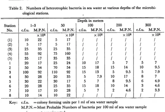
Table 2. Numbers of heterotrophic bacteria in sea water at various depths of the microbi
ological stations.
Depth in meters
Station 1-3 50 100 200 300
No. c.f.u. M.P.N. c.f.u. M.P.N. c.f.u. M.P.N. c.f.u. M.P.N. c.f.u. M.P.N.
Xl0« XlO* X102 X102 X102 (1) 10 22 5 17 / / / / / (2) 5 17 5 17 / / / / / (3) 25 35 25 35 / / / / / (4) 55 92 75 160 / / / / / (5) 35 17 35 35 / / / / / 1 20 17 25 24 10 17 5 7 5 7 2 50 54 70 54 15 18 15 14 10 9.5 3 100 92 110 92 15 13 5 9.5 5 7.9 4 30 28 20 35 5 7.9 10 17 8 7 5 30 22 30 17 5 7 8 9.5 6 4.9 6 20 28 25 35 15 18 10 14 3 4.6 7 10 17 10 28 5 7 2 4.6 2 3.3 8 10 14 10 22 / / / / / /
Key: c.f.u. = colony forming units per 1 ml of sea water sample
M.P.N. = Most Probable Numbers of bacteria per 100 ml of sea water sample / =not experimented
(colony forming units, c.f.u.) per 1 ml of sample and most probable number (M.
P. N.) of bacteria per 100 ml of sample.
The measured values by two defferent me
thods almost agree.
It is decided in Table 2 that the heterotrophs are found at every
depth below the surface to 300m at the stations and usually more abundant at a
depth of 50 m than in water near the surface.
Even at' a depth of 50 m, the range
of the numbers of bacteria per ml of sea water is from a few to a hundred. The sam
ples from the 100 m depth contain from 5 to 18 bacteria per ml. Beginning with the
data recorded in Table 2 suggest that the bacterial population is generally most abun
dant in the photosynthetic zone near depth 50 m and then decreases with depth at
various stations. Table 2 also shows that the quantity of heterotrophs is more abun dant at three stations (4), 2, and 3 than at other stations. An increase in the num
bers of heterotrophs usually occurred at the upper layers of water at the junction of
currents, where the amount of organic substances was relatively high. One must
assume that these conditions are reproduced to some extent at the interface of some
layers in a water mass.
Detection of marine bacteriophage systems. The marine bacteriophages in
the sea water samples were detected by lytic action for the bacteria isolated from the
same samples. A total of 576 cultures of bacteria isolated from the sea water sam
ples, of which 72 cultures were sensitive to the bacteriophage lysates enriched from
the sea water. However, it was difHcalt to detect of phage action to the isolated bacteria by direct method from the sea water samples. These 72 bacteriophage sys tems were divided broady into seven systems (tentative names: A, B, C, D, E, F, and G group) by host-phage cross infection method. The distribution of the seven marine bacteriophage systems in the every stations are shown in Table 3. As in
Table 3. Distribution of marine bacteriophage systemsisolated.
^\Depth (meters) 1-3 50 100 200 300 Station No. (1) A, D A, D / (2) A, D, E A, D, E / (3) C, D C, D / (4) D, E C, D, E / (5) D C, D / 1 A, D, G A, D G — — 2 A, D, E A, C, D, E A — — 3 A, D, E A, D, E F — — 4 A, B, C A, B, C, E G C — 5 B, C, E B, C, E, F — — — 6 A, C A, C, E — — — 7 D A, D — — — 8 A, D A, D / / /
A and B, Pseudomonas; C, D and E, Vibrio; F, Photobacterium; G, Lucibacterium. —, not detected; /, not experimented.
Table 3, bacteriophages are detected in the layers from surface to 50 m depth at all of the stations, and the composition of them is almost same in the layers, but it doesn't always follow that it is same at the every stations. In the layers below 100 m depth at the majority of the station bacteriophages are not found. It is indicated that the bacteriophage is not present in high consentrations in the ocean or that it occurs only sporadically in water layers harboring large bacterial populations.
60 Mem. Fac. Fish., Kagoshima Univ. Vol. 26 (1977)
Table 4. Brief characterization of the representative host bacterium strain of each seven groups.
Group A B C D E F G 6YK-strain No. 3212 5201 1102 2202 3210 3306 1303 Cell form R R R R R R R Gram's stain — — — — — . . — . — Flagellation M M M M M M P Kovacs' oxidase + + ' 4- + + — +
Hugh & Leifson test NC O F F F F F
Sensitivity to 0/129 - - + + + + — Arginine dihydrolase + + — — + + — Gelatin hydrolysis + + + + + — + Starch hydrolysis — + + — + — + Growth in 7.5% NaCl + + + + + + + Growth at 37°C — — — — — — — Luminescence — — — — — + + Pigments - - - — Indole production — — — — + — + Nitrate reduction + + + + + + + H2S production - + - — - - — V. P. test — — — — — + — M. R. test - - + + - - +
Key: R, rods; M, monotrichous; P, peritrichous; —, negative; +, positive; O, oxida tive; F, fermentative; NC, growth with no change in reaction.
brief characterization of the representative host bacterium strain of each seven groups is shown in Table 4. All of them are aerobic or facultative anaerobic Gram-negative rods and psychrophilic bacteria. They are typical marine bacteria. The two strains of them, 6YK-3212 and 6YK-5201, are motile by polar monotrichous; oxidase (Kovacs5), positive; and carbohydrate metabolism, respiratory. Other three strains, 6YK-1102, 6YK-2202, and 6YK-3210, are motile by a single polar flagellum; oxi dase (Kovacs'), positive; carbohydrate metabolism, fermentative; and sensitive to vibriostatic compound (0/129). The rest, 6YK-3306 and 6YK-1303, are charac
terized by luminescence. And one of the two strains, 6YK-3306 is motile by polar flagella; oxidase (Kovacs5), negative; sensitive to 0/129. On the other hand, 6YK-1303 is motile by peritirichous flagella; oxidase (Kovacs'), positive; and insensitive to 0/129. They belong to the following genera: 6YK-3212 and 6YK-5201,
Pseudo-monas; 6YK-1102, 6YK-2201, and 6YK-3210, Vibrio; 6YK-3306, Photobacterium; and 6YK-1303, Lucibacterium.
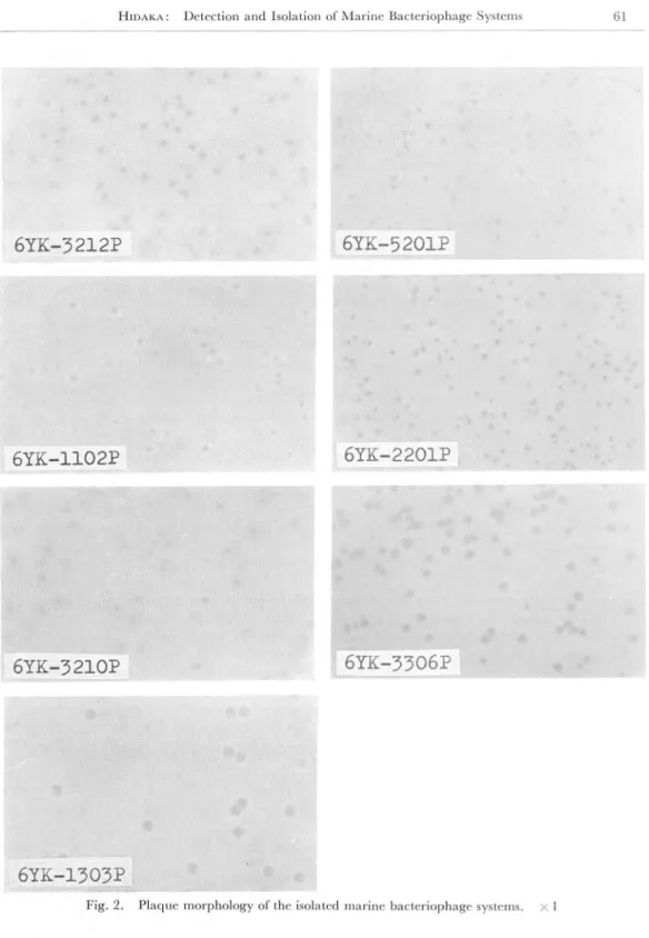
The plaque morphorogy of the isolated marine bacteriophage systems was examin ed by the double agar layer technique. The appearances of plaque formed by the seven representative bacteriophage systems are shown in Fig. 2. The bacteriophage systems produce clear or turbid plaque of about 1-2 mm in diameter.
The titers of these phage lysates are of 108"9 plaque forming units (p.f.u.) per ml
6YK-3212P
6YK-5201P
6YK-1102P
6YK-2201P
.„.m
6YK-3210P
6YK-3306P
6YK-1303P
62 Mem. Fac. Fish., Kagoshima Univ. Vol. 26 (1977)
Acknowledgement
The author thanks to all members of the survey team for their many helpful discus sions. The author also wishes to express gratitude to Captain, Mr. T. Henmi and crews of Keiten Maru, a research ship of Kagoshima University, for their very able and kind assistance with the sampling of sea water.
References
Bain, N. and J. M. Shewan (1968): Identification of Aeromonas, Vibrio and related organisms, in
"Identification Methods for Microbiologists, Part B" (B. M. Gibbs and D. A. Shapton, ed.),
79-84, Academic Press, New York.
Buchanan, R. E. and N. E. Gibons (1974): "Bergey's Manual of Determinative Bacteriology, 8th
Ed.', The Williams and Willkins Co. Baltimore.
Harrigan, W. F. and M. E. McCance (1966): "Laboratory Methods in Microbiology", Academic
Press, New York.
Hendrie, M. S. and J. M. Shewan (1966): The identification of certain Pseudomonas species, in "Identification Methods for Microbiologist, Part A" (B. M. Gibbs and F. A. Skinner, ed.),
1-7, Academic Press, New York.
Hidaka, T. (1971): Isolation of marine bacteriophages from sea water. Bull. Jap. Soc. Fish., 37, 1199-1206.
Hidaka, T. and T. Fujimura (1971a): A morphological study of marine bacteriophages. Mem.
Fac. Fish. Kagoshima Univ., 20 (1), 141-154.
Hidaka, T, and T. Fujimura (1971b): On the heat- and chloroform-resistance of marine bacterio
phages. Mem. Fac. Fish. Kagoshima Univ., 20 (1), 155-158.
Hidaka, T. (1972): On the stability of marine bacteriophages. Bull. Jap. Soc. Sci. Fish., 38, 517-523.
Hidaka, T. and K. Ichida (1972): On the undesirable effects of filtration through membrane filter of some marine bacteriophages. Mem. Fac. Fish. Kagoshima Univ., 21 (1), 97-102.
Hidaka, T. (1973): Characterization of marine bacteriophages newly isolated. Mem. Fac. Fish.
Kagoshima Univ., 22 (1), 47-61.
Hidaka, T. and T. Shirahama (1974): Preliminary characteristics of a temperate phage system isolated from marine mud. Mem. Fac. Fish. Kagoshima Univ., 23, 137-148.
Hidaka, T. (1975): Identification of the type of nucleic acid in marine bacteriophages with acridine
orange staining. Mem. Fac. Fish. Kagoshima Univ., 24, 133-138.
Hidaka, T. and K. Ichida (1976): Properties of a marine RNA-containing bacteriophage. Mem.