F-18-FDG Positron Emission Tomography
Findings Correlate Pathological Proliferative Activity
of Oral Squamous Cell Carcinoma
Osamu Toyoizumi,
Noboru Oriuchi,
Mitsuyuki Miyakubo
Tomohiro Ishikita,
Yoshiki Nakasone,
Akihide Negishi
Keigo Endo,
Takashi Nakajima,
Kenji Mogi
and Satoshi Yokoo
Background: It is still controversial whether FDG uptake is correlated with cellular proliferation and prognosis of oral squamous cell carcinoma(OSC). In this study,we performed PET study and immuno-histochemical analysis to elucidate the relationship between FDG uptake and expression of cellular proliferative markers and pathological prognostic markers in patients with OSC. M ethods: FDG PET and immunohistochemical staining have been carried out in sixteen patients with OSC. Tumor uptake of FDG was expressed with standardized uptake value(SUV). The expression of Ki-67,Topoisomerase IIα(Topo IIα),p53,and p63 in cancer cells was quantitatively assessed with positivity of the immunohis-tochemical staining. SUV was compared with the results of immunohisimmunohis-tochemical analysis. Results: FDG PET study revealed that SUV ranged from 3.6 to 22.1 with average of 10.4. Average positive rate of Ki-67,Topo IIα,p53,and p63 was 68.9%,58.9%,72.0%,and 65.2%,respectively. Pearson product-moment correlation coefficient analysis revealed that SUV was significantly correlated with Ki-67 (r= 0.616, p=0.01), Topo IIα (r=0.677, p=0.004), p53 (r=0.613, p=0.01), and p63 (r=0.710, p=0.002), respectively. Conclusion : The present preliminary study indicated that FDG uptake was closely correlated with pathological cellular proliferative and prognostic markers in patients with OSC.
(Kitakanto Med J 2010;60:1∼8)
Key Words: Oral squamous cell carcinoma,FDG PET,Cellular proliferation,p53,Topoisomerase IIα, p63
Introduction
Oral squamous cell carcinoma (OSC) is an ordi-nary type of oral cancer. It is usually found by clini-cal examinations such as simple inspection and palpa-tion ; however, it is difficult to evaluate the precise state of tumor progression. The accurate assessment of the tumor is important because the treatment of OSC is based on the accurate clinical TNM classification. Recent advances in diagnostic imaging methods such as CT, MRI and PET have improved the diagnostic accuracy of OSC and the prognosis of patient with
OSC.
In the field of head and neck tumors including OSC, 2-deoxy-2-[F-18] fluoro-D-glucose (FDG) PET and CT/MRI have shown increased diagnostic accuracy, but FDG PET has been more advantageous than CT/MRI to detect tumor recurrence and lymph node metastasis, secondary or occult malignancy, and distant metastasis. FDG PET is a functional imaging which demonstrates increased glucose uptake as a metabolic marker for malignant tumor. FDG uptake in the tumor is influenced by the expression of glucose transporters and hexokinase,which play a key
1 Departments of Oral and Maxillofacial Surgery 2 Diagnostic Radiology and Nuclear Medicine,Gunma University Graduate School of Medicine,3-39-22 Showa-machi,Maebashi,Gunma 371-8511,Japan 3 Pathology Division,Shizuoka Cancer Center Hospital, Nagaizumi-cho, Sunto-gun, Shizuoka 411-8777, Japan
Received : December 28, 2009 Accepted : January 12, 2010
Address: NOBURO ORIUCHI Department of Diagnostic Radiology and Nuclear Medicine,Gunma University Graduate School of Medicine, 3-39-22 Showa-machi, Maebashi, Gunma 371-8511, Japan
role in glucose uptake and glycolysis in the cell. Malignant cells are known to have increased glucose consumption as their substrate for energy production or the maintenance of accelerated proliferative activity. Although FDG uptake is correlated with cellular proliferation and prognosis in many types of cancer,it is still controversial whether FDG uptake is correlated with the histological markers of cellular proliferation and prognosis in OSC. In this study, we aimed to elucidate the relationship between FDG PET and immunohistochemical markers for cellular prolifera-tion and prognosis in patients with OSC.
Patients and methods
PatientsWe studied 16 patients (10 females and 6 males, age from 52 to 82 year-old) with OSC at our institu-tion from May 2003 to June 2006. Primary site of OSC in all patients was shown in Table 1. The pathological tumor staging was based on the TNM-classification by the International Union against Can-cer.
The origin of OSC was gingiva in eight, floor of mouth in four,tongue in two,and cheek and palate in one patient each. Ten patients showed marked ker-atinization and histologically classified into well dif-ferentiated carcinoma. All patients underwent resec-tion of the tumor within one month after the FDG PET study, and histological examination was perfor-med on the tumor specimen. Postoperative clinical course was assessed for more than 24 months in all patients. Existence or absence of recurrence was cen-sored at 24 months after the surgery.
FDG PET study
Fluorine-18 was produced in an in-house cyclo-tron (BC1710, Japan Steel Works, Muroran, Japan), and FDG was synthesized by the method that has been reported. PET images were obtained with a whole-body PET scanner (SET 2400W, Shimadzu Corpora-tion, Kyoto, Japan) with a 59.5 cm transaxial field of view, 20 cm axial field of view, which produced 63 image planes,spaced 3.125 mm apart. Transaxial spa-tial resolution was 4.2 mm full width at half maximum (FWHM) at the center of the field of view and axial resolution was 5.0 mm FWHM. A whole-body imag-ing by the simultaneous emission-transmission method with a rotating external source (Ge-68/Ga-68) was started at 60 min after the injection of 5 MBq (185 mCi)/kg (body weight) FDG. Four to five bed posi-tions from the head to the thigh were imaged for 8 min per position. Patients fasted for at least 6 hours before FDG injection. The study protocols of FDG PET were approved by the Institutional Review Board, and all the patients gave informed consent to be in-cluded in the study and undergo the examination.
Attenuation-corrected transaxial images were reconstructed by the ordered subsets expectation max-imization (OS-EM) algorithm into 128×128 matrix with pixel dimensions of 4.0 mm in a plane and 3.125 mm axially. Finally, 3 consecutive slices were added to generate transaxial images with slice thickness of 9.8 mm for visual interpretation and quantitative analysis by using standardized uptake value (SUV). Coronal images with slice thickness of 9.8 mm were also recon-structed from transaxial images.
PET data were visually interpreted by two nuclear physicians independently, and statistical analysis was performed using SUV.
Table 1 Characterictics of Patients with Squamous Cell Carcinoma of the Oral Cavity
No. of
Patients Sex Age Localization TNM Classification(pStage) DifferentiationHistological Prognosis
1 M 82 Tongue T2N0M0 (Ⅱ) Well No recurrence
2 F 81 Gingiva, Mandible T4N0M0 (Ⅳ) Well Dead
3 M 73 Gingiva, Mandible T2N0M0 (Ⅱ) Well No recurrence 4 M 52 Floor of mouth T1N0M0 (Ⅰ) Well No recurrence 5 M 64 Floor of mouth T2N0M0 (Ⅱ) Well No recurrence 6 F 61 Gingiva, Maxilla T3N0M0 (Ⅲ) Well Recurrent
7 M 75 Tongue T2N0M0 (Ⅱ) Well No recurrence
8 F 68 Gingiva, Mandible T2N0M0 (Ⅱ) Well Recurrent 9 F 66 Floor of mouth T1N0M0 (Ⅰ) Well No recurrence
10 F 65 Cheek T1N1M0 (Ⅲ) Well No recurrence
11 M 76 Gingiva, Maxilla T4N0M0 (Ⅳ) Moderate Dead 12 F 72 Gingiva, Mandible T2N1M0 (Ⅲ) Moderate No recurrence 13 M 64 Gingiva, Mandible T2N2bM0 (Ⅳ) Moderate Dead 14 F 71 Gingiva, Mandible T2N0M0 (Ⅱ) Moderate Dead 15 F 70 Floor of mouth T2N0M0 (Ⅱ) Moderate No recurrence
16 F 81 Palate T2N2bM0 (Ⅳ) Poor Dead
No recurrence: Alive without recurrence at 24 months after operation,Dead : Died of OSC within 24 months,Recurrence: Recurrence of OSC within 24 months.
Immunohistochemical study
After histopathological diagnosis was established, immunohistochemical staining of Ki-67, Topoisomer-ase IIα(Topo IIα),products of p53 and p64(p53,and p63, respectively) was carried out using 4μm-thick paraffin sections from the resection samples in all OSC cases. The paraffin sections were dewaxed with xylene and endogenous peroxidase activity was block-ed using 3% hydrogen peroxide in methanol solution for 30 min at room temperature. Then the sections were rehydrated in a degraded ethanol series and foll-owed by the treatment of antigen retrieval with micro-wave, if necessary, which was detailed in Table 2. After the treatment with normal horse serum to prevent non-specific staining,the sections were incubated with each primary antibody at 4℃ overnight. Details of primary antibodies used in this study were shown in Table 2. Then, the sections were incubated with biotinylated secondary antibody for 30 min at room temperature, followed by avidin-biotin-peroxidase complex solution according to the manufacturers instructions (VECTASTAIN, Vector Laboratories, Burlingame, CA, USA). The peroxidase was visual-ized with 0.02% 3-3-diaminobenzidine tetrahydroch-loride containing 0.005% H O in 0.01M tris-phosphate buffer, pH7.4. Finally, the sections were counterstained lightly with hematoxylin.
Immunohistochemical staining for Ki-67, Topo IIα, p53, and p63 was quantitatively evaluated by counting 200 to 500 tumor cells in the area where positive nuclei were gathered. Labeling index (LI) was defined as the percentage of immunoreactive tumor cells in totally counted tumor cells. Immunor-eactive tumor cells were defined as nuclear staining for each antibody.
Statistical Analysis
The relationship between LI of Ki-67,Topo IIα, p53, p63 and SUV was statistically assessed using Pearson product-moment correlation coefficient analy-sis. P<0.05 was considered significant.
Results
All patients showed FDG accumulation in the primary tumor of the oral cavity. A representative case is shown in Figure 1. As shown in Table 3,SUV in 10 patients with well differentiated squamous cell carcinoma showed SUV ranged from 3.6 to 22.1 (10.7±6.2). SUV in five patients with moderately differentiated squamous cell carcinoma ranged from 6.1 to 21.2 (10.2±6.2). SUV in a patient with poorly differentiated squamous cell carcinoma was 8.9. SUV showed no correlation with tumor size, lymph node metastasis, nor pathological stage.
Immunoreactivity for Ki-67, Topo IIα, p53, and p63 was detected only in the nuclei, not in the cyto-plasm (Fig.2). In well differentiated squamous cell carcinoma, these markers were usually seen in the periphery of the alveolus. LI of Ki-67 ranged from 36.1% to 90.0% with the average of 68.9%. In 10 patients with well differentiated squamous cell car-cinoma, LI of Ki-67 ranged from 36.1% to 87.3% (63.1%±15.7%). Five patients with moderately dif-ferentiated squamous cell carcinoma showed LI of Ki-67 ranged from 70.5% to 90.0% (78.1%±7.3%). Positive rate of Ki-67 in a patient with poorly differ-entiated squamous cell carcinoma was 81.0%. No significant difference in the LI was noted between well differentiated squamous cell carcinoma and moderate-ly differentiated squamous cell carcinoma.
LI of Topo IIα,p53,and p63 in all patients were from 20% to 86.2% with the average of 58.9%, from 58.6% to 89.8% with the average of 72%, and from 16.2% to 93.5% with the average of 65.2%,respectively (Table 3). Comparison of the results of immunohisto-chemical staining revealed that no significant differ-ence in the expression of Topo IIα, p53, and p63 was noted between well differentiated squamous cell car-cinoma and moderately differentiated squamous cell carcinoma.
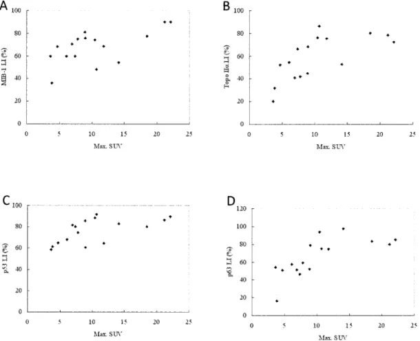
The correlation between SUV and LI of Ki-67 was statistically analyzed. There is a significant corre-lation with the correcorre-lation coefficient (r) of 0.616 and p value of 0.01 (Fig.3A). SUV was also significantly
Table 2 Primary Antibodies and Antigen Retrieval Method for Immunohistochemical Staining
Antibodies Clone Dilution RetrievalAntigen Source
Anti-Ki-67 MIB-1 ×100 B DakoCytomation , Glostrup, Denmark Anti-Topo IIα 3F6 ×50 B Novocastra Lab., Newcastle upon Tyne, UK Anti-p53 DO7 ×25 A Novocastra Lab., Newcastle upon Tyne, UK Anti-p63 Ab-4 ×100 B NeoMarkers, Fremont, CA, USA
A : Antigen retrieval with 20% ZnSO solution for 20 min at 98℃
B : Antigen retrieval with 0.1M citric acid buffer, pH 6.0, for 20 min at 98℃ Topo IIα: Topoisomerase IIα
Fig. 1 A representative case (Patient no. 1) with oral squamous cell carcinoma (A) CT shows an enhanced mass in the tongue on the right side (arrow).
(B) An axial section of FDG PET image shows an increased accumulation of FDG with maximum SUV value of 22.1 (arrow).
(C) Hematoxylin and eosin staining of the specimen obtained by the surgical removal of the tumor shows histological diagnosis of squamous cell carcinoma.
Table 3 Results of F-FDG PET (maximal SUV) and Immunohistochemical Staining (% positivity of stained cells) No. of Patients F-FDG maxSUV Ki-67 (%) Topo-IIα (%) p53 (%) p63 (%)
1 22.1 90.0 72.5 89.8 85.3 2 18.5 77.8 80.4 80.0 83.0 3 14.1 54.2 52.7 83.0 97.7 4 11.8 68.6 75.2 64.5 74.6 5 10.7 48.2 86.2 91.7 75.1 6 10.4 74.2 76.2 88.3 93.5 7 7.4 60.0 66.3 80.0 46.2 8 4.7 68.2 52.3 65.0 50.9 9 3.8 36.1 32.0 61.3 16.2 10 3.6 60.0 20.0 58.6 54.0 11 21.2 90.0 78.5 86.6 79.8 12 9.0 75.7 68.2 60.6 78.9 13 7.8 75.0 41.9 74.7 59.2 14 6.9 70.5 40.8 81.5 51.3 15 6.1 60.0 54.5 68.0 57.4 16 8.9 81.0 44.8 85.7 52.2
correlated with Topo IIα (r=0.677, p=0.004), p53 (r=0.613,p=0.01),and p63 (r=0.710,p=0.002)(Fig. 3B, C, D).
Clinical follow-up disclosed that 5 patients (No. 2,11,13,14,and 16) died of OSC and 2 patients(No. 6 and 8) recurred within 24 months after operation. Although SUV of died patients(12.7±6.7)was higher than that of alive patients(9.4±5.4),the difference was not statistically significant.
Discussion
The results of the present study revealed that the uptake of FDG was correlated with the proliferative activity as determined by the expression of Ki-67, Topo IIα,and also with p53 and p63 as pathological prognostic markers in OSC.
To assess proliferative activity of OSC, we used two different immunohistochemical markers such as Ki-67 and Topo IIα. Proliferative markers can be classified into three main categories: (i) growth frac-tion markers; (ii)markers of specific phases of the cell cycle; and (iii) cell cycle time markers. Ki-67 anti-bodies identify an antigen expressed in late G1,S,G2, and M phases of the cell cycle. They are growth fraction markers and now widely used to evaluate
proliferative activity by immunohistochemical analy-sis. Topo IIα is a cell cycle related protein and expressed in normal and neoplastic cells in the S, G2, and M phases. Topo IIαis an enzyme that exerts an important role in DNA topology, repair, and replica-tion by breaking and rejoining the DNA double helix. Previous studies have shown that the expression of Topo IIαis closely related with that of Ki-67 in many tumors. In the present study,the expression of Topo II αand Ki-67 was weakly correlated but no statistical significance was observed in OSC (r=0.348)
As markers of cellular proliferation relating the cell cycle, tritiated thymidine and bromodeoxyuridine are used for the indices for the S-phase. Duration of the cell cycle can be evaluated by the potential dou-bling time and argyrophilic nucleolar organizer regions (AgNORs) are known to be a histochemical marker of the cell cycle. AgNORs count has been known as a significant prognostic indicator in OSC. Cellular proliferation is regarded not only as one of the most important biological markers in oncogenesis, but also as a prognostic marker of patients.
In the present study,we analyzed the expression of p53 and p63. The expression of both p53 and p63 does not usually reflect proliferative activity which is
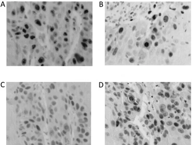
Fig. 2 Results of the immunohistochemical staining with Ki-67 (A), Topo IIα (B), p53 (C), and p63 (D) in a representative case (Patient no. 1) with oral squamous cell carcinoma.
determined by the expression of Ki-67 or Topo IIα. The present study indicated that the expression of p53 and p63 showed weak correlation with the expression of Ki-67, but no statistical significance was observed (r=0.354, 0.476, respectively). Both p53 and p63 are multifunctional proteins that control cell cycle, apoptosis, DNA repair, and cellular proliferation and differentiation. In squamous cell carcinoma of the head and neck, strong nuclear expression of p53 and p63 are closely related with proliferative activity of the tumor as well as prognosis of the patient. Both p53 and p63 are highly expressed in cancer cells and act in close cooperation with each other to stop cell division. Products of p53 act in G1/S check point of the cell cycle, respectively. The results of the present study revealed that the expression of p53 and p63 was weakly correlated but no statistical significance was observed (r=0.488). In approximately 50% of squamous cell carcinomas of the head and
neck,func-tion of wild type p53 is abrogated by the mutaneck,func-tion. Consequently, nonfunctioning and more stable p53 proteins are generated and accumulated in the nucleus of the tumor cells. On the other hand,p63 expressed in at least six isoforms and supposed to play diverse roles in the differentiation and proliferation of the keratinocyte.
Based on these knowledge and the results obtained in the present study, cellular proliferation as deter-mined by the FDG uptake might reflect tumor progres-sion and may have prognostic value in OSC.
The association of FDG uptake and cellular proliferation has been examined in many types of cancers such as lung cancer and brain tumor,but there are a few reports of the associations in head and neck cancer, especially in OSC. Minn et al. showed a clear correlation between the proliferative activity and the uptake of FDG in thirteen patients with malignant head and neck tumors. In addition, another report
Fig. 3 Correlation between FDG accumulation with markers of cellular proliferation and p53/p63 of oral squamous cell carcinoma as determined by immunhistochemical staining.
(A) A significant correlation is noted between maximum SUV and labeling index of Ki-67 (r=0.616, p= 0.01).
(B) A significant correlation is noted between maximum SUV and labeling index of Topo IIα(r=0.677,p= 0.004).
(C) A significant correlation is noted between maximum SUV and labeling index of p53 (r=0.613,p=0.01). (D) A significant correlation is noted between maximum SUV and labeling index of p63(r=0.710,p=0.002).
indicated that tumors with higher SUV and higher AgNORs score had significantly higher incidence of residual viable tumor cells after chemoradiotherapy. The study suggests a close association between SUV and proliferative activity in OSC, because previous studies demonstrated significant association between AgNORs score and proliferative activity. A recent study by Linecker et al. disclosed that FDG uptake was significantly correlated with the prognosis of patients with OSC. Histological marker would pos-sibly evaluate noninvasively the clinical course of patients. However, no study has been proved the correlation of FDG uptake with histological markers including Ki-67, Topo IIα, p53, and p63 in patients with OSC.
In this study with small number of patients, nei-ther the uptake of FDG nor the immunohistochemical expression of proliferative and prognostic markers showed correlation with the actual prognosis of patients with OSC.
The limitations of the present study were that the number of patients included in the study was small. Although FDG uptake in the tumor was correlated with the proliferative markers and pathological prog-nostic markers such as p53 and p63 in the present study,further study with increasing number of patients would be preferable to confirm the results. Another limitation is that neither the uptake of FDG nor the expression of pathological proliferative and prognostic markers showed significant correlation with the actual prognosis of patients. The prognosis of patients should be correlated with the results of FDG PET and pathological markers in a large number of patients in the further studies. Moreover, metastasis of cervical lymph nodes affects the prognosis of patients with OSC. In the present study,lymph node metastasis was noted in four patients (No.10, 12, 13, 16). However, SUV of the primary tumor of patient No.10 was as low as 3.6, and the other two patients (No.12, 13) with moderately differentiated OSC and a patient (No.16) with poorly differentiated OSC had SUV of 9.0, 7.8, and 8.9, respectively. These values in patients with well and moderately differentiated OSC were not rela-tively higher than average SUV of each histological type. We should confirm prognostic factors for OSC by the multivariate analysis with FDG uptake in the primary tumor, pathological lymph node metastasis, and expression of immunohistochemical markers.
In conclusion,significant correlation was indicat-ed between FDG accumulation and cellular prolifera-tion determined by the immunohistochemical expres-sion of Ki-67, Topo IIα,and pathological prognostic markers such as p53 and p63 in OSC. FDG PET may be useful for evaluating clinical status and for
consider-ing clinical management of patients with OSC.
References
1. Schwartz DL, Rajendran J, Yueh B, et al. FDG-PET prediction of head and neck squamous cell cancer outcomes. Arch Otolaryngol Head Neck Surg 2004; 130: 1361-1367 2. Ng SH, Yen TC, Chang JT, et al. Prospective study of
[ F] fluorodeoxyglucose positron emission tomography and computed tomography and magnetic resonance imaging in oral cavity squamous cell carcinoma with palpably negative neck. J Clin Oncol 2006; 24: 4371-4376 3. Lowe VJ, Boyd JH, Dunphy FR, et al. Surveillance for
recurrent head and neck cancer using positron emission tomography. J Clin Oncol 2000; 18: 651-658
4. Schoder H, Carlson DL, Kraus DH, et al. F-FDG -PET/CT for detecting nodal metastasis in patients with oral cancer stage N0 by clinical examination and CT/MRI. J Nucl Med 2006; 47: 755-762
5. Yen TC, Chang JT, Ng SH, et al. Staging of untreated squamous cell carcinoma of buccal mucosa with F-FDG PET : comparison with head and neck CT/MRI and his-topathology. J Nucl Med 2005; 46: 775-781
6. Nakasone Y,Inoue T,Oriuchi N,et al. The role of whole -body FDG-PET in preoperative assessment of tumor stag-ing in oral cancers. Ann Nucl Med 2001; 15: 505-512 7. Kitagawa Y, Nishizawa S, Sano K, et al. Whole-body
F-fluorodeoxyyglucose positron emission tomography in patients with head and neck cancer. Oral Surg Oral Med Oral Pathol Oral Radiol Endod 2002; 93: 202-207 8. Miller FR, Hussey D, Beeram M, et al. Positron emission
tomography in the management of unknown primary head and neck carcinomas. Arch Otolaryngol Head Neck Surg 2005; 131: 626-629
9. Tian M,Zhang H,Higuchi T,et al. Hexokinase-II expres-sion in untreated oral squamous cell carcinoma: compari-son with FDG PET imaging. Ann Nucl Med 2005; 19 : 335-338
10. Kunkel M, Reichert TE, Benz P, et al. Overexpression of Glut-1 and increased glucose metabolism in tumors are associated with a poor prognosis in patients with oral squamous cell carcinoma. Cancer 2003; 97: 1015-1024 11. UICC. TNM classification of malignant tumours,5th eds.
Geneva: UICC 1997.
12. Ishiwata K,Kawashima K,Ido T. Metabolism of 2-deoxy-2-[18F] fluoro-D-glucose: presence of 2-deoxy-2-[18F] fluoro-D-glucose 6-phosphate in plasma of mice, rats, and humans. Ann Nucl Med 1987; 1: 51-54
13. Gerdes J, Schwab U, Lemke H, Stein H. Production of a monoclonal antibody reactive with a human nuclear antigen associated with cell proliferation. Int J Cancer 1983; 31: 13-20
14. Cattoretti G, Becker MHG, Key G et al. Monoclonal antibodies against recombinant parts of the MIB-1 antigen (MIB-1 and MIB-3) detect proliferating cells in microwave-processed formalin-fixed paraffin sections. J Pathol 1992; 168: 357-363
15. Brustmann H,Naude S. Expression of topoisomerase IIα, Ki-67, proliferating cell nuclear antigen, p53, and argyro-phillic nucleolar organizer regions in vulvar squamous lesions. Gynecol Oncol 2002; 86: 192-199
16. Hirabayashi S. Immunohistochemical detection of DNA topoisomerase type II and Ki-67 in adenoid cystic car-cinoma and pleomorphic adenoma of the salivary gland. J
Oral Pathol Med 1999 ; 28: 131-136
17. Nakamura M, Sano K, Kitagawa Y, et al. Diagnostic significance of FDG-PET and argyrophillic nucleolar orga-nizer regions (AgNORs) in oral squamous cell carcinoma. Oral Oncol 2004; 40: 190-198
18. de Oliveira LR, Ribeiro-Silva A, Zucoloto S. Prognostic impact of p53 and p63 immunoexpression in oral squamous cell carcinoma. J Oral Pathol Med 2007; 36: 191-197 19. Lo Muzio L, Santarelli A, Caltabiano R,et al.p63
overex-pression associates with poor prognosis in head and neck squamous cell carcinoma. Hum Pathol 2005; 36: 187-194 20. Couture C, Raybaud-Dioge ne H,Tetu B,et al. p53 and MIB-1 as markers of radioresistance in head and neck carcinoma. Cancer 2002; 94: 713-722
21. Takeda T, Sugihara K, Hirayama Y, et al.
Immunohisto-chemical evaluation of Ki-67, p63, CK19 and p53 expres-sion in oral epithelial dysplasia. J Oral Pathol Med 2006; 35: 369-375
22. Bading JR, Shields AF. Imaging of cell proliferation : status and prospects. J Nucl Med 2008; 49 Suppl 2: 64S-80S
23. Minn H,Joensuu H,Ahonen A,et al. Fluorodeoxyglucose imaging : a method to assess the proliferative activity of human cancer in vivo. Comparison with DNA flow cytometry in head and neck tumors. Cancer 1998; 61: 1776-1781
24. Linecker A, Kermer C, Sulzbacher I, et al. Uptake of 18F-FLT and 18F-FDG in primary head and neck cancer correlates with survival. Nuklearmedizin 2008; 47: 80-85