Content from this work may be used under the terms of theCreative Commons Attribution 3.0 licence. Any further distribution of this work must maintain attribution to the author(s) and the title of the work, journal citation and DOI.
Published under licence by IOP Publishing Ltd 1
Effects of the solvent during the preparation of MoS
2nanoparticles by laser ablation
Makoto Kanazawa1, Pankaj Koinkar1*,, Kei-ichiro Murai2, Toshihiro Moriga2 ,
Akihiro Furube1*
1Department of Optical System Engineering, Tokushima University, 2-1
Minamijosanjima-cho, Tokushima, Tokushima, 770-8506, Japan
2Department of Applied chemistry, Tokushima University, 2-1 Minamijosanjima-cho,
Tokushima, Tokushima, 770-8506, Japan
Email: [email protected]*, [email protected]*
Abstract. Pulsed laser ablation in liquid is a well-known and effective method which can be used to prepare the various nanostructures. However, ablated samples have various problems such as wide size distribution, and effect of solvent to sample during laser ablation in liquid has not been well understood. In response to these problems, in this study, we prepared nanoparticles by irradiating nanosecond laser to samples using different solvents. The experimental results of prepared samples were compared, and we evaluated how the different solvents affect to their morphological and optical properties. The morphology, crystal structures and optical properties of the MoS2 nanoparticle were characterized by Scanning electron microscopy, X-ray diffraction, and UV-Vis absorption spectroscopy. Upon the laser ablation of the samples, the absorbance of UV-Vis spectra increased as approaching the shorter wavelength side. From the SEM images, it confirmed that the particle size became smaller for laser ablated MoS2 sample, which is good agreement with the result of UV-Vis spectra. The XRD spectra shows the appearance of new peak for laser ablated MoS2 in methanol as compared to those samples ablated in ethanol and N-methyl-2-pyrrolidone. It can be said that the crystal structure of the sample has changed after ablation. It suggested that because the particle size became smaller after ablation and the band gap increased. Such MoS2 nanostructure has its own importance for optoelectronics devices.
1. Introduction
Nanomaterials have characteristics that are noticeable different from bulk materials. Two-dimensional (2D) atomically thin inorganic layered materials have attracted much attention as new nano-functional materials due to its outstanding electrical and optical properties [1-3]. Currently, the significant interest in planar devices have attracted great attention in transition metal dichalcogenide (TMDC) in this context, TMDC materials such as molybdenum disulfide (MoS2), which has two-dimensional
structure similar to graphene, is considered as promising material for future planer devices. The MoS2
has been investigated and its shows that properties exhibited can be potential candidates for planar device technologies and to improve the performance of transistors, LED, solar cells and so on. In addition, few reports suggested methods to produce nanostructure of MoS2 as well as nanoparticles of
another material with good efficiency, which can be used practical application for the fabrication of technological various devices [4-6]. Among these various ways to produce nanostructure, pulsed laser
2
ablation in liquid (PLAL) is considered as one of useful, simple and easy ways to synthesize nanostructure. As compared to other experimental methods, PLAL can obtain stable suspensions of various nanostructures in a wide range of liquids with a one-step, simple and economic procedure [7,8]. However, ablated samples have various problems such as wide size distribution and effect of solvent during PLAL process is not completely elucidated yet. To addressed these problems, in the present study, we have prepared MoS2 nanoparticles by irradiating nanosecond laser using the
different solvents. The morphology, crystal structures and optical properties of the MoS2 nanoparticle
were characterized by scanning electron microscopy, X-ray diffraction, and UV-Vis absorption spectrum. After laser ablation, the experimental results of the all prepared samples have been compared, and we tried to evaluate how the solvents used affect the morphological changes and optical property.
2. Experimental procedure
2.1. Materials
Commercial MoS2 particles (average size less than 2μm) were purchased from Sigma-Aldrich and
they were used without any prior treatment and purification treatment. Methanol, ethanol, and N-methyl-2-pyrrolidone (NMP) were purchased from Sigma-Aldrich.
2.2. Synthesis of MoS2 nanoparticle
Commercial MoS2 particles of 80 mg (from Sigma-Aldrich, average size less than 2μm) were
magnetically stirred in methanol, ethanol, and NMP of 40 ml and its aqueous solution samples were sonicated using bath-type sonicator for 1 hour to obtain well dispersed solution. After sonification, these samples were exposed to the laser pulses from the second harmonic of a Spectra Physics Nd:YAG laser (wavelength 532 nm, repetition frequency 10 Hz , 10 ns fwhm/pulse, 55 mJ pulse-1) for 120 minutes. After laser ablation, a colloidal suspension was heated to evaporate solvent to get the resulted MoS2 in the form of dry powder. These samples were characterized using UV-visible
spectrophotometry, X-ray diffraction (XRD) and field emission scanning electron microscopy (FE-SEM) to confirm nanostructure formation and reveal the surface morphology changes and structural features. XRD spectra of synthesized MoS2 nanostrcuture were measured by a X-ray diffractrometer
(Rigaku SmartLab 9 kW) with CuKα (λ = 1.54184 Å) radiations operated at the voltage of 45 kV and the current of 15 mA in the 2θ range from 20° to 80°. The surface morphology was investigated using a FE-SEM (S4700, Hitachi High-Technologies) while the UV-Visible absorption spectra were recorded by means of the UV-Visible spectrophotometer (V-670, JASCO Corporation).
3. Results and discussion
3.1. XRD pattern analysis
To identify the crystal structures of different MoS2 powders prepared using different solvents, XRD
measurements were carried out for each sample. Figure 1 shows the X-Ray diffraction pattern of the laser ablated MoS2 nanostructures for 120 min in different solvents, namely ethanol, methanol, and
NMP. In the sample before ablation, a characteristic peak was confirmed in bulk MoS2. In case of
laser ablated samples, it is clearly observed that there is a systematic decrease in intensity of characteristics peak and this could be due to the structural reflection from the crystal plane disappearing when irradiated with X-rays. Also, the diffraction patterns show that all major peaks present a broadening depend on the nature of the solvent. As ccompared to the sample before ablation, new peaks appeared for laser ablated samples and were confirmed their presence at 31.25, 34.8, 35.94, 42.45, 40.55 degrees. In addition, only peak at 40.5 degrees was observed for MoS2 prepared using
methanol as a solvent. According to the prior literature, the MoS2 structure has been changed after the
3
at 44 degrees (106) is stronger in the samples treated with the laser ablation. Thus, the XRD pattern results indicate that MoS2 size changes after the laser ablation with solvent medium.
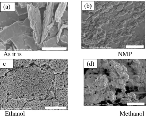
3.2. Scanning electron microscopy analysis
Typical SEM images of the bulk and as prepared MoS2 nanoparticles in different solvents are
illustrated in in Figure 2(a-d). The scale bars indicated in all images are 1 µm. Fig. 2 (a) displays SEM image of bulk MoS2 with micron-scale sheets (microflake) having the size about 2 µm . It is found that
that the morphology and the size of MoS2 nanoparticles depend on the nature of solvent used during
the laser ablation. In case of NMP used as a solvent, the formation of nanosheets of approximately several hundred nm to less than 50 nm could be observed as shown in Fig. 2(b). When ethanol was used as a solvent, the uniform formation and large amounts of spherical particles having the diameter about 20 nm to 50 nm were observed as shown in Fig 2(c). However, when methanol was used as a solvent, nanosheets from 500 nm to less than 50 nm as well as spherical particles having a diameter of 20 nm to several hundred nm as illustrated in Fig. 2(d). These results indicate the formation of spherical or nanosheet-like nanoparticles as confirmed by the SEM image and becomes sufficiently smaller than the bulk MoS2. In particular, it is reported that 2D and 3D nanosheet MoS2 can be formed
by laser ablation using methanol [15]. Focusing only on the result of methanol, the sheet-like structure in SEM images seems to be 2D nanosheet MoS2, while the spherical shape one obtained in this
experiment seems to be 3D nanosheet MoS2 as indicated with XRD patterns, which showed the
characteristic of morphology with new peak. Furthermore, SEM image of bulk MoS2 show
microsheets with sharp corner whereas for the MoS2 samples, laser-treated in different solvents,
[004] [102] [100] [103] [110] [006] [008] [101] [106] [105]
Figure 1. XRD patterns of (a) bulk MoS2 and laser ablated MoS2 in different
solvents (b) ethanol (c) methanol and (d) NMP. 2θ (degree)
4
revealed that most of the nanosheets and nanoparticles are found with the rounded. This could be caused by the heat generated during the ablation process.
3.3. UV-visible spectroscopy analysis
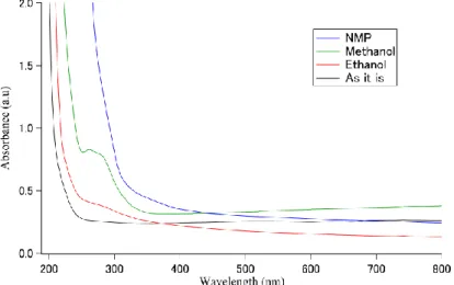
In order to measure UV-vis spectrum spectra of nanoparticles, each sample of 2 ml taken out from ablated samples was centrifuged at 6000 r.p.m for 10 min. and then UV-vis spectra of precipitate liquid and the supernatant liquid of each samples were measured as shown in Fig. 3 and Fig.4 respectively. The UV-Vis optical absorption spectra were recorded at room temperature in the wavelength region of 200–800 nm. Fig.3 shows UV-vis spectra of bulk and laser ablated MoS2. Each
sample is diluted to 100 times so that the sample peak can be confirmed. In case of bulk MoS2 sample
i.e. before ablation, characteristic peaks at about 630 nm and 690 nm in the form of a small noticeable hump were observed. According to earlier literature, the separation energy (about 59 nm) between A and B excitonic transitions can be explained with spin-orbit splitting, and their characteristic energy can be assigned to A and B excitonic transitions on the high energy side of first absorption threshold splitting value is approximately 60 nm (0.17 eV) [11-14]. In case of laser ablated MoS2 using NMP as
a solvent, a noticeable peak was not observed around 260 nm in the sample after ablation. However, the value of the absorbance became larger as approaching the shorter wavelength side and this could be because of the particle size reduced by laser ablation and the band gap is increased by the quantum size effect. [10]. In order to confirm this, only the supernatant was collected from the sample set at a speed of 6000 rpm and subjected to centrifugation for 10 minutes. In this way, we have obtained a high concentration of exfoliated particles in the supernatant and thus the absorbance of the sample containing only exfoliated particles was measured as displayed in Fig. 4. As a result, unlike the spectrum before applying the sample to the centrifugal separator, it showed significant absorption on the short wavelength side. While, in the sample using ethanol, the effect of miniaturization by laser ablation was confirmed as there are increase in the absorbance on the shorter wavelength side as well as a small hump of two characteristic peaks at 260 nm and 280 nm observed as shown in Fig. 3. In addition, when the absorption spectrum of the supernatant liquid of the sample employed to the centrifuge was measured, it was clearly seen that that the peak intensities at 260 nm and 280 nm
As it is NMP
Figure 2. FE-SEM images of bulk MoS2 and laser ablated MOS2 in different solvents .
Ethanol Methanol
(a)
c
(b)
5
became stronger as depicted in Fig 4. Even with a sample prepared using methanol as a solvent, the effect of miniaturization by laser ablation was confirmed due to the increase in absorbance on the shorter wavelength side. This result is well in agreement with the earlier reported data [15]. Similarly to ethanol, when the absorption spectrum of the supernatant liquid taken from centrifuged sample was measured, it was found that the peak intensities at 260 nm and 280 nm became stronger as indicated in Fig. 4. Therefore, it suggested that the particle size became smaller after laser ablation and also the band gap increased due to the quantum size effect. This result well explains that the absorbance of the absorption spectrum of the sample has greatly increased on the short wavelength side.
4. Conclusions
In this study, we have prepared MoS2 nanoparticles by irradiating nanosecond laser to samples in
different solvents and compared the experimental results of the prepared samples to evaluate the effect of the solvent based on their morphological, structural and optical properties. The XRD pattern results indicate that MoS2 size changes after the laser ablation with different solvent medium and the presence
of new peaks were confirmed in laser ablated samples. Particularly in samples using methanol, presence of particles of MoS2 having different shapes was confirmed. From the results of the SEM
Figure 3. UV-vis spectra of precipitated bulk MoS2 and laser ablated MoS2 ablated in various solvents
Figure 4. UV-vis spectra of supernatant bulk MoS2 and laser ablated MoS2 ablated in various
6
image, it was seen that there was a difference in shape of particles produced by ablation using different solvents. Furthermore, UV-Vis spectra confirm the effect of miniaturization by laser ablation due to the increase in absorbance on the shorter wavelength side. There are still various problems even for the synthesis of nanoparticles by laser ablation in liquid medium. Comparing and examining the results obtained by actually changing the conditions of the sample will help to understand the effect of laser ablation on the preparation of nanoparticles. In the future, we will plan to study and clarify how to effectively and efficiently produce MoS2 nanoparticles with desired shape and optical properties
using PLAL. It may accelerate application to planar devices such as LEDs and solar panels. References
[1] Castellanos-Gomez, A.; Poot, M.; Steele, G. A.; van der Zant, H. S. J.; Agraït, N.; Rubio-Bollinger, G. Elastic Properties of Freely Suspended MoS2 Nanosheets. Adv. Mater. 2012, 24, 772−775.
[2] Gaur, A. P. S.; Sahoo, S.; Ahmadi, M.; Guinel, M. J.-F.; Gupta, S. K.; Pandey, R.; Dey, S. K.; Katiyar, R. S. Optical and Vibrational Studies of Partially Edge-Terminated Vertically Aligned Nanocrystalline MoS2 Thin Films. J. Phys. Chem. C 2013, 117, 26262−26268.
[3] Ruinart de Brimont, M.; Dupont, C.; Daudin, A.; Geantet, C.; Raybaud, P. Deoxygenation Mechanisms on Ni-Promoted MoS2 Bulk Catalysts: A Combined Experimental and Theoretical Study. J. Catal.
2012, 286, 153−164.
[4] Late, D. J.; Liu, B.; Matte, H. S. S. R.; Rao, C. N. R.; Dravid, V. P. Rapid Characterization of Ultrathin Layers of Chalcogenides on SiO2/Si Substrates. Adv. Funct. Mater. 2012, 22, 1894−1905.
[5] Late, D. J.; Liu, B.; Matte, H. S. S. R.; Dravid, V. P.; Rao, C. N. R. Hysteresis in Single-Layer MoS2 Field Effect Transistors. ACS Nano 2012, 6, 5635−5641.
[6] Kashid, R. V.; Late, D. J.; Chou, S. S.; Huang, Y. K.; De, M.; Joag, D. S.; More, M. A.; Dravid, V. P. Enhanced Field-Emission Behavior of Layered MoS2 Sheets. Small 2013, 9, 2730−2734.
[7] S. Barcikowski, F. Devesa, K. MoldenhauerImpact and structure of literature on nanoparticle generation by laser ablation in liquids J. Nanopart. Res., 11 (2010), pp. 1883-1893
[8] G.W. YangLaser ablation in liquids: application in the synthesis of nanocrystals, Progr. Mater. Sci., 52 (2007), pp. 648-698
[9] Shuichi Hashimoto. Pulsed Laser-Ablation-Induced Fabrication of Metal Nanoparticles in Liquids.Volume 45, Number 5 2017, The Review of Laser Engineering.
[10] Herme G. Baldovı , Marcos Latorre-Sanchez . Ivan Esteve-Adell . Anish Khan . Abdullah M. Asiri . Samia A. Kosa . Hermenegildo Garcia (2016) Generation of mos2 quantum dots by laser ablation of mos2 particles in suspension and their photocatalytic activity for H2 generation. (J Nanopart Res) 18:240
[11] Wilcoxon, J. P.; Newcomer, P. P.; Samara, G. A. Synthesis and Optical Properties of MoS2 and Isomorphous Nanoclusters in the Quantum Confinement Regime. J. Appl. Phys. 1997, 81, 7934−7944.
[12] Abrams, B. L.; Wilcoxon, J. P. Nanosize Semiconductors for Photooxidation. Crit. Rev. Solid State Mater. Sci. 2005, 30, 153−182.
[13] Thurston, T. R.; Wilcoxon, J. P. Photooxidation of Organic Chemicals Catalyzed by Nanoscale MoS2. J. Phys. Chem. B 1999, 103, 11−17.
[14] Wilcoxon, J. P.; Newcomer, P. P.; Samara, G. A.; Venturini, E. L.; Williamson, R. L. Fundamental Science of Nanometer-Size Clusters; Sandia National Labs.: Albuquerque, NM (USA), October 1995; pp 19−30.
[15] Tugba Oztas,Huseyin Sener Sen, Engin Durgun, and B lend Orta .Synthesis of Colloidal 2D 3D MoS2 Nanostructures by Pulsed Laser Ablation in an Organic Liquid Environment. J. Phys.