Study on novel physiological function of
1-deoxynojirimycin derived from mulberry
leaves
著者
Shung E
学位授与機関
Tohoku University
学位授与番号
11301甲第18742号
Study on novel physiological function of
1-deoxynojirimycin derived from mulberry leaves
(桑葉由来 1-デオキシノジリマイシンの新規生理機能に
関する研究)
Table of contents
Chapter 1: 1-Deoxynojirimycin attenuates high
glucose-accelerated senescence in human umbilical vein endothelial
cells
1.
Abstract ... 4
2.
Abbreviations ... 6
3.
Introduction ... 7
4.
Materials and methods ... 10
4.1. Materials ... 10
4.2. Cells and cell culture ... 10
4.3. Cytotoxicity of DNJ ... 11
4.4. Preparation of senescent cells ... 11
4.5. Senescence-associated ß-galactosidase (SA-ß-gal) staining ... 13
4.6. mRNA expression analysis ... 13
4.7. Monocyte adhesion assay ... 14
4.8. NF-kB activity assay ... 15
4.9. Reactive oxygen species (ROS) detection ... 16
4.10. Statistical analysis ... 17
5.
Results ... 19
5.1. Cytotoxicity of DNJ ... 19
5.2. Effects of high glucose and DNJ on cell proliferation ... 19
5.3. Effects of high glucose and DNJ on cellular senescence ... 20
5.4. Effects of high glucose and DNJ on monocyte adhesion ... 21
5.5. Effects of high glucose and DNJ on NF-kB activity and ROS generation ... 21
6.
Discussion... 29
Chapter 2: Intake of mulberry 1-deoxynojirimycin prevents
colorectal cancer in mice
1.
Abstract ... 39
2.
Abbreviations ... 40
3.
Introduction ... 41
4. Materials and Methods ... 43
4.1. Materials ... 43
4.2. Animals and diets ... 43
4.3. Histological analysis of colon tissue ... 44
4.5. Biochemical analyses of serum and liver ... 45
4.6. mRNA expression analyses... 45
4.7. Determination of lipid peroxides ... 46
4.8. DNJ concentration in colorectal cancer and normal tissue ... 47
4.9. Statistical analysis ... 48
5.
Results ... 50
5.1. Effects of caloric restriction and DNJ on growth parameters ... 50
5.2. Effects of caloric restriction and DNJ on tumor formation ... 50
5.3. Effects of caloric restriction and DNJ on serum and liver parameters ... 51
5.4. Effects of caloric restriction and DNJ on apoptosis ... 52
5.5. DNJ concentration in colorectal cancer and normal tissues ... 52
6.
Discussion... 59
7.
References ... 62
1-Deoxynojirimycin attenuates high glucose-accelerated senescence in
human umbilical vein endothelial cells
1. Abstract
The influence of 1-deoxynojirimycin (DNJ) derived from mulberry on senescence of endothelial
cells was examined with the goal of discovery of a method for prevention of senescence of blood
vessels. The effect of DNJ on senescence of human umbilical vein endothelial cells (HUVECs)
promoted under high glucose condition was determined. HUVECs were cultured in normal glucose
(5.6 mmol/L, NG group), normal glucose plus DNJ (10 μmol/L, DNJ group), high glucose (30
mmol/L, HG group), or high glucose plus DNJ (10 μmol/L, HG + DNJ group) and passaged until
they reached senescence. The proliferation rate was markedly decreased in the HG group compared
with the NG group, and this phenomenon was reversed by DNJ. The frequency of senescent
(SA-ß-Gal-positive) cells and the expression level of senescence genes (PAI-1 and p21) were
significantly higher in the HG group compared with the NG group, and these changes were blocked
by DNJ. Monocyte adhesion, NF-kB activity, and reactive oxygen species production, all of which
group, and again these changes were blocked by DNJ. Therefore, these results show that DNJ delays
2. Abbreviations
DNJ: 1-Deoxynojirimycin
HUVEC: human umbilical vein endothelial cells
ROS: reactive oxygen species
HILIC-QTRAP MS/MS: hydrophilic interaction liquid chromatography with hybrid quadrupole/linear
ion trap tandem mass spectrometry
hEGF: human epidermal growth factor
hFGF-B: human basic fibroblast growth factor
PDL: Population doubling level
CPDL: cumulative population doubling level
ICAM1: intercellular adhesion molecule 1
SELE: selectin E
VCAM1: vascular cell adhesion molecule 1
3. Introduction
Lifestyle-related diseases such as diabetes mellitus and arteriosclerosis are increasing yearly and can
progress to cerebral and cardiac diseases (10, 11). Elderly persons are mainly affected and these conditions
are referred to as aging-related diseases. Thus, prophylaxis for these diseases is important for maintenance
of a healthy life. Aging progresses gradually and a method of delaying of senescence through food intake
may be effective for prevention of aging-related disease. Senescent vascular endothelial cells have been
found in arteriosclerotic sites in humans, which indicates a possible relationship of these cells with an
aging-related disease (12). We recently found that senescence of vascular endothelial cells is promoted by
monocyte adhesion (30). Senescence of blood vessels is also promoted by reactive oxygen species (ROS),
the production of which increases when the blood glucose level is high, such as in patients with diabetic
mellitus (6, 17, 30, 31). Therefore, suppression of intracellular ROS production is likely to be effective for
senescence delay.
1-Deoxynojirimycin (DNJ) may be a food constituent that delays senescence of blood vessels. DNJ is
derived from mulberry (Moraceae) and has a chemical structure like that of glucose (Figure 1) (29). DNJ
inhibits α-glucosidase in the small bowel mucosa, which reduces absorption of sugar and suppresses
plasma in rodents (8, 28). Therefore, DNJ may also delay senescence of endothelial cells induced by
oxidative stress, but this effect of DNJ is not clear. In this study, we examined whether DNJ can delay
senescence of endothelial cells promoted under high glucose condition, with the goal of discovering a
method for prevention of aging-related diseases.
Senescent cells were prepared by culturing human umbilical vein endothelial cells (HUVECs) until they
reached the Hayflick limit (30). Senescence of HUVECs is promoted under high glucose condition, and thus
a culture in high glucose was used (19, 31, 32). Since the plasma DNJ level reaches 3.2 μmol/L in human
receiving a small amount of DNJ (6.3 mg) (16) and 100 μmol/L in rat receiving DNJ (110 mg/kg of body
weight) (15), it was thought that DNJ can reach 10 μmol/L in human plasma. Therefore, DNJ in the culture
medium was adjusted to 10 μmol/L. HUVECs were cultured in normal glucose (NG), normal glucose plus
DNJ (DNJ), high glucose (HG), or high glucose plus DNJ (HG + DNJ), and changes in cellular
proliferative potential, proportion of senescent cells, and expression of senescence genes were evaluated.
HUVECs also have increased expression of proteins that promote monocyte adhesion (30), which occurs in
the first stage of arteriosclerosis and is promoted by NF- kB and ROS (17, 18, 21). Thus, the effects of DNJ
Figure 1. Chemical structures of 1-deoxynojirimycin (DNJ) and D-glucose.
OH
HO
HO
OH
NH
1-Deoxynojirimycin (DNJ)
OH
HO
HO
OH
O
OH
D-glucose
4. Materials and methods
4.1. Materials
DNJ was extracted from mulberry leaves (Morus alba) and purified using ion-exchange
chromatography followed by recrystallization (8, 15, 16). The purity of DNJ was shown to be N98%
by hydrophilic interaction liquid chromatography with hybrid quadrupole/linear ion trap tandem
mass spectrometry (HILIC-QTRAP MS/MS) (16).
4.2. Cells and cell culture
HUVECs were purchased from Kurabo (Osaka, Japan) and cultured in HuMedia-EG2 growth
medium (Kurabo, Osaka, Japan) at 37 °C in a humidified atmosphere of 5% CO2 in air (25, 30).
HuMedia-EG2 medium consists of base medium (HuMedia-EB2) supplemented with 2% FBS, 0.5
mg/L human epidermal growth factor (hEGF), 2 mg/L human basic fibroblast growth factor
(hFGF-B), 5 g/L insulin, 50 g/L gentamicin, and 50 mg/L amphotericin B. HUVEC monolayers of
passage 9 were used in the experiments. THP-1 monocytes were obtained from the Cell Resource
Center for Biomedical Research at Tohoku University School of Medicine (Sendai, Japan) and
μg/mL streptomycin at 37 °C in a humidified atmosphere of 5% CO2.
4.3. Cytotoxicity of DNJ
Cytotoxicity of DNJ was assessed using a WST-8 assay (20, 24, 30). HUVECs at 80% confluency were
trypsinized and transferred to 96-well plates (4000 cells/well). Stock solutions of DNJ were
prepared in HuMedia- EG2 medium. DNJ test media were prepared from the stock solutions and
diluted to final concentrations of 0–200 μmol/L in HuMedia-EG2 medium. After incubation for 24
h at 37 °C, the cells were placed in 200 μL of fresh HuMedia-EG2 medium with various
concentrations of DNJ. After 24 or 48 h, 10 μL WST-8 solution (Dojindo, Kumamoto, Japan) was
added to each well. After incubation for 3 h at 37 °C, cytotoxicity was measured using a microplate
reader (Infinit F200; Tecan Japan, Kawasaki, Japan) at a wavelength of 450 nm and a reference
wavelength of 655 nm.
4.4. Preparation of senescent cells
Senescent cells were prepared by further culturing of HUVECs (30) using a culture condition in high
glucose (19, 31, 32). HUVECs were subcultured upon reaching 80% confluence using 0.25%
were determined when subcultured by passing a 1/50 dilution of cells through a Coulter Counter (2,
30). Population doubling level (PDL) was estimated at each passage using the following equation: n
= (log2X − log2Y) (with n = PD, X = number of cells at the end of one passage, Y = number of cells
that were seeded at the beginning of one passage) (19). For each passaging, the resulting PDL
estimation was added to the sum of PDL from the previous passages to achieve the cumulative
population doubling level (CPDL), which was plotted against time to obtain a growth curve (19).
Since the plasma DNJ level reaches 3.2 μmol/L in human receiving a small amount of DNJ (6.3 mg)
(16) and 100 μmol/L in rat receiving DNJ (110 mg/kg of body weight) (15), it was thought that DNJ
can reach 10 μmol/L in human plasma. Therefore, DNJ in the culture medium was adjusted to 10
μmol/L. HUVECs were cultured in normal glucose (5.6 mmol/L, NG), normal glucose plus DNJ
(10 μmol/L, DNJ), high glucose (30 mmol/L, HG), or high glucose plus DNJ (10 μmol/L, HG +
DNJ) at 37 °C in a 5% CO2 atmosphere and passaged until they reached the Hayflick limit, based
on no observation of cell division for 30 days (30). Since HUVECs at PDL 28.2 reached the Hayflick
limit in the HG group, in all experiments, four groups of cells were used: HUVECs at PDL28-29
exposed to NG (NG group), HUVECs at PDL28-29 exposed to NG and 10 μmol/L DNJ (DNJ group),
10 μmol/L DNJ (HG + DNJ group).
4.5. Senescence-associated ß-galactosidase (SA-ß-gal) staining
HUVECs of the NG, DNJ, HG, and HG + DNJ groups at 80% confluency were trypsinized,
transferred to 6-well plates (8000 cells/well), and preincubated in HuMedia-EG2 medium for 24 h.
The cells were detected with a senescence detection kit (BioVision Inc., Mountain View, CA, USA)
(3, 30). After SA-ß-Gal staining, cells were washed once with PBS (-), 1.5 mL of Hoechst 33342
(Dojindo) (1 μg/mL) was added to each well, and the cells were incubated at room temperature for
30 min. Alteration of cellular morphology was observed by fluorescent microscopy (Biozero,
Keyence, Osaka, Japan). The data were shown by the ratio to young cells (normal-glucose exposed
HUVECs at PDL8-9) to indicate a change with age.
4.6. mRNA expression analysis
A quantitative reverse transcriptase-PCR assay was conducted on total RNA extracted from the liver
using an RNeasy mini kit (Qiagen, Valencia, CA, USA) (25, 27, 30). Expression levels of PAI-1, p21,
intercellular adhesion molecule 1 (ICAM1), selectin E (SELE) and vascular cell adhesion molecule
(Takara Bio, Shiga, Japan), which allowed real-time quantitative detection of the PCR products by
measuring the increase in fluorescence caused by binding of SYBR green to double-stranded DNA.
In brief, cDNA was made from the total RNA in HUVECs of the NG, DNJ, HG, and HG + DNJ
groups using a PrimeScript RT Master Mix (Perfect Real Time) kit (Takara), and subjected to PCR
amplification with SYBR Premix Ex Taq (Takara) and a gene-specific primer for ICAM1, PAI-1,
p21, SELE, VCAM1 or ß-actin (Table 1). Amplification was performed with an activation step at
95 °C for 10 s, followed by 40 cycles at 95 °C for 5 s (denaturation) and 60 °C for 31 s (extension),
and a dissociation stage at 95 °C for 15 s, 60 °C for 30 s and 95 °C for 15 s. The ß-actin content in
each sample was used to normalize the results. The data were shown by the ratio to young cells
(normal-glucose exposed HUVECs at PDL8-9) to indicate a change with age.
4.7. Monocyte adhesion assay
Monocyte adhesion assays were carried out essentially as previously described (7, 9, 30). HUVECs of
the NG, DNJ, HG, and HG + DNJ groups at 1 × 10⁴ cells/mL were seeded in 96-well plates in
complete medium (200 μL/well) and incubated at 37 °C in a 5% CO2 atmosphere for 6 h. After
incubation, the cells were washed twice with RPMI-1640 medium and the medium was added to
105 cells/mL were added to each well (100 μL/well) containing HUVECs and incubated at 37 °C
in 5% CO2 for 10 min. After incubation, the wells were filled with RPMI-1640 medium, sealed,
inverted, and centrifuged at low speed (1300 rpm) for 5 min. After nonadherent THP-1 cells were
removed, 100 μL of Hoechst 33342 (1 μg/mL) was added to each well and the cells were incubated
at room temperature for 30 min (26). The numbers of HUVECs and adherent THP-1 cells were
counted. The data were shown by the ratio to young cells (normal-glucose exposed HUVECs at
PDL8-9) to indicate a change with age.
4.8. NF-kB activity assay
NF-kB activity was assessed using a Trans-AM NF-κB p65 transcription factor assay kit (Active
Motif, Carlsbad, CA, USA) (33). Nuclear extracts in HUVECs of the NG, DNJ, HG, and HG + DNJ
groups were prepared by nuclear lysis after cell lysis. Cells were suspended in 30 μl of buffer
containing 10 mM HEPES (pH 7.9), 1.5 mM MgCl2, 10 mM KCl, 0.5 mM dithiothreitol, and 0.2
mM phenylmethylsulfonyl fluoride. The cells were subjected to vigorous vortex for 15 s, allowed
to stand at 4 °C for 10 min, and centrifuged at 2000 rpm for 2 min. Pelleted nuclei were resuspended
in a buffer containing 20 mM HEPES (pH 7.9), 25% glycerol, 420 mM NaCl, 1.5 mM MgCl2, 0.2
and then the lysates were centrifuged at 15,000 rpm for 2 min. Supernatants containing solubilized
nuclear proteins were used for the NF-kB activity assay. The data were shown by the ratio to young
cells (normal-glucose exposed HUVECs at PDL8-9) to indicate a change with age.
4.9. Reactive oxygen species (ROS) detection
2′,7′-Dichlorodihydrofluorescein diacetate (H2DCFDA, Wako) is a specific molecular probe for
H2O2(4, 5, 13, 30). H2DCFDA diffuses through cell membranes and is enzymatically hydrolyzed by
intracellular esterases to non-fluorescent dichlorofluorescein, which reacts with H2O2 to form a
fluorescent compound. HUVECs of the NG, DNJ, HG, and HG + DNJ groups at 4 × 10⁴ cells/mL
were seeded in 24-well plates and preincubated in HuMedia-EG2 medium for 6 h. After incubation,
cells were washed once with PBS (-) and fixed with 10% formaldehyde for 15 min at room
temperature. Cells were washed once with PBS (-), 250 μL of Hoechst 33342 (1 μg/mL) was added
to each well, and the cells were incubated at room temperature for 30 min (26). Cells were washed
once with PBS (-), and then 250 μL of H2DCFDA (10 μM) was added to each well and incubated
at 37 °C in a 5% CO2 atmosphere for 30 min. After incubation, the fluorescence intensity at 485
nm excitation and 535 nm emission was measured using an Infinite 200 spectrometers. The data
a change with age.
4.10. Statistical analysis
Results are expressed as means ± SD. To test the significance of the effects of glucose and DNJ
concentrations, and their interaction, two-way ANOVA was used. When a significant interaction (P
< 0.05) was found, individual comparisons were made by a Tukey honestly significant difference
test. Significant difference was expressed as follows: *P < 0.05, **P < 0.01 (vs. NG); #P < 0.05,
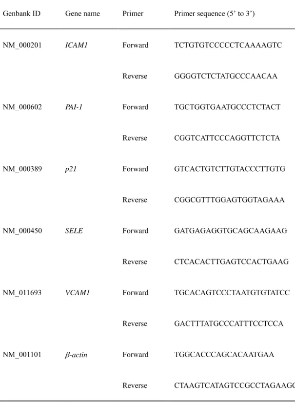
Table 1. Primer pairs used for qRT-PCR
Genbank ID Gene name Primer Primer sequence (5’ to 3’)
NM_000201 NM_000602 NM_000389 NM_000450 NM_011693 NM_001101 ICAM1 PAI-1 p21 SELE VCAM1 -actin Forward Reverse Forward Reverse Forward Reverse Forward Reverse Forward Reverse Forward Reverse TCTGTGTCCCCCTCAAAAGTC GGGGTCTCTATGCCCAACAA TGCTGGTGAATGCCCTCTACT CGGTCATTCCCAGGTTCTCTA GTCACTGTCTTGTACCCTTGTG CGGCGTTTGGAGTGGTAGAAA GATGAGAGGTGCAGCAAGAAG CTCACACTTGAGTCCACTGAAG TGCACAGTCCCTAATGTGTATCC GACTTTATGCCCATTTCCTCCA TGGCACCCAGCACAATGAA CTAAGTCATAGTCCGCCTAGAAGCA
5. Results
5.1. Cytotoxicity of DNJ
To examine the cytotoxicity of DNJ, the survival of HUVECs at PDL8-9 exposed to DNJ (0-200
μmol/L) was examined (Figure 2). The survival rate of HUVECs exposed to 0 μmol/L DNJ for 24
or 48 h was defined as 100%. DNJ did not influence the survival of HUVECs.
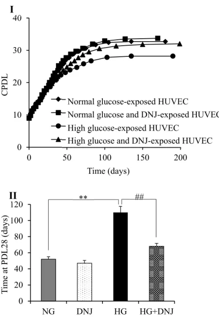
5.2. Effects of high glucose and DNJ on cell proliferation
To examine the effects of high glucose and DNJ on cell proliferation in HUVECs, HUVECs exposed
to high glucose and DNJ were cultured until they reached the Hayflick limit. HUVECs increased
logarithmically up to PDL10 and then cell proliferation decreased with increasing PDL (Figure 3I).
A particularly marked decrease in cell proliferation was observed in HUVECs exposed to high
glucose compared with those exposed to normal glucose, and this change was reversed by DNJ.
HUVECs in the NG, DNJ, HG, and HG + DNJ groups reached the Hayflick limit at PDL 32.7, 33.7,
28.2 and 32.0, respectively. The number of days required for HUVECs to reach PDL28 was
compared (Figure 3II). Cell proliferation was significantly decreased in the HG group compared
between NG and DNJ groups.
5.3. Effects of high glucose and DNJ on cellular senescence
HUVECs were evaluated by a SA-ß-Gal staining assay to detect senescent cells and observe the
progression of senescence with increasing PDL. HUVECs at PDL28-29 in the NG group were larger
than the cells at PDL8-9, and the rates of SA-ß-Gal-positive cells at PDL28-29 were significantly
higher than at PDL8-9 (data not shown). The rate of SA-ß-Gal-positive cells was significantly higher
in the HG group compared with the NG group and this increase was suppressed by DNJ (Figure 4).
Additionally, the rate of SA-ß-Gal-positive cells was significantly lower in the DNJ group compared
with the NG group. mRNA levels for PAI-1 and p21, which are upregulated by aging in HUVECs,
were also significantly higher in HUVECs at PDL28-29 than at PDL8-9 in a quantitative RT-PCR
assay (data not shown). The mRNA levels of PAI-1 and p21 were also significantly higher in the
HG group compared with the NG group and this increase was also suppressed by DNJ (Table 2).
Additionally, the mRNA level of PAI-1 was also significantly lower in the DNJ group compared
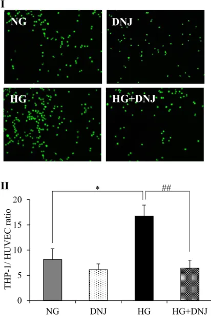
5.4. Effects of high glucose and DNJ on monocyte adhesion
To examine the effects of high glucose and DNJ on monocytic adhesion, HUVECs and calcein
AM-labeled THP-1 cells were co-cultured. Monocytic adhesion with HUVECs at PDL28-19 in the NG
group was significantly higher than that with cells at PDL8-9 (data not shown). Monocytic adhesion
was significantly greater in the HG group compared with the NG group and this increase was
suppressed by DNJ (Figure 5). There was no significant difference between NG and DNJ groups.
mRNA levels of ICAM1, SELE, and VCAM1, which are cell adhesion molecules, were
significantly higher in HUVECs at PDL30 than at PDL9 in a quantitative RT-PCR assay (data not
shown). These mRNA levels were also significantly higher in the HG group compared with the NG
group and these increases were suppressed by DNJ (Table 2). Additionally, the mRNA levels of
ICAM1 and VCAM1 were significantly lower in the DNJ group compared with the NG group.
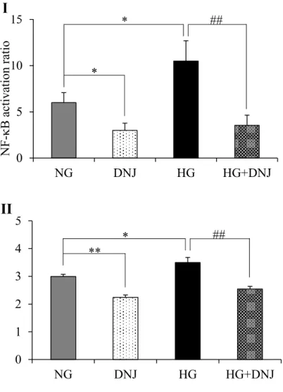
5.5. Effects of high glucose and DNJ on NF-kB activity and ROS generation
Monocyte adhesion in HUVECs is promoted by NF-kB activation and ROS generation. NF-kB
activity in HUVECs at PDL28-29 was significantly higher than that at PDL8-9 (data not shown)
and was significantly higher in the HG group compared with the NG group, with this increase was
group compared with the NG group. An H2DCFDA assay showed that ROS generation in HUVECs
at PDL28-29 was also significantly higher than that at PDL8-9 (data not shown). ROS generation
was also significantly higher in the HG group compared with the NG group and this increase was
suppressed by DNJ (Figure 6II). Additionally, ROS generation was also significantly lower in the
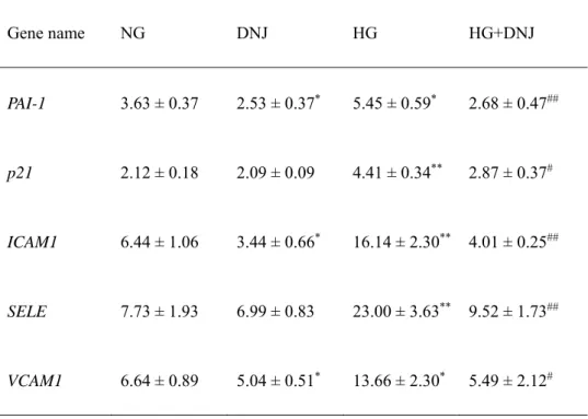
Table 2. Effects of DNJ on mRNA expression for genes related to cellular senescence and monocyte adhesion. Gene name NG DNJ HG HG+DNJ PAI-1 p21 ICAM1 SELE VCAM1 3.63 ± 0.37 2.12 ± 0.18 6.44 ± 1.06 7.73 ± 1.93 6.64 ± 0.89 2.53 ± 0.37* 2.09 ± 0.09 3.44 ± 0.66* 6.99 ± 0.83 5.04 ± 0.51* 5.45 ± 0.59* 4.41 ± 0.34** 16.14 ± 2.30** 23.00 ± 3.63** 13.66 ± 2.30* 2.68 ± 0.47## 2.87 ± 0.37# 4.01 ± 0.25## 9.52 ± 1.73## 5.49 ± 2.12#
The mRNA expression of 5 genes related to cellular senescence and adhesion molecules in NG,
DNJ, HG, and HG+DNJ groups was determined by using qRT-PCR. The data were shown by the
ratio to young cells (normal-glucose exposed HUVECs at PDL8-9). Values are expressed as the
mean ± SD (n=10). *P < 0.01, **P < 0.01 (vs. NG); #P <0.05, ##P <0.01 (vs. HG). NG;
normal-glucose exposed HUVECs at PDL28-29, DNJ; normal-normal-glucose and 10 mol/L DNJ exposed
HUVECs at PDL28-29, HG; high-glucose exposed HUVECs at PDL28-29, HG+DNJ; high-glucose
Figure 2. Cytotoxicity of DNJ in HUVECs at PDL8-9. Cell survival rates of HUVECs exposed to
DNJ for 24 h (I) and 48 h (II) were examined by WST-8 assay. Values are expressed as the mean ±
SD (n = 6).
0
20
40
60
80
100
120
0
100
200
Cell survival rate (% of 0
M)
24h
I
0
20
40
60
80
100
120
0
100
200
DNJ (M)
Cell survival rate (% of 0
M)
48h
Figure 3. Effects of DNJ on cell proliferation in HUVECs exposed to high glucose. (I) Growth
curve showing the cumulative population doubling level (CPDL) during culturing from young to
senescent. HUVECs exposed to normal glucose (NG), NG + DNJ (10 μmol/L), high glucose
(HG), and HG + DNJ (10 μmol/L) were cultured until they became senescent. (II) The number of
days required for HUVEC to reach PDL28 was compared. Values are expressed as the mean ± SD
(n = 10). *P < 0.05, **P < 0.01 (vs. NG). #P b 0.05, ##P b 0.01 (vs. HG).
0
10
20
30
40
0
50
100
150
200
NG
DNJ
HG
HG+DNJ
I
CPDL
Time (days)
Normal glucose-exposed HUVEC
Normal glucose and DNJ-exposed HUVEC
High glucose-exposed HUVEC
High glucose and DNJ-exposed HUVEC
0
20
40
60
80
100
120
NG
DNJ
HG
HG+DNJ
T
im
e at PDL28 (days)
II
**
##
Figure 4. Effects of DNJ on senescence of HUVECs exposed to high glucose. (I) Photographs of
typical SA-ß-Gal-stained HUVECs in the NG, DNJ, HG, and HG + DNJ groups. (II) Percentages
of SA-ß-Gal-positive HUVECs in the NG, DNJ, HG, and HG + DNJ groups. The data were
shown by the ratio to young cells (normal-glucose exposed HUVECs at PDL8-9). Values are
expressed as the mean ± SD (n = 10). *P < 0.05, **P < 0.01 (vs. NG). #P < 0.05, ##P < 0.01 (vs.
HG).
I
NG
HG
HG+DNJ
DNJ
0
20
40
60
80
100
NG
DNJ
HG
HG+DNJ
SA--Gal p
ositiv
e ce
lls
(%)
II
*
*
##
Figure 5. Effects of DNJ on monocyte adhesion. (I) Photographs of typical fluorescence- stained
THP-1 cells in HUVECs in the NG, DNJ, HG, and HG + DNJ groups. (II) Percentages of THP-1
cells in HUVECs in the NG, DNJ, HG, and HG + DNJ groups. The data were shown by the ratio
to young cells (normal-glucose exposed HUVECs at PDL8-9). Values are expressed as the mean ±
SD (n = 10). *P < 0.05, **P < 0.01 (vs. NG). #P < 0.05, ##P < 0.01 (vs. HG).
I
NG
HG
HG+DNJ
DNJ
0
5
10
15
20
NG
DNJ
HG
HG+DNJ
THP-1/ HUVEC ratio
II
*
##
Figure 6. Effects of DNJ on NF-kB activation and ROS generation in high-glucose exposed
HUVECs. (I) Proportion of NF-kB activation in the NG, DNJ, HG, and HG + DNJ groups. (II)
ROS generation in the NG, DNJ, HG, and HG + DNJ groups based on a H2DCFDA assay. The
data were shown by the ratio to young cells (normal-glucose exposed HUVECs at PDL8-9).
Values are expressed as the mean ± SD (n = 10). *P < 0.05, **P < 0.01 (vs. NG). #P < 0.05,
##P < 0.01 (vs. HG).
0
5
10
15
NG
DNJ
HG
HG+DNJ
I
N
F-B activ
ation
ratio
*
*
##
0
1
2
3
4
5
NG
DNJ
HG
HG+DNJ
II
**
*
##
6. Discussion
Delayed senescence of endothelial cells promoted by DNJ may be effective for prevention of
aging-related diseases. In this study, we showed for the first time that DNJ can delay senescence of
HUVECs that is promoted under high glucose condition. Since there has been no report of the
cytotoxicity of DNJ in HUVECs, we first showed that DNJ up to 200 μmol/L did not influence
survival of HUVECs. We have also shown previously that the DNJ level in human plasma can reach
3.2 μmol/L (16) and in rat plasma about 100 μmol/L (15), and therefore a level of 10 μmol/L was used
to evaluate the effects of DNJ on the senescence of endothelial cells.
A marked decrease in cell proliferation was observed in HUVECs exposed to high glucose
compared to those cultured with normal glucose. SA-ß-Gal-positive cells in these groups were
significantly more frequent than in young cells, and significantly more frequent in the HG group
compared with the NG group. mRNA levels of PAI-1 and p21, which are upregulated by senescence
in HUVECs, were significantly higher in the NG group compared to young cells, and significantly
higher in the HG group compared with the NG group. The decrease in cell proliferation and
increases in SA-ß-Gal-positive cells and mRNA levels of PAI-1 and p21 were reversed by DNJ.
may be effective for prevention of aging-related diseases. Additionally, the rate of SA-ß-
Gal-positive cells and the mRNA level of PAI-1 were significantly lower in the DNJ group compared
with the NG group. Therefore, DNJ might be effective also for delay senescence.
We have previously shown that senescence HUVECs have increased levels of genes related to
monocyte adhesion (30). In this study, senescence of HUVECs promoted monocyte adhesion and this
effect was markedly increased by high glucose and suppressed by DNJ. Monocyte adhesion occurs
in the first stage of arteriosclerotic (21). Therefore, intake of DNJ might be effective for prophylaxis
of arteriosclerosis. Expression of monocyte adhesion molecules is induced by NF-kB and ROS (17,
18), and we found that senescence of HUVECs increased ROS production which activated NF-kB,
with these changes promoted by high glucose and suppressed by DNJ. We previously showed that
DNJ reduced oxidative stress in the liver and plasma of mice and rats (22, 23). Additionally, in this
study, ROS generation was significantly lower in the DNJ group compared with the NG group.
These findings suggest that DNJ behaves as an antioxidant, decreases ROS production, and delays
cellular senescence. Attenuation of high glucose-accelerated senescence by decreasing ROS levels
may make DNJ useful as a health food supplement and medicine. Further studies are needed to
is quickly eliminated from the body (15, 16). Hence, in the case of considering the application to
humans, it is necessary to ingest many times, to obtain the effect of DNJ. For example, it may be
7. References
(1) Asano, N., Nash, R.J., Molyneux, R.J., Fleet, G.W.J., 2000. Sugar-mimic glycosidase inhibitors:
natural occurrence, biological activity and prospects for therapeutic application.
Tetrahedron-Asymmetry 11, 1645–1680.
(2) Boisen, L., Drasbek, K.R., Pedersen, A.S., Kristensen, P., 2010. Evaluation of endothelial cell culture
as a model system of vascular ageing. Exp. Gerontol. 45, 779–787.
(3) Dimri, G.P., Lee, X., Basile, G., Acosta, M., Scott, G., Roskelley, C., Medrano, E.E., Linskens, M.,
Rubelj, I., Pereira-Smith, O., 1995. A biomarker that identifies senescent human cells in culture and
in aging skin in vivo. Proc. Natl. Acad. Sci. U. S. A. 92, 9363–9367.
(4) Eu, J.P., Liu, L., Zeng, M., Stamler, J.S., 2000. An apoptotic model for nitrosative stress.
Biochemistry 39, 1040–1047.
(5) Hanada, S., Harada, M., Kumemura, H., Bishr Omary, M., Koga, H., Kawaguchi, T., Taniguchi, E.,
Yoshida, T., Hisamoto, T., Yanagimoto, C., Maeyama, M., Ueno, T., Sata, M., 2007. Oxidative stress
induces the endoplasmic reticulum stress and facilitates inclusion formation in cultured cells. J.
Hepatol. 47, 93–102.
Kamalanathan, S., Hattori, Y., Ignarro, L.J., Iguchi, A., 2006. Endothelial cellular senescence is
inhibited by nitric oxide: implications in atherosclerosis associated with menopause and diabetes.
Proc. Natl. Acad. Sci. U. S. A. 103, 17018–17023.
(7) Hayashida, K., Kume, N., Minami, M., Kita, T., 2002. Lectin-like oxidized LDL receptor-1
(LOX-1) supports adhesion of mononuclear leukocytes and a monocyte-like cell line THP-1 cells under
static and flow conditions. FEBS Lett. 511, 133–138.
(8) Kimura, T., Nakagawa, K., Kubota, H., Kojima, Y., Goto, Y., Yamagishi, K., Oita, S., Oikawa, S.,
Miyazawa, T., 2007. Food-grade mulberry powder enriched with 1-deoxynojirimycin suppresses the
elevation of postprandial blood glucose in human. J. Agric. Food Chem. 55, 5869–5874.
(9) Kume, N., Cybulsky, M.I., Gimbrone Jr., M.A., 1992. Lysophosphatidylcholine, a component of
atherogenic lipoproteins, induces mononuclear leukocyte adhesion molecules in cultured human and
rabbit arterial endothelial cells. J. Clin. Invest. 90, 1138–1144.
(10) Lakatta, E.G., Levy, D., 2003. Arterial and cardiac aging: major shareholders in cardiovascular
disease enterprises: Part I: aging arteries: a “set up” for vascular disease. Circulation 107, 139–146.
(11) Minamino, T., Komuro, I., 2007. Vascular cell senescence: contribution to atherosclerosis. Circ. Res.
(12) Minamino, T., Miyauchi, H., Yoshida, T., Ishida, Y., Yoshida, H., Komuro, I., 2002. Endothelial cell
senescence in human atherosclerosis: role of telomere in endothelial dysfunction. Circulation 105,
1541–1544.
(13) Molina-Jiménez, M.F., Sánchez-Reus, M.I., Andres, D., Cascales, M., Benedi, J., 2004. Neuro-
protective effect of fraxetin and myricetin against rotenone-induced apoptosis in neuroblastoma cells.
Brain Res. 1009, 9–16.
(14) Mudra, M., Ercan-Fang, N., Zhong, L., Furne, J., Levitt, M., 2007. Influence of mulberry leaf extract
on the blood glucose and breath hydrogen response to ingestion of 75 g sucrose by type 2 diabetic
and control subjects. Diabetes Care 30, 1272–1274.
(15) Nakagawa, K., Kubota, H., Kimura, T., Yamashita, S., Tsuzuki, T., Oikawa, S., Miyazawa, T., 2007.
Occurrence of orally administered mulberry 1-deoxynojirimycin in rat plasma. J. Agric. Food Chem.
55, 8928–8933.
(16) Nakagawa, K., Kubota, H., Tsuzuki, T., Kariya, J., Kimura, T., Oikawa, S., Miyazawa, T., 2008.
Validation of an ion trap tandem mass spectrometric analysis of mulberry 1-deoxynojirimycin in
human plasma: application to pharmacokinetic study. Biosci. Biotechnol. Biochem. 72, 2210–2213.
induces monocyte-endothelial cells adhesion and transmigration by increasing VCAM-1 and
MCP-1 expression in human aortic endothelial cells. Atherosclerosis MCP-193, 328–334.
(18) Redmond, E.M., Morrow, D., Kundimi, S., Miller-Graziano, C.L., Cullen, J.P., 2009. Acetaldehyde
stimulates monocyte adhesion in a P-selectin- and TNFalpha-dependent manner. Atherosclerosis 204,
372–380.
(19) Rogers, S.C., Zhang, X., Azhar, G., Luo, S., Wei, J.Y., 2013. Exposure to high or low glucose levels
accelerates the appearance of markers of endothelial cell senescence and induces dysregulation of
nitric oxide synthase. J. Gerontol. A Biol. Sci. Med. Sci. 68, 1469–1481.
(20) Shinohara, N., Tsuduki, T., Ito, J., Honma, T., Kijima, R., Sugawara, S., Arai, T., Yamasaki, M.,
Ikezaki, A., Yokoyama, M., Nishiyama, K., Nakagawa, K., Miyazawa, T., Ikeda, I., 2012. Jacaric
acid, a linolenic acid isomer with a conjugated triene system, has a strong antitumor effect in vitro
and in vivo. Biochim. Biophys. Acta 1821, 980–988.
(21) Takahashi, M., Ikeda, U., Masuyama, J., Kitagawa, S., Kasahara, T., Shimpo, M., Kano, S., Shimada,
K., 1996. Monocyte-endothelial cell interaction induces expression of adhesion molecules on human
umbilical cord endothelial cells. Cardiovasc. Res. 32, 422–429.
Intake of 1-deoxynojirimycin suppresses lipid accumulation through activation of the beta-oxidation
system in rat liver. J. Agric. Food Chem. 57, 11024–11029.
(23) Tsuduki, T., Kikuchi, I., Kimura, T., Nakagawa, K., Miyazawa, T., 2013a. Intake of mulberry
1-deoxynojirimycin prevents diet-induced 1 obesity through increases in adiponectin in mice. Food
Chem. 139, 16–23.
(24) Tsuduki, T., Kuriyama, K., Nakagawa, K., Miyazawa, T., 2013b. Tocotrienol (unsaturated vitamin E)
suppresses degranulation of mast cells and reduces allergic dermatitis in mice. J. Oleo Sci. 62, 825–
834.
(25) Tsuzuki, T., Kawakami, Y., 2008. Tumor angiogenesis suppression by α-eleostearic acid, a linolenic
acid isomer with a conjugated triene system, via peroxisome proliferator-activated receptor g.
Carcinogenesis 29, 797–806.
(26) Tsuzuki, T., Tanaka, K., Kuwahara, S., Miyazawa, T., 2005. Synthesis of the conjugated trienes
5E,7E,9E,14Z,17Z-eicosapentaenoic acid and 5Z,7E,9E,14Z,17Z-eicosapentaenoic acid, and their
induction of apoptosis in DLD-1 colorectal adenocarcinoma human cells. Lipids 40, 147–154.
(27) Tsuzuki, T., Kambe, T., Shibata, A., Kawakami, Y., Nakagawa, K., Miyazawa, T., 2007. Conjugated
colorectal adenocarcinoma human cells. Biochim. Biophys. Acta 1771, 20–30.
(28) Watson, A.A., Fleet, G.W.J., Asano, N., Molynerux, R.J., Nash, R.J., 2001. Polyhydroxylated
alkaloids—natural occurrence and therapeutic applications. Phytochemistry 56, 265–295.
(29) Yagi, M., Kouno, T., Aoyagi, Y., Murai, H., 1976. Structure of moranoline, a piperidine alkaloid from
Morus specises. Nippon Nogei Kagaku Kaishi 50, 571–572.
(30) Yanaka, M., Honma, T., Sato, K., Shinohara, N., Ito, J., Tanaka, Y., Tsuduki, T., Ikeda, I., 2011.
Increased monocytic adhesion by senescence in human umbilical vein endothelial cells. Biosci.
Biotechnol. Biochem. 75, 1098–1103.
(31) Yokoi, T., Fukuo, K., Yasuda, O., Hotta, M., Miyazaki, J., Takemura, Y., Kawamoto, H., Ichijo, H.,
Ogihara, T., 2006. Apoptosis signal-regulating kinase 1 mediates cellular senescence induced by high
glucose in endothelial cells. Diabetes 55, 1660–1665.
(32) Zhong, W., Zou, G., Gu, J., Zhang, J., 2010. L-arginine attenuates high glucose-accelerated
senescence in human umbilical vein endothelial cells. Diabetes Res. Clin. Pract. 89, 38–45.
(33) Zou, L., Yang, S., Champattanachai, V., Hu, S., Chaudry, I.H., Marchase, R.B., Chatham, J.C., 2009.
Glucosamine improves cardiac function following trauma-hemorrhage by increased protein
H523.
(34) E S, Kijima R, Honma T, Yamamoto K, Hatakeyama Y, Kitano Y, Kimura T, Nakagawa K, Miyazawa
T, Tsuduki T.1-Deoxynojirimycin attenuates high glucose-accelerated senescence in human
Intake of mulberry 1-deoxynojirimycin prevents colorectal cancer in mice
1. Abstract
The effect of 1-deoxynojirimycin (DNJ), a caloric restriction (CR) mimetic, was examined in ICR mice
with AOM (azoxymethane)/DSS (Dextran sodium sulfate)-induced colorectal cancer. AOM is a
carcinogen (10mg/kg body weight), and 2%DSS (w/v) used as a colitis-inducing agent. Mice were
separated into 5 groups: a group without colorectal cancer fed a normal diet (CO- group), and groups with
colorectal cancer fed a normal diet (CO+ group), a calorie-restricted diet (CR group), and diets including
0.02% and 0.1% DNJ (L-DNJ and H-DNJ groups). The tumor incidence and number were reduced
significantly in the CR group compared to the CO+ group, and were also suppressed in a dose-dependent
manner by 1-deoxynojirimycin. mRNA for anti-apoptotic Bcl-2 was decreased and that for pro-apoptotic
Bax was increased in the carcinoma tissue of CR, L-DNJ and H-DNJ groups. These results suggest that
CR and 1-deoxynojirimycin inhibit growth of colorectal cancer by inducing apoptosis in an induced cancer
2. Abbreviations
DNJ: 1-Deoxynojirimycin
CRC: Colorectal Cancer
CR: Caloric Restriction
AOM: Azoxymethane
DSS: Dextran Sodium Sulfate
Bax: Bcl-2 associated X protein
3. Introduction
Cancer is the leading cause of morbidity and mortality worldwide, with approximately 14 million new
cases and 8.2 million cancer-related deaths in 2012. (1) More than 60% of new cases annually occur in
Africa, Asia and Central and South America, and among these, colorectal cancer is a major cause of
tumor-related morbidity and mortality. (1,2) This disease develops due to long-term exposure to environmental
factors. (3) In Japan, rapid Westernization of diet has increased the incidence and mortality of colorectal
cancer, and this suggests that dietary treatment, especially caloric restriction (CR), may be effective for
disease prevention. CR has beneficial effects on cancer prevention, with one study showing that the
incidence of neoplasia with CR was significantly lower than that with an ad libitum diet. (4,5) However, CR
is accompanied by considerable stress in humans, which makes it difficult to use as a method for cancer
prevention.
1-Deoxynojirimycin (DNJ) is a D-glucose analogue that is a characteristic constituent of mulberry
(Moraceae) leaves. Dietary mulberry DNJ may be beneficial for suppression of abnormally high blood
glucose. (6) In addition, we have shown anti-obesity and anti-lipid peroxidation effects of DNJ, with
decreased serum insulin and glucose, improved carbohydrate metabolism, and decreased lipid peroxide
have a CR effect. Moreover, DNJ intake showed changes in lipid metabolic parameters like CR. (7) Cancer
cells require larger amounts of glucose than normal cells, which suggests that growth of these cells might
be inhibited by DNJ. (8,9)
Screening for agents for colorectal cancer prevention has been carried out in mouse models using the
potent carcinogen azoxymethane (AOM), which induces colorectal cancers at a high incidence. (10) Dextran
sodium sulfate (DSS), a colitis-inducing agent, can be used after AOM to make a two-stage mouse
colorectal cancer model. (11,12) In this study, we used this two-stage model to examine the effect of DNJ on
colorectal cancer. We also examined the mechanism of the DNJ effect by measuring the levels of
4. Materials and Methods
4.1. Materials
DNJ was extracted from mulberry leaves (Morus alba) and purified using ion-exchange chromatography
followed by recrystallization. (13) The purity of DNJ was shown to be >98% by hydrophilic interaction
liquid chromatography with hybrid quadrupole/linear ion trap tandem mass spectrometry (HILIC-QTRAP
MS/MS). (7) NaCl, AOM, DSS, miglitol and 10% formalin were purchased from Wako Pure Chemicals
Industries (Osaka, Japan).
4.1. Animals and diets
All procedures were performed in accordance with the Animal Experiment Guidelines of Tohoku
University. The animal protocol was approved by the Animal Use Committee at Tohoku University. Male
ICR mice (n=100, 3 weeks of age) and CE-2 (a control diet) were obtained from Japan Clea (Tokyo, Japan).
Mice were housed with ten in each cage and with free access to the respective diets and distilled water in
a temperature- and humidity-controlled room with light cycles of 12 h on and 12 h off. (14) After being
acclimatized to the control diet for one week, the 100 mice were randomly divided into 5 groups: control
diet-fed mice without colorectal cancer inducement (CO-); and control (CO+), calorie-restricted (CR), low
The experimental protocol is shown in Figure 1. The CO+, CR, L-DNJ and H-DNJ groups received a
single intraperitoneal injection of AOM in sterile saline at a dose of 10 mg/kg body weight to induce
colorectal cancer. Starting one week after the injection, animals received 2% DSS in drinking water for
one week to promote tumor progression. The CO- group received a single intraperitoneal injection of
sterile saline only. The CO- and CO+ groups were fed CE-2 diet only. The CR, L-DNJ and H-DNJ groups
were fed CE-2 diet for three weeks from the start of the experiment. Then, the CR group was fed every
other day with CE-2 diet for 12 weeks, starting 1 week after cessation of DSS exposure. The L-DNJ and
H-DNJ groups were fed CE-2 diet containing 0.02% and 0.1% DNJ, respectively, for 13 weeks, starting
one week after cessation of DSS exposure. At the end of the 16-week period (21 weeks old), the mice were
weighed and blood samples were collected after decapitation. Liver, kidney, pancreas, epididymis adipose
tissue, and colon tissue were removed and weighed. The number of colorectal tumors detectable with the
naked eye was measured. Serum was isolated by cold centrifugation at 1000×g for 15 min at 4°C
(CAX-370 Hybrid Refrigerated Centrifuges, Tomy Digital Biology, Tokyo, Japan). Serum and tissue were stored
at −80°C until use.
4.2. Histological analysis of colon tissue
sections (5 μm) were cut, mounted on glass slides, stained with hematoxylin and eosin (H&E), and
observed using a microscope (BZ-9000; Keyence, Osaka, Japan).
4.4 Biochemical analyses of serum and liver
To confirm a CR effect, biochemical analyses of serum and liver samples were performed as described
previously. (7, 16) Triacylglycerol (TG) and total cholesterol (TC) levels in serum and liver, and phospholipid
(PL), aspartate aminotransferase (AST), alanine aminotransferase (ALT), and glucose in serum were
measured using commercial kits (Wako Pure Chemical, Osaka, Japan). The PL content in liver was
determined using the method described by Rouser (1970). (17) Insulin levels in serum were determined
using an ELISA kit (Shibayagi, Shibukawa, Japan).
4.5. mRNA expression analyses
For real-time quantitative reverse transcriptase PCR (qRT-PCR), total RNA was isolated from colon (tumor
and normal tissues) using a RNeasy Mini Kit (Qiagen, Valencia, CA), after elution with 30 μl of
RNase-free water, and stored at −80 °C until use. (18) To quantify expression of genes, the mRNA levels of β-actin,
B-cell lymphoma 2 (Bcl-2), Bcl-2 associated X protein (Bax) and sirtuin 1 (Sirt1) were determined using
quantitative detection of PCR products by measuring the increase in fluorescence caused by binding of
SYBR green to double-stranded DNA. cDNA was synthesized from the total RNA in colon using a
Ready-To-Go T-Primed First-Strand Kit (GE Healthcare, UK). The cDNA was subjected to PCR amplification
using SYBR® Premix Ex Taq™ (Perfect Real Time, Takara Bio) and gene-specific primers for β-actin,
Bcl-2, BAX or Sirt1. Primer sequences were as follows: β-actin (forward) 5′-AGT GTG ACG TTG ACA
TCC GTA-3′, β-actin (reverse) 5′-GCC AGA GCA GTA ATC TCC TTC T - 3′, Bcl-2 (forward) 5′-TGT
GGT CCA TCT GAC CCT CC-3′, Bcl-2 (reverse) 5′-ACA TCT CCC TGT TGA CGC TCT-3′, BAX
(forward) 5′-TGA AGA CAG GGG CCT TTT TG-3′, BAX (reverse) 5′-AAT TCG CCG GAG ACA CTC
G-3′, Sirt1 (forward) 5′-GAC GAT GAC AGA ACG TCA CAC-3′, Sirt1 (reverse) 5′-CGA GGA TCG GTG
CCA ATC A-3′. The PCR conditions were 95°C for 10 s, and then 95°C for 5 s and 60°C for 31 s over 40
cycles for each gene. Melting curve analysis was performed following each reaction to confirm the
presence of a single reaction product. The cycle threshold (CT) represents the PCR cycle at which the
reporter fluorescence increased above a baseline signal. The ratio between the β-actin content in standard
and test samples was defined as the normalization factor.
4.6. Determination of lipid peroxides
substances (TBARS) in serum and liver were determined. (7, 14) To examine oxidative stress caused by
AOM and DSS in colon tissue, TBARS level in normal colon tissue was determined. (19)
4.7. DNJ concentration in colorectal cancer and normal tissue
DNJ concentrations in colon tissue (tumor or normal) were determined using HILIC MS/MS. (17) In brief,
a 1:10 dilution of colorectal cancer or normal tissue homogenate (500 μL), 100 μL of 0.1 μg/mL miglitol
(internal standard) and 600 μl of acetonitrile were mixed by sonicating for 1 min and vortexing for 30 s.
After centrifugation at 8,000×g for 10 min at 4°C (CAX-370), the supernatant was collected. A 5-μL
aliquot of the resulting extract was subjected to HILIC-MS/MS using a Shimadzu liquid chromatograph
and a 4500-tandem mass spectrometer (AB Sciex, Tokyo, Japan). Under positive ion electrospray
ionization conditions, MS/MS parameters were optimized with DNJ and miglitol. Samples (5 μL each)
were separated on a HILIC column (TSK gel Amide-80, 4.6 mm × 150 mm; Tosoh, Tokyo, Japan), eluted
with a mixture of acetonitrile and water (675:325, v/v) containing 6.5 mM ammonium formate (pH 5.5) at
a flow rate of 0.2 ml/min and a temperature of 40°C. Post-column, DNJ was detected by HILIC-MS/MS
with multiple reaction monitoring for transition of the parent ion to the product ion. DNJ concentrations
4.8. Statistical analysis
Results are expressed as the mean ± standard error of the mean (SE). Data were analyzed using a one-way
ANOVA with a Tukey-Kramer test for multiple comparisons among three or four groups. A difference was
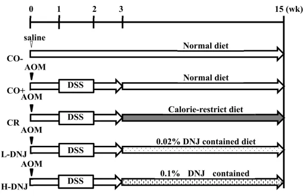
Figure 1. Study design. The CO− group received a single intraperitoneal injection of saline and a normal
diet with no more treatment. The CO+, CR, L DNJ and H DNJ groups received a single intraperitoneal
injection of AOM and 2% DSS in drinking water for 7 days, starting 1 week after the injection. From the
third week, the groups received normal, caloric restricted, 0.02% DNJ and 0.1% DNJ diets, respectively.
After 16 weeks, all mice were sacrificed. AOM, azoxymethane; DNJ, 1 deoxynojirimycin; DSS, dextran
sulfate sodium.
saline
Normal diet
Normal diet
Calorie-restrict diet
0.02% DNJ contained diet
0.1% DNJ contained
CO-
CO+
CR
L-DNJ
H-DNJ
0
1 2 3 15 (wk)
AOM
AOM
AOM
AOM
DSS
DSS
DSS
DSS
5. Results
5.1. Effects of caloric restriction and DNJ on growth parameters
There were significant decreases in food and energy intake in the CR group compared to the CO- and CO+
groups. The CR group had caloric restriction of about 20% compared to the CO+ group. In contrast, there
were significant increases in food and energy intake in the H-DNJ group compared to the CO- group (Table
1). There were no significant differences in body weight and tissue weights among the groups with induced
colorectal cancer.
5.2. Effects of caloric restriction and DNJ on tumor formation
The number of colonic tumors visible with the naked eye was counted after sacrifice (Figure 2-I). H&E
staining of colon tissues to confirm the occurrence of a tumor (adenoma and adenocarcinoma) showed
colon inflammation (Figure 2-IIB), adenoma (Figure 2-IIC) and adenocarcinoma (Figure 2-IID) in mice
with induced cancer, but normal tissue (Figure 2-IIA) in the control group (CO-). Tumor incidences and
numbers are shown in Figure 3. Compared to the CO+ group, there was a 28% decrease in tumor incidence
in the CR group (Figure 3-I), and 5.2% and 31.5% decreases in the L-DNJ and H-DNJ groups, respectively.
There were also significant decreases in the number of tumors in the CR, L-DNJ, and H-DNJ groups
manner, and the results in the H-DNJ group were like those in the CR group.
5.3. Effects of caloric restriction and DNJ on serum and liver parameters
To investigate the effects of caloric restriction and DNJ on lipid and carbohydrate metabolism, serum levels
of TG, TC, PL, glucose and insulin were determined (Table 2). There were significant increases in TC and
PL in the L-DNJ group compared to the CO+, CR and H-DNJ groups. There was a significant decrease in
insulin in the CR, L-DNJ and H-DNJ groups compared to the CO+ group. To evaluate the safety of caloric
restriction and DNJ, serum and liver TBARS and serum ALT and AST levels were measured. There were
significant decreases in serum and liver TBARS in the CR and H-DNJ groups compared to the CO- and
CO+ groups, and TBARS decreased dose-dependently with DNJ. There were significant decreases in
serum AST in the CR and H-DNJ group compared to the CO+ group. To investigate the effects of caloric
restriction and DNJ on lipid metabolism, liver levels of TG, TC, and PL were determined (Table 2). There
were no significant differences in liver parameters among the groups with induced colorectal cancer. There
were no significant differences in serum and liver parameters between the CO- and CO+ groups. To
investigate the effects of caloric restriction and DNJ on the oxidative stress caused by AOM and DSS in
normal colon tissue, TBARS levels in the colon were determined (Table 2). There was significant increase
TBARS in CR, L-DNJ and H-DNJ groups compared to the CO+ group, and TBARS decreased
dose-dependently with DNJ.
5.4. Effects of caloric restriction and DNJ on apoptosis
To examine the tumor suppression mechanism, the mRNA levels of two apoptosis-related genes (Bcl-2
and BAX) were measured. There were significant increases in mRNA for pro-apoptotic Bax in the CR and
H-DNJ groups compared to the CO+ and L-DNJ groups (Figure 4-I), and significant decreases in mRNA
for the anti-apoptotic gene Bal-2 in the CR group compared to the CO+ group, with a tendency for
decreases in the L-DNJ and H-DNJ groups (Figure 4-II). The mRNA levels for the two genes varied
dose-dependently with DNJ. These findings suggest that caloric restriction and DNJ induce apoptosis of cancer
cells. In addition, the caloric restriction-related gene Sirt1 was examined as a caloric restriction marker
(Figure 4-III). There were significant increases in mRNA for Sirt1 in the CR group compared to the CO+
group, and tendencies for increases in the L-DNJ and H-DNJ groups.
5.5 DNJ concentration in colorectal cancer and normal tissues
To determine whether DNJ is absorbed in colon tissue, DNJ was measured in normal and tumor colon
significantly higher than that in the L-DNJ group in normal tissue (0.448 ± 0.114 vs. 0.089 ± 0.013 ng/g)
and tumor tissue (1.03 ± 0.09 vs. 0.204 ± 0.048 ng/g). The DNJ concentration in tumor tissue was
Table1. Effect of calorie restriction and DNJ on growth parameters in colon cancer-induced male
mice.
Values are means ±SE, n = 18-20. Means in a row with different letters are significantly different at P <
0.05.
CO- CO+ CR L-DNJ H-DNJ
Food intake (g/d) 5.41 ± 0.04 b 5.69 ± 0.08 bc 4.45 ± 0.07 a 5.66 ± 0.09 bc 5.72 ± 0.08 c
Energy intake (kcal/day) 18.7 ± 0.15 b 19.6 ± 0.28 bc 15.4 ± 0.20 a 19.5 ± 0.31 bc 19.7 ± 0.27 c
Final body weight (g) 44.5 ± 0.91 43.8 ± 0.79 41.4 ± 0.32 43.0 ± 0.39 41.7 ± 0.67
Tissue weight (g/100g body weight)
Liver 4.09 ± 0.06 4.49 ± 0.13 4.33 ± 0.15 4.47 ± 0.09 4.38 ± 0.13
Pancreas 0.85 ± 0.03 0.93 ± 0.04 0.90 ± 0.04 0.93 ± 0.04 0.92 ± 0.04
Kidney 1.82 ± 0.03 a 1.92 ± 0.05 ab 1.90 ± 0.04 ab 2.03 ± 0.04 b 1.89 ± 0.06 ab
Table2. Effect of calorie restriction and DNJ on plasma, liver and colon parameters in colon cancer-induced male mice.
Values are means ± SE, n = 18-20. Means in a row with different letters are significantly different at P <
0.05. HOMA-IR, homeostasis model assessment-insulin resistance; TBARS, thiobarbituric acid reactive
substances; ALT, alanine transaminase; AST, aspartate aminotransferase.
CO- CO+ CR L-DNJ H-DNJ
Serum
Triacylglycerol (mmol/L) 1.51 ± 0.09 1.57 ± 0.09 1.50 ± 0.10 1.72 ± 0.14 1.45 ± 0.11 Total cholesterol (mmol/L) 2.32 ± 0.06 ab 2.19 ± 0.04 a 2.11 ± 0.06 a 2.55 ± 0.08 b 2.23 ± 0.07 a
Phospholipid (mmol/L) 2.24 ± 0.04 a 2.15 ± 0.05 a 2.14 ± 0.07 a 2.51 ± 0.09 b 2.24 ± 0.08 a Glucose (mmol/L) 4.57 ± 0.20 5.03 ± 0.26 5.00 ± 0.18 4.69 ± 0.21 5.04 ± 0.28 Insulin (mg/L) 0.24 ± 0.03 ab 0.26 ± 0.04 a 0.15 ± 0.03 b 0.15 ± 0.01 b 0.15 ± 0.01 b HOMA-IR 1.00 ± 0.15 1.20 ± 0.20 0.66 ± 0.06 0.65 ± 0.06 0.66 ± 0.07 TBARS (μmol/L) 5.14 ± 0.21 a 5.47 ± 0.48 a 4.17 ± 0.23 b 4.90 ± 0.29 ab 4.27 ± 0.25 b ALT (UI/L) 9.75 ± 0.42 10.4 ± 0.52 10.0 ± 0.54 9.50 ± 0.92 11.5 ± 0.57 AST (UI/L) 53.0 ± 5.44 a 61.0 ± 7.89 b 46.5 ± 5.28 a 44.7 ± 3.32 a 43.5 ± 2.41 a Liver Triacylglycerol (μmol/g) 12.2 ± 1.13 8.82 ± 0.84 11.0 ± 1.21 13.0 ± 1.34 10.7 ± 1.39 Total cholesterol (μmol/g) 7.68 ± 0.65 7.84 ± 0.73 7.02 ± 0.20 6.45 ± 0.50 6.55 ± 0.41
Phospholipid (μmol/g) 34.5 ± 0.8 32.7± 1.4 35.4 ± 1.0 35.5 ± 1.2 34.6 ± 1.4
TBARS (nmol/g) 68.9 ± 5.3a 63.6 ± 3.9a 44.7 ± 2.8b 59.5 ± 3.1a 41.1 ± 3.3b
Colon
Figure 2 Effect of caloric restriction and DNJ on colon tissue in male mice with induced colorectal cancer.
(II) Colon tissue in each group. Arrows indicate tumors. (I) Representative histology images from
hematoxylin and eosin stained colon specimens (magnification ×4): (A) normal colon, (B) colon tissue
with mild pathology, (C) dysplastic crypts, and (D) well differentiated tubular adenocarcinoma. Bars
indicate 100 μm. CO-CO+ CR L-DNJ H-DNJ
I
1 00 μ m 1 0 0 μ m 1 00 μ m 1 0 0 μ mII
A
B
C
D
Figure 3 Effect of caloric restriction and DNJ on tumor incidence (I) and number (II) in male mice with
induced colorectal cancer. Values are means ± SE, n = 18–20. a, b p<0.05.
0%
(0/20)
94.7%
(19/20)
66.7%
(12/18)
89.5%
(17/19)
63.2%
(12/19)
0%
20%
40%
60%
80%
100%
CO-
CO+
CR
L-DNJ H-DNJ
(r
at
e)
I
a
b
b
b
0
2
4
6
8
10
CO+
CR
L-DNJ
H-DNJ
(/
m
ou
se
)
II
Figure 4 Effect of caloric restriction and DNJ on BAX (I), Bcl 2 (II), and Sirt 1 (III) mRNA levels in
male mice with induced colorectal cancer. Values are means ± SE, n = 18–20. a, b p<0.05.
a
ab
a
b
0.0
1.0
2.0
3.0
4.0
5.0
6.0
CO+
CR
L-DNJ
H-DNJ
Bax
m
RN
A
r
el
at
iv
e
cont
en
t
I
a
b
ab
ab
0.0
0.5
1.0
1.5
2.0
2.5
CO+
CR
L-DNJ
H-DNJ
B
cl-2
m
R
N
A
r
el
at
ive
co
nt
en
t
II
a
b
a
ab
0.0
0.5
1.0
1.5
2.0
2.5
3.0
CO+
CR
L-DNJ
H-DNJ
S
ir
t 1
m
R
N
A
r
el
at
iv
e
co
nt
en
t
III
6. Discussion
In this study, we showed that caloric restriction inhibits AOM/DSS-induced colorectal cancer in ICR mice,
and that DNJ suppresses this disease through a caloric restriction-like mechanism. This is the first report
of DNJ on colorectal cancer.
Cancer cells require more energy, and especially more glucose, for growth compared to normal cells. This
is referred to as the Warburg effect. (8, 20) Therefore, we speculated that caloric restriction can inhibit the
growth of tumor cells. In this study, the growth of mice was not particularly affected by caloric restriction,
since there was only 20% restriction in the CR group, but colorectal tumors were significantly reduced in
the CR group compared to the CO+ group. This suggests that caloric restriction can have an inhibitory
effect on colorectal cancer. Similarly, Reddy et al. (1987) reported that 30% caloric restriction reduced the
growth of colorectal cancer significantly. Similar results were also found by Olivo-Marston et al. (2014).
(4,15) The suppressive effect of DNJ on development of colorectal cancer occurred in a dose-dependent
manner, and DNJ had no significant effect on mouse growth.
Sirt1 is involved in acute and chronic energy limitation, such as fasting and diet restriction, and controls
metabolism by deactivating many transcriptional regulatory factors and affecting gene expression. (21, 22)
Sirt1 was increased in the CR group, also we found that sirt1was increased in a DNJ dose-dependent
manner. In addition, since the serum insulin concentration and insulin restriction marker HOMA-IR trend
was lower in DNJ groups, confirmed in the earlier report, it was objectively shown that DNJ has a
metabolism regulation effect. (7) Thus, caloric restriction appears to be involved in one of the tumor
suppressor mechanisms of DNJ.
Tumor suppression through caloric restriction occurs through induction of apoptosis in cancer cells. (23)
To confirm this mechanism, we measured mRNA levels of the anti-apoptotic gene Bcl-2 and pro-apoptotic
gene Bax. (24-26) In caloric restriction, mRNA expression for Bax increased and mRNA for Bcl-2 decreased
in cancer cells. Similar results were obtained with DNJ intake, it suggests that DNJ induces apoptosis in
cancer cells through the Bcl-2/Bax signaling pathway. These findings are also consistent with the role of
DNJ as a caloric restriction mimetic.
Absorption of DNJ was verified in normal and tumor colon tissues, it indicates that DNJ can act directly
on cancer cells. In addition, the DNJ concentration in colorectal cancer tissue was higher than that in
normal colon tissue in both L-DNJ and H-DNJ groups. The similarity of the structure of DNJ to that of
glucose may allow DNJ to be taken into cancer cells because these cells have a high demand for glucose
As found previously, the levels of TBARS, an oxidative stress indicator, in serums and livers were reduced
by DNJ intake. (7) Moreover, in this study, TBARS level in colon was also reduced by DNJ intake.
Oxidative stress promotes cancer, and thus DNJ may inhibit cancer growth by reducing oxidative stress.
(27, 28) In addition, oxidative stress greatly affects the immune system such as promotion of inflammation.
(11, 12) And, the immune system is closely related to the onset of colorectal cancer, like inflammation. (29, 30)
Therefore, DNJ may inhibit cancer growth by affecting the immune system through suppressing oxidative
stress.
Caloric restriction is a potential approach to prevention of colorectal cancer, but eliminating food intake is
also stressful. Therefore, a caloric restriction mimetic such as DNJ would be ideal for cancer prevention.
In this study, we showed the efficacy of DNJ for this purpose. Determination of the proper dose of DNJ
and understanding of the detailed mechanism of colorectal cancer development suppression effect, will
7. References
(1) Bernard WS, Christopher PW. (2014). World cancer report 2014. Lyon: IARC, (Chapter 1).
(2) Half E, Arber N. Colon cancer: preventive agents and the present status of chemoprevention. Expert
Opin Pharmacother 2009; 10: 211-219.
(3) Tanaka T. Colorectal carcinogenesis: review of human and experimental animal studies. J Carcinog
2009; 8: 5.
(4) Olivo-Marston SE, Hursting SD, Perkins SN, et al. Effects of calorie restriction and diet-induced
obesity on murine colon carcinogenesis, growth and inflammatory factors, and microRNA
expression. PloS one. 2014; 9: e94765.
(5) Colman RJ, Anderson RM, Johnson SC, et al. Caloric restriction delays disease onset and mortality
in rhesus monkeys. Science 2009; 325: 201-204.
(6) Kimura T. Development of mulberry leaf extract for suppressing postprandial blood glucose
elevation. In: Rigobelo EC, eds. Rijeka: InTech, 2011: 25-36.
(7) Tsuduki T, Kikuchi I, Kimura T, Nakagawa K, Miyazawa T. Intake of mulberry 1-deoxynojirimycin
prevents diet-induced obesity through increases in adiponectin in mice. Food Chem 2013; 139: