Study on structural changes of growth plate
and primary cancellous bone in growing rats
著者
?橋 将人, 中井 真悟, 大迫 正文
雑誌名
東洋大学大学院紀要
巻
51
ページ
183-201
発行年
2014
URL
http://id.nii.ac.jp/1060/00007306/
Study on structural changes of growth plate and primary cancellous bone in growing rats
Abstruct
Purpose of this study was to investigate structural characteristics of a growth plate and a primary cancellous bone in growing period, and their functional roles in bone remodeling. Thirty six male rats(wistar strain, 3, 7 and 13-week-old)were used as materials, structures of their tibiae were observed, and various parameters were measured by bone-morphometric method.
Each cell layers were thicker at middle portion of the growth plates, and were thinner at anterior and posterior portions, in 3 and 7-week-old. A starting portion of bone formation approached gradually to the growth plate in the primary cancellous bone with growth. Those changes were recognized at the posterior portion first, and were also found at middle and anterior portion after that. Osteoblasts having poor organelles were found at the bone matrix that consisted of loose fibers, on the other hand, those cells having developed rough endoplasmic reticulum and Golgi apparatus existed at the bone matrix that were composed of dense fibers.
From these findings, it was suggested that maturation of the growth plate proceeded toward different direction from the primary cancellous bone. Furthermore, it was suggested that the differentiation degree of the osteoblasts affected the density and the arrangements of the bone matrix fibers in growing period.
Keywords:: rats’ tibiae, growth plate, primary cancellous bone
* Graduate School of Welfare Society Design, Master’s Course of Human Centered Life Design, first Year Grade, Toyo
University.
**Professor of the Faculty of Human Life Design, Toyo University.
Study on structural changes of growth plate and
primary cancellous bone in growing rats
TAKAHASHI, Masato*
NAKAI, Shingo*
OHSAKO, Masafumi**
1. Introduction
Growth plate plays an important role of lengthening of long bone in growing period. Thickness of the growth plate decreases by decline of cell proliferation ability accompanied with growing1), and as a result, lengthening rate of the long bone decreases2). It is thought that proportions of chondrocytes’ number in each cell layers are constant even if growing up3). On the other hand, it was reported that the number of cells at resting and proliferating layers of the growth plate decreased remarkably from birth to 6-week-old, in rat4-7). It was shown that a site difference in arrangements of bone trabeculae appeared with growth at the secondary cancellous bone in metaphysis of long bone, they arranged straight toward the inferior direction at the anterior portion of the secondary cancellous bone, and arranged in the oblique direction at the middle and posterior portion in that cancellous bone8). It was also reported that thickness of an articular cartilage decreased gradually from birth, but a site difference in rate of the decrease was found9). Thus, growing processes of the secondary cancellous bone and the articular cartilage varies according to portions, and this is related to acquiring functions of each portion. However, it hasn’t been cleared whether the site differences exist or not in the growth plate and the primary cancellous bone.
Then, purpose of this study was to compare and investigate characteristics of growing processes of the growth plate and the cancellous bone, using rats’ tibiae.
2. Materials and methods
2-1. Animals and Sampling
In this study, twelve male rats(wister strain, 3, 7 and 13-week-old)were used as materials. Pentobarbital Na of the fatal dose(2-4 times of the quantity of normal anesthesia)was shot to them. Rats’ tibiae were excised, and soft tissues were removed from them as much as possible, after confirming their death. These samples were divided in the sagittal direction by a dental hand motor, and were immersed immediately in Karnovsky fixation fluid containing 4% paraformaldehyde or 4% paraformaldehyde and 5% glutaraldehyde.
2-2. Histological analyses
Fixed samples were decalcified by 8% EDTA solution(pH 7.2~7.4), were embedded in paraffin wax, and were sectioned(thickness of about 4μm). Those paraffin sections were stained by Masson-trichrome and Polychrome stain methods and were observed with light
Study on structural changes of growth plate and primary cancellous bone in growing rats
― 185 ―
decalcification were ground until thickness of about 100 μm. Those specimens were stained by dye of toluidine blue and were observed with a light microscope and were measured morphometrically. Furthermore, the other specimens were embedded in an Epon resin. Ultrathin sections were cut, were stained by a uranyl acetate and a lead citrate, and were observed by a transmission electron microscope(TEM).
2-3. Morphometric measurements
The number and size of chondrocytes, and thickness of the growth plate were measured, in each week-old, using ground specimens embedded in Rigolac resin described above. Furthermore, the area of the calcified cartilage trabeculae just under the growth plate and the distance from a growth plate to the starting portion of bone formation were also measured using those specimens. The data were assayed by Mann-Whitney U test for independent samples using the SPSS application.
3. Observations
3.1 Macroscopic observations
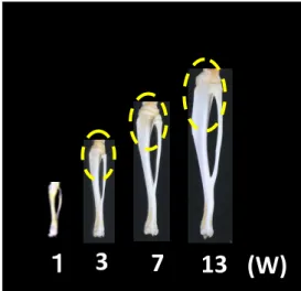
A remarkable lengthening of rat’s tibia was seen in the period from 1 to 7-week-old(early growing period), compared to the period from 7 to 13-week-old(late growing period). Shape of tibia was like a stick at 1-week-old. However, it showed a sigmoid curve when observing from the side, because a proximal portion of the tibia leaned to posterior direction and a distal portion leaned to anterior direction.(Fig. 1)
Fig 1. Morphological Changes in tibiae with growth
1
3
7
13 (W)
3
5
10 (W)
Fig 2. Arrangements of bone trabeculae at the
proximal cancellous bone in each week‐old
Fig. 3 Changes of thickness of proximal growth plate of
1
2
3
The bone trabeculae were short and arranged straight toward the inferior direction at the proximal cancellous bone of tibia in immature rat. They became thicker and site difference in their arrangements was observed, in mature rat. That is, they arranged in the same direction as the immature rats, at the anterior portion of the secondary cancellous bone. And, they arranged in the oblique direction at the middle and posterior portion, and their inferior ends fused to the posterior cortical bone.(Fig. 2)
Fig 1. Morphological Changes in tibiae with growth
1
3
7
13 (W)
3
5
10 (W)
Fig 2. Arrangements of bone trabeculae at the
proximal cancellous bone in each week‐old
Fig. 3 Changes of thickness of proximal growth plate of
tibiae with growth
1: 3 week‐old, 2: 7 week‐old, 3: 13week‐old
left side: anterior, right side: posterior
1
2
3
3.2 Histological Observations 3.2.1 Growth plateThe proximal growth plates of tibiae were stained color of pale blue, when observing their sagittal sections that were decalcified and stained by Masson-trichrome stain method. Their sizes increased in anterioroposterior direction, but thickness of them decreased, with growth.(Fig. 3)
Fig 1. Morphological Changes in tibiae with growth
1
3
7
13 (W)
3
5
10 (W)
Fig 2. Arrangements of bone trabeculae at the
proximal cancellous bone in each week‐old
Fig. 3 Changes of thickness of proximal growth plate of
tibiae with growth
1: 3 week‐old, 2: 7 week‐old, 3: 13week‐old
left side: anterior, right side: posterior
1
2
3
Study on structural changes of growth plate and primary cancellous bone in growing rats
Thickness of Resting, proliferating and hypertrophic cell layers of the growth plate showed significant decrease(p<0.05 or p<0.01)at the period from 3 to 13-week-old, except for hypertrophic cell layers in late growing period.(Fig. 4-A-C)
*2 *1 *3 *1: P<0.01 (3W:7W) *2: P<0.05 (7W:13W) *3: P<0.01 (3W:13W) (μm) F=24.82 P=.000 *2 *3 *1 (μm ) *1: P<0.01 (3W:7W) *2: P<0.05 (7W:13W) *3: P<0.01 (3W:13W) F=87.14 P=.000 *2 *1 (μm) *1: P<0.01 (3W:7W) *2: P<0.01 (3W:13W) F=47.26 P=.000 A C B
Fig. 4 Changes in thickness of each cell layers
with growth
each week‐old and each portions
A: resting layer, B: proliferating layer
C: hypertrophic layer
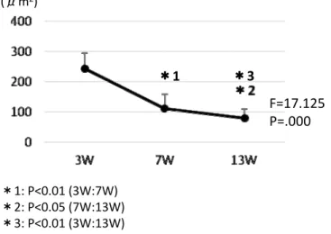
The sizes of hypertrophic chondrocytes significantly decreased(p<0.01 and p<0.05, respectively), and as a result, intracellular matrixes oppositely increased.(Fig. 5-7)The thickness of each portions(anterior, middle and posterior )of the growth plates decreased drastically in the early growing period, and decreased gently in the late growing period. (Fig. 8) The thickness of the cell proliferation layer decreased in the growth plate, and
the number and size of chondrocytes at that cell layer also decreased in the early growing period, compared to the late growing period. When comparing the thickness of resting and proliferating layers between each portions of the growth plate, those of the middle portion was thick but those of marginal portions(anterior and posterior portions)were thin. The same results were observed at the resting cell layer in 7-week-old, too.(Fig. 9-A-C)
Fig. 5 Changes in size of chondrocytes with growth
*1 (μm2) *3 *1: P<0.01 (3W:7W) *2: P<0.05 (7W:13W) *3: P<0.01 (3W:13W) F=17.125 P=.000Fig. 6 Changes in shape of the hypertrophic
chondrocytes with growth (light microscopic
images)
A‐C: anterior portion, D‐E: middle portion,
G‐I: posterior portion
A, D, G: 3week‐old, B, E, H: 7week‐old
C, F, I: 13week‐old
A
I
H
G
F
E
D
C
B
Study on structural changes of growth plate and primary cancellous bone in growing rats
Fig 8. Changes in the number of proliferating
chondrocytes with growth
*2 *1 (N) *1: P<0.01 (3W:7W) *2: P<0.01 (3W:13W) F=15.714 P=.000Fig. 7 Changes in thickness of each cell layers with growth
A, D, G: 3week‐old, B, E, H: 7week‐old
C, F, I: 13week‐old
P: proliferating layer, H: hypertrophic layer
Anterior Middle Posterior
H
P
A B C D E F G H IFig 8. Changes in the number of proliferating
chondrocytes with growth
*2 *1 (N) *1: P<0.01 (3W:7W) *2: P<0.01 (3W:13W) F=15.714 P=.000Fig. 7 Changes in thickness of each cell layers with growth
A, D, G: 3week‐old, B, E, H: 7week‐old
C, F, I: 13week‐old
P: proliferating layer, H: hypertrophic layer
Anterior Middle Posterior
H
P
(μm) N.S. N.S. N.S. A M P F=1.526 P=.291 N.S. * ** (μm) A M P F=7.562 P=.023 (μm) * N.S. N.S. A M P F=20.398 P=.002 A C B
Fig 9: Comparison of thickness of each cell layers at different portions
A: resting layer in 3week‐old
B: proliferating layer in 3‐week‐old.
C: resting layer in 7‐week‐old.
A: anterior portion, M: middle portion
P: posterior portion
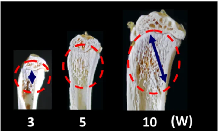
3.2.2 Cancellous boneMany thin calcified cartilage trabeculae existed just under the growth plate at 3-week-old, and they became thicker after that.(Fig. 10) The cancellous bone of proximal metaphysis stained color of blue at 3-week-old, when observing the sections stained by Masson-trichrome staining method. A range that stained color of red by dye of panceau fuchsin, extended with growth.(Fig. 11)
Study on structural changes of growth plate and primary cancellous bone in growing rats (μm) *2 *1 *1: P<0.01 (3W:7W) *2: P<0.01 (3W:13W) F=24.065 P=.000
Fig 11. Changes in thickness of calcified cartilage
trabeculae with growth
Fig 10. Changes in the calcified cartilage trabeculae
with growth
A: 3week‐old, B: 13week‐old
Red arrow: calcified cartilage matrix
GP: growth plate
A
B
GP
GP
The calcified cartilage trabeculae of the primary cancellous bone stained by color of blue, and the starting portion of bone formation was far from the growth plate at every portions of 3-week-old. The calcified cartilage trabeculae indicated the same stainability as those of 3-week-old at the anterior portion of the primary cancellous bone in 7-week-old. However, in 7-week-old, the bones stained color of red by dye of panceau fuchsin were added to surface of the calcified cartilage trabeculae at middle and posterior portions of the primary cancellous bone, and the starting portion of bone formation approached to growth plate. The bones stained by color of red were found just under the growth plate at every portions of the primary cancellous bone, and the starting portion of bone formation approached
considerably to a lower margin of the growth plate.(Fig. 12-14)
Fig. 12 Comparison of stainability of the cancellous bone in each week‐old
1: 3week‐old, 2: 7week‐old, 3: 13week‐old
left side: anterior, right side: posterior
1 2 3 A I H E D C B G FFig. 13 Magnified images of the primary cancellous bone in each portions
A‐C: 3week‐old, D‐E: 7week‐old, G‐I: 13week‐old
A, D, G: anterior portion, B, E, H: middle portion
C, F, I: posterior portion
Yellow arrow: calcified cartilage matrix
GP GP GP GP GP GP GP GP GPStudy on structural changes of growth plate and primary cancellous bone in growing rats (μm) *1 *2 *1:P<0.05 (3W : 7W) *2:P<0.01 (3W : 13W) F=13.22 P=.000
Fig 14. Comparison of distance from growth plate to the
starting portion of bone formation in each week‐old
** * ** (μm) A M P F=17.945 P=.003Fig 15. Comparison of distance from growth plate to the
starting portion of bone formation in each week‐old
A: anterior portion, M: middle portion
P: posterior portion
(μm) *1 *2 *1:P<0.05 (3W : 7W) *2:P<0.01 (3W : 13W) F=13.22 P=.000Fig 14. Comparison of distance from growth plate to the
starting portion of bone formation in each week‐old
** * ** (μm) A M P F=17.945 P=.003Fig 15. Comparison of distance from growth plate to the
starting portion of bone formation in each week‐old
A: anterior portion, M: middle portion
P: posterior portion
The calcified cartilages trabeculae just under the growth plate were stained by color of blue and the bone added to surface of those trabeculae were stained by color of red, when observing sections stained by Polychrome staining method. The bones formed at the secondary cancellous bone were stained color of orange or yellow. The bones having lamellar structure were also recognized at the same portions.(Fig. 15) It was recognized, by observation with TEM in this study, that the bone matrix stained by color of red was consisted by loose and irregular arrangement matrix fibers, and the bone matrix stained by color of orange or yellow was consisted of dense and regular arrangement matrix fibers. Furthermore, dense matrix fiber bundles that cut longitudinally and transversally were
found, in the case of the bone having lamellar structure.(Fig. 16, 17) Cytoplasm of osteoblasts that existed at the surface of bone stained by color of red were pale, and rough endoplasmic reticulum was poor and they arranged irregular in their cytoplasm. On the other hand, the osteoblasts were large and slightly dark, and had rich rough endoplasmic
Fig 16. Structures of primary cancellous bone just under growth plate and secondary cancellous bone in 3‐week‐old (decalcified specimens, polychrome stain) GP: Growth plate B: Magnified image of ①, C: Magnified image of ②, D: Magnified image of ③ A B C 2 D 3
L
L
T
4 1 G P ① ② ③ Fig 17. TEM images of immature and lamellar bone matrix 1: immature bone matrix near the calcified cartilage matrix, 2: magnified image of 1 3: mature bone matrix 4: bone matrix showed lamellar structure L: fiber bundles cut Longitudinally T: fiber bundles cut TransversallyFig 16. Structures of primary cancellous bone just under growth plate
and secondary cancellous bone in 3‐week‐old (decalcified
specimens, polychrome stain)
GP: Growth plate
B: Magnified image of ①, C: Magnified image of ②,
D: Magnified image of ③
A
B C 2 D 3L
L
T
4 1G
P
①
②
③
Fig 17. TEM images of immature and lamellar bone matrix
1: immature bone matrix near the calcified cartilage matrix,
2: magnified image of 1
3: mature bone matrix
4: bone matrix showed lamellar structure
L: fiber bundles cut Longitudinally
T: fiber bundles cut Transversally
Study on structural changes of growth plate and primary cancellous bone in growing rats
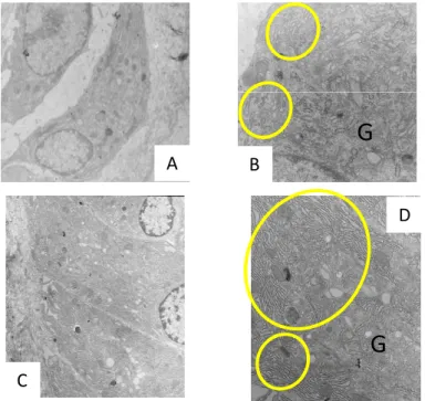
Fig 18. TEM images of the osteoblasts at each portions in 3‐week‐old.
A: synthesized the matrix fiber like Fig 17‐2
B: synthesized the matrix fiber like Fig 17‐3, 4
Yellow circle: Rough endoplasmic reticulum
G: Golgi area
G
G
D
C
B
A
4. DiscussionThe growth plate concerned to lengthening of a long bone, and is divided into some cell layers from the point of view of shape of cells. It was reported that proportion of the number of chondrocytes in each cell layers was constant for the period from 21 to 80-day-old at the proximal growth plate3). However, it was shown that cell division ability of the resting cell layer declined for the period from birth to 6-week-old, and the cells decreased remarkably after that4,5). To the contrary, it was reported that cell division ability declined at the proliferating cell layer, but the number of cells maintained after 6-week-old yet6,7).
In this study, the number and size of the cells decreased at the proliferating and hypertrophic cell layers in the early growing period, compared to the late growing period. Lengthening rate of tibia was higher in the early growing period than the late growing period. The bone trabeculae were formed by addition new bone around the calcified cartilage trabeculae. And, the bone lengthening was accomplished by incorporating those
bone trabeculae into a cortical bone at metaphysis. Decrease in the thickness of the growth plate means decline in cell division ability at there1), and relates to rate forming the calcified cartilage trabeculae existed at the inferior margin of the growth plate. Therefore, it is supposed that the decline in the cell division ability at the growth plate causes decrease in lengthening rate of tibia in the early growing period2).
It was found, in this study, that thickness of the calcified cartilage trabeculae increased with growth. Furthermore, it was also recognized that large size cells existed densely at the hypertrophic cell layer in 3-week-old, but they miniaturized after that. Then, it is thought that this gave increase in thickness of the calcified cartilage trabeculae.
Thickness of an articular cartilage decreases after birth, but the decreasing rate depends on the portion9). In this study, it was found that the superficial and middle cell layers of the articular cartilage were thick at the middle portion, but was thin at the anterior and posterior portions, that is, at peripheral portions, in three week-old. And, the same site difference in the thickness of cell layers was recognized at the resting cell layer in 7-week-old. It was supposed that those site differences in the thickness of the articular cartilage meant differences in maturating degree of the chondrocytes between middle and peripheral portions, and the maturation of the chondrocytes progresses from the middle portion toward the peripheral portions in the growth plate.
It was shown that the site differences were also found about the arrangements of the bone trabeculae at the secondary cancellous bone8). In this study, the bone trabeculae arranged in the superior-inferior direction at the anterior portion, but they arranged in the oblique direction at the middle and posterior portions in the secondary cancellous bone. It was thought that mechanical loadings from femur were dispersed and transmitted to posterior cortical bone of tibia by the bone trabeculae at middle and posterior portions, and were received finally by whole tibia that showed shape of sigmoid curve8).
Ohsako10)reported that loose matrix fibers arranged irregularly at the bone matrix that was stained by color of blue, on the other hand, dense matrix fibers arranged regularly at the bone matrix that stained by color of red, based on the results of stainability by Masson-trichrome staining method and observations by TEM. In this study, observing sections stained by Masson-trichrome method the cancellous bone of the proximal metaphysis was stained by color of blue, but the portions that stained by color of red extended. When applying the results of this study to the report of Ohsako10), it is supposed that the bone matrix of the metaphysis of 3-week-old consisted of loose and irregular arrangement fibers,
Study on structural changes of growth plate and primary cancellous bone in growing rats
In this study, it was found that the calcified cartilage trabeculae stained color of blue at the primary cancellous bone just under the growth plate, when staining by Polychrome staining method. Furthermore, there were the bone matrix that stained color of orange or yellow, and had lamellar structures that stained by colors of orange and yellow, alternately, at the secondary cancellous bone. It was speculated, based on Ohsako’s report10)described above, that the area that stained by color of red was consisted by loose fibers, and the area stained color of orange was consisted by dense fibers. As described above, it was found that the area that stained color of red was consisted by loose fibers, and the area stained color of orange was consisted by dense fibers. In fact, relationships between stainabilities by Polycrhome staining and observation by TEM were recognized and their relevancies were confirmed in this study, too. Then, it was thought that the matrix fibers of the bone trabeculae were going to be dense and to arrange regularly at the primary cancellous bone of tibial proximal metaphysis, and the structures that could resist to more mechanical loading were obtained, with growth.
The primary cancellous bone was constructed by only the calcified cartilage trabeculae in each portions of anterior, middle and posterior of the primary cancellous bone, and distance among the growth plate and the starting portion of bone formation was far, in 3-week-old. The starting portion of bone formation approached to the growth plate at middle and especially posterior portion in 7-week-old, and the bones were already added to the calcified cartilage trabeculae from just under the growth plate at every portions in 13-week-old. Thus, the starting portions of bone formation approached the growth plate gradually, and those changes were appeared from the posterior portion. Then, it was supposed that maturation of the bone was proceeded from the posterior portion toward the anterior portion sequentially, when considering early bone addition to the calcified cartilage as the maturation of bone.
In this study, the osteoblasts having poor organelles were found at the surface of the bone consisted of the matrix fibers that were loose and irregular arrangement just under the growth plate. To the contrary, the osteoblasts having developed rough endoplasmic reticulum and Golgi apparatus were recognized at the surface of the bone consisted of the matrix fibers that were dense and regular arrangement or of lamellar structure. Thus, differentiated osteoblasts appeared with growth, and it was thought that these changes of the cells affected both the differentiation of bone tissues and resistance against mechanical loading. Therefore, from these observations, it was understood that processes of tissue differentiation of the growth plate differ from that of the primary cancellous bone, the
degree of the differentiation of osteoblasts also differ depends on the portion and those cell differentiation affected the maturation of the bone matrix at the primary cancellous bone.
5. Conclusion
It was suggested that maturation of the growth plate proceeded toward different direction from the primary cancellous bone, and the differentiation degree of the osteoblasts affected the density and the arrangements of the bone matrix fibers in growing period.
Committee of Animal Experiment and Ethics
This study was approved by Committee of Animal Experiment and Ethics for the research, Graduate School of Welfare Society design, Toyo University.
Acknowledgments
This work thanks to the support of graduate and undergraduate laboratory member and the cooperation with you for guidance and encouragement.
References
1) Thurston M. N., et al.: Cell kinetics of growth cartilage in stumpy: a new chondrodystrophic mutant in the mouse. J. Anatomy 136:407-415, 1983.
2) Marino R., et al.: Catch-up growth after hypothyroidism is caused by delayed growth plate senescence. Endocrinol. 149:1820-1828, 2008.
3) Hunziker E., Schenk R., et al.: Physiological mechanisms adopted by chondrocytes in regulating longitudinal bone growth in rats. J. physiol. 414:55-71, 1989.
4) Lenneke S., et al.: Depletion of resting zone chondrocytes during growth plate senescence. J.Endocrinol. 189:27-36, 2006.
5) Julian C. L., et al.: Growth plate senescence and catch-up growth. Endocrinol. Develop. 21:23-29, 2011.
6) Nilsson O., et al.: Growth plate senescence is associated with loss of DNA methylation. J.Endocrinol. 186:241-249, 2005.
7) Yazaki Y., et al.: Immunohistochemical localization of bone morphogenetic proteins and the receptors in epiphyseal growth plate.Anticancer Res. 18:2339-2344, 1998.
8) Morita T., Obuchi N., Ohsako M.: Study of morphologic change and remodeling of tibia in growing rat. J. Human Life Design 6:73-84, 2011.
Study on structural changes of growth plate and primary cancellous bone in growing rats
cartilage cells in growing rat. Bull. Grad. Sch. Toyo Univ. 48:117-131, 2012.
10) Ohsako M.: Histological studies on growth and function of condylar process in rat mandible. J. Stomatol Socie. 60:475-524, 1993.
要 約 長骨骨幹端における二次海綿骨や関節軟骨では、前方、中央および後方部における機能獲 得に関連して、発育に伴う構造変化に部位差が見られることが示されている。しかし、一次 海綿骨や骨端板では、部位によって異なる成熟過程をたどるか否かは明らかにされていない。 本研究はラット脛骨を用いて、近位骨幹端における骨端板とその直下の一次海綿骨の発育変 化を観察することにより、それらの成熟過程の特徴について検討することを目的とした。 材料として、3、7および13週齢の雄性ウィスター系ラットを用いた。それらを安楽死させ た後、脛骨を摘出し、矢状割断して固定液に浸漬した。それらの標本を用いて種々な標本を 作製し、肉眼的および組織学的に観察するとともに、骨形態計測学的に計測した。 脛骨骨端板の各細胞層の厚さは、3~13週齢の間に有意(p<0.05またはp<0.01)に減少し た。肥大軟骨細胞の大きさは3~7週齢および7~13週齢のいずれの期間においても有意な(そ れぞれ、p<0.01およびp<0.05)低下を示し、反対に細胞間基質は増加した。3週齢の静止細 胞層および増殖層の厚さを部位別に比較すると、中央部は厚いが前方および後方の辺縁部が 薄く、同様な部位差が7週齢の静止細胞層においても観察された。骨端板直下の石灰化軟骨 梁は発育に伴って太くなった。3週齢の一次海綿骨では、前方、中央および後方のいずれの 部位においても石灰化軟骨梁のみで構成されたが、発育に伴って後方部から徐々に骨形成開 始部位が骨端板に近づいた。また、添加される骨の基質線維は密となり、規則的な配列を示 した。基質線維の疎な部位には、細胞小器官の乏しい骨芽細胞が認められ、基質線維の密な
Study on structural changes of growth plate and
primary cancellous bone in growing rats
福祉社会デザイン研究科ヒューマンデザイン専攻博士前期課程1年
髙橋 将人
福祉社会デザイン研究科ヒューマンデザイン専攻博士前期課程1年
中井 真悟
ライフデザイン学部健康スポーツ学科教授
大迫 正文
Study on structural changes of growth plate and primary cancellous bone in growing rats 以上のことから、骨端板と一次海綿骨では成熟の過程に違いがあることが示唆された。ま た、海綿骨の中でも骨端板直下とそれより下方の領域とでは、骨芽細胞の分化度が異なり、 このことがそれらの領域の一次海綿骨における基質の成熟度の違いをもたらしているであろ うことが理解された。 キーワード:ラット脛骨、骨端板、一次海綿骨