エタネルセプト投与中に発症した食道・腸結核の 1 例
1,2穴澤 梨江
1,3鈴木 優毅
1,4三輪 秀樹
1三木 良浩
1冨田 和宏
1中村 秀範
は じ め に 食道結核は肺外結核の中で稀な疾患であり,腸結核と の合併例は世界的にもごく少数の報告例しかない。今回 われわれはエタネルセプト投与中の関節リウマチ(RA) 患者において胸水貯留と食道壁の肥厚を認め精査を行っ た結果,食道結核・腸結核の合併と確定診断した 1 例を 経験したので文献的考察を加えて報告する。 症 例 患 者:88 歳,女性。 主 訴:なし(胸部異常陰影精査目的)。 既往歴:2011 年に関節リウマチの診断。 喫煙歴:なし。 家族歴:結核罹患者なし。 現病歴:2000 年ごろから両膝関節痛があり近医整形 外科にて変形性関節症と診断された。2011 年 3 月に多 関節炎を認め炎症反応および MMP-3 が高値であったた め,他院にて関節リウマチと診断された。サラゾスルフ ァピリジンやプレドニン(PSL)の内服を開始したが関 節症状の増悪を認めたため,PSL 6 mg ⁄日にメトトレキ セート 6 mg ⁄週が追加投与された。症状の改善が得られ なかったため 2011 年 7 月よりエタネルセプト(ETN)25 mg ⁄週が導入された。ETN 導入前のツベルクリン反応は 弱陽性(12 mm×17 mm,硬結なし)であったが,Quanti FERON®TB Gold(QFT-3G)検査は未施行でありイソニ アジド(INH)の治療は行われていなかった。2012 年 1 月に近医で胸部 X 線と単純 CT を施行したところ,右胸 水貯留と右上葉から中葉にかけて炎症性変化を認めたた め,精査目的に 2012 年 2 月 6 日当科へ紹介となった。 初診時現症:体温 36.7 度,血圧 143/66 mmHg,脈拍 87 回 ⁄分(整),呼吸数 12 回 ⁄分,SpO2 98%(室内気),右背 側に fi ne crackles を聴取した。 初診時血液検査所見:WBC 11,560/μl,CRP 7.0 mg/dl と炎症反応高値を認めた。Hb 11.1 g/dl,TP 7.1 g/dl,Alb 3.4 g/dl,BUN 17 mg/dl,Cre 0.55 mg/dl,Na 135 mEq/l, HbA1c(NGSP)6.5% であり,重度の低栄養や糖尿病は 認 め な か っ た。QFT 検 査 は 陽 性(TB 抗 原 4.71 IU/ml, 1聖隷浜松病院呼吸器内科,2現:東京労災病院呼吸器内科,3現: 君津中央病院呼吸器内科,4現:千葉大学医学部附属病院呼吸 器内科 連絡先 : 穴澤梨江,東京労災病院呼吸器内科,〒 143 _ 0013 東 京都大田区大森南 4 _ 13 _ 21(E-mail : [email protected]) (Received 28 Feb. 2014 / Accepted 5 Jun. 2014)要旨:症例は 88 歳女性。2011 年に他院にて関節リウマチと診断され,同年 7 月よりエタネルセプト を開始した。2012 年 2 月に右胸水貯留にて当科紹介となった。胸部単純 CT では右胸水と一部石灰化 を伴う縦隔リンパ節の腫脹のほかに中部食道壁の肥厚を認めた。上部消化管内視鏡検査では,中部食 道に潰瘍性病変があり同部位の生検病理で乾酪壊死を伴わない肉芽腫とその内部に Langhans 巨細胞 を認めた。病理組織の Ziehl-Neelsen 染色で陽性を示す抗酸菌菌体も数カ所で観察した。胃液の Myco-bacterium tuberculosis PCR 法が陽性であり併せて食道結核と診断した。腹部単純 CT にて腸管膜リンパ 節の腫脹があり,下部消化管病変の合併の可能性を考慮し下部消化管内視鏡検査を行った。回盲部の 多発性びらんから生検した結果は食道と同様であり,食道結核と腸結核の合併と診断した。抗結核化 学療法を 12 カ月間施行し胸水,消化管病変はいずれも改善した。確定診断ができた食道結核・腸結 核の合併例はこれまで世界的にも報告が少なく,きわめて貴重であると考え報告する。 キーワーズ:食道結核,腸結核,エタネルセプト
Fig. 1 (a) Plain chest X-ray revealed right-sided pleural effusion. No opacities nor cavitary lesion were shown in the both
lung fi eld. (b) (c) Computed tomography showed pleural effusion, thickening and nodules of the fi ssures in the right lung, lymphadenopathy of mediastinal lymph node with partial calcifi cation, and the thickened wall on the middle esophagus.
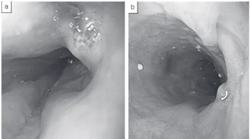
Fig. 2 (a) Before anti-tuberculosis therapy, gastroendoscopy revealed the ulcerative lesion on the
middle esophagus. (b) After treatment, ulcerative lesion and narrowing were improved. Nil 0.27 IU/ml,Mitogen 1.00 IU/ml)であった。
画像検査(Fig. 1):胸部 X 線,CT で右胸水を認めた。 右上葉から中葉にかけては葉間胸膜の肥厚と多発する胸 膜結節を認めた。縦隔条件では中部食道壁が軽度肥厚し ており,周囲のリンパ節は腫脹し一部石灰化を認めた。 経 過:肺野病変に関しては抗酸菌感染症の可能性が 考えられたため外来にて 3 日間の喀痰抗酸菌検査を行っ たが,塗抹検査,Mycobacterium tuberculosis PCR 法は共 に陰性だった。食道壁の肥厚があり食道癌も考えられた ため 2 月 13 日に上部消化管内視鏡検査(GIF)を施行し た。門歯より 27 cm の中部食道 12 時方向に辺縁不整な隆 起性病変を認め,中心部には潰瘍を伴っていた(Fig. 2a)。他に食道炎(ロサンゼルス分類 grade M)の所見も 認めた。潰瘍周堤部から生検を施行したところ,食道粘 膜全体にリンパ球の浸潤があり乾酪壊死を伴わない類上 皮肉芽腫を認めた。肉芽腫組織内には Langhans 巨細胞が 散見された。Ziehl-Neelsen 染色陽性の抗酸菌菌体を数カ 所で認めた(Fig. 3)。胃液の Mycobacterium tuberculosis PCR 法が陽性であり併せて食道結核と診断した。他の消 化管病変の合併を考慮し腹部 CT を施行したところ腹膜 の肥厚と腸間膜リンパ節腫脹を認めた(Fig. 4a,4b)。下 部消化管内視鏡検査(CF)では回腸末端に小びらんが散 見された(Fig. 4c)。同部位からの生検組織は食道の生 検組織と同様で回腸粘膜全体のリンパ球浸潤と乾酪壊死 を伴わない類上皮肉芽腫があり,Ziehl-Neelsen 染色陽性 の抗酸菌菌体を認めた。2 月 24 日より INH 300 mg ⁄日,リ ファンピシン(RFP)450 mg ⁄日,エタンブトール(EB) 750 mg ⁄日の 3 剤にて結核治療を開始した。治療開始 2
Fig. 3 Histological examination of the esophageal biopsy showed (a) non caseating granuloma with
Langhans giant cells (10×150), and (b) Ziehl-Neelsen stain was positive for acid-fast bacilli (10×600).
Fig. 4 Computed tomography showed (a) thickening of the peritoneum and (b) swelling of
mesenteric lymph nodes. (c) Colonoscopy showed multiple erosions on the ileum. カ月で胸部 X 線上右胸水は消失した。同時期に施行した GIF では消化管の潰瘍性病変は瘢痕化していた(Fig. 2 b)。胃液の培養が 8 週目で陽性となり INH,RFP,EB への感受性が良好であったため,EB は 6 カ月で終了と し INH,RFP にて合計 12 カ月治療を行った。治療中に副 作用や合併症は認めず,治療終了後の GIF でも潰瘍性病 変は瘢痕化しており経過は良好であった。高齢のため CF の再検は行わなかった。 考 察 関節リウマチ患者は免疫機能異常に伴う疾患そのもの の影響や,ステロイド・メトトレキセートなどの免疫抑 制剤による治療を必要とするため,結核発症のリスクが 高いと考えられてきた。さらに近年関節リウマチの治療 において劇的な進歩をもたらした生物学的製剤は,tumor necrosis factor(TNF)を阻害することで結核発症リスク を増加させることは世界各国から報告されている。結核 罹患率が日本と同程度のスペインからの報告ではインフ リキシマブ(IFX)または ETN を使用した 1,540 例中 17 例に結核発症を認めており,そのうち 65% が肺外結核で あった1)。日本における ETN の市販後調査では 13,894 例 中 12 例に結核発症を認めており,そのうち 30% が肺外 結核であった2)。国内外の報告からも分かるように TNF 阻害薬を使用した際には肺外結核が多くみられることが 特徴で,大半は潜在性結核感染(LTBI)からの内因性 再燃と考えられている。2008 年に米国リウマチ学会から 生物学的製剤使用前の結核スクリーニングに関しての勧 告がなされ,2012 年に改訂されたものと同様の主旨が日 本でも日本呼吸器学会から発表された。その中で LTBI のスクリーニング検査としてツベルクリン反応検査だけ
Table Cases of the esophageal tuberculosis in Japan, 2000 _ 2012. Case Age Sex Medical past history Other lesion/Lower
gastrointestinal tract Bacteriology Histopathology
Clinical course
1 71 M CL Unknown Caseating granuloma
2 30 M ML Esophageal tissue
culture (+)
Granuloma
3 30’s M ML/Normal (CF) Negative Epithelioid granuloma
4 27 M During treatment of Tb ML, miliary Tb Unknown Epithelioid granuloma
5 76 F ML Unknown Epithelioid granuloma
6 20 F ML Gastric juice PCR (+) Epithelioid granuloma
7 39 M ML Negative Epithelioid granuloma
8 64 M ML Negative Epithelioid granuloma
9 42 F Primary/Normal
(barium enema)
Esophageal tissue PCR (+)
Caseating necrosis
10 55 M HCV ML, pulmonary Tb Sputum smear (+) Granuloma
11 57 M Esophageal cancer ML Unknown Caseating granuloma
12 23 M During treatment of Tb ML Sputum PCR &
culture (+)
Granuloma
13 33 M Tuberculous pleurisy Unknown Epithelioid granuloma
14 52 M Diabetes, hemodialysis Pulmonary Tb Gastric juice smear & PCR (+)
Granuloma 15 56 M Diabetes, hemodialysis CL & ML, pulmonary Tb Sputum culture (+) Normal 16 35 F During treatment of Tb ML, pulmonary Tb Sputum smear &
culture (+)
Not done
17 72 M HCC ML, tuberculous pleurisy Gastric juice PCR (+) Langhans giant cellls, Z-N stain (+)
18 59 M Schizophrenia ML Gastric juice PCR (+) Epithelioid granuloma
19 80’s F ML/Normal (unknown) Negative Caseating granuloma
20 73 M SLE Pulmonary Tb Sputum smear (+) Epithelioid granuloma,
Z-N stain (+)
21 40’s M ML Gastric juice culture (+) Epithelioid granuloma
Well after anti-tuberculous treatment. (Case 8 & 11 unknown)
M=male. F = female. Tb=tuberculosis. HCV=hepatitis C virus. HCC=hepatocellular carcinoma. SLE=systemic lupus erythematosus. CL=cervical lymphadenitis.
ML=mediastinal lymphadenitis. CF=colonoscopy. Z-N stain=Ziehl-Neelsen stain.
でなく IGRA 検査を行うことの重要性や,疑わしければ 胸部 X 線検査のみならず胸部 CT 検査を積極的に併用す ること,LTBI の診断となった際の INH 予防投薬などに ついて述べられている3) 4)。本症例では ETN 導入前に行 ったスクリーニング検査はツベルクリン反応検査と胸部 X 線のみであり,ツベルクリン反応は弱陽性であったが それ以上の精査は行わなかったため LTBI を発見できな かった可能性がある。ETN を導入したことで LTBI が再 燃し,肺外結核である食道結核,腸結核,結核性胸膜炎 が発症したと考えられる。 食道結核は肺外結核の中では稀な疾患であり,その頻 度は結核死亡患者の剖検の解析ではわずか 0.15% と報告 されている5)。Tania らの報告では,肺外結核の中で消化 管結核は 6 番目に多く全体の 1.2% 程度だが,その中で食 道結核は約 0.2% であった6)。また Momin らは 1995 年か ら 2010 年の 16 年間で結核患者 3,123 人のうち食道結核は 2 例(0.06%)のみであったと報告している7)。その理由 としては食道の内腔は平滑で彎入も少ないこと,嚥下運 動により菌の付着に至るまでの時間的余裕が少ないこと, 組織学的にも扁平上皮からなること,他の消化管粘膜と 比較してリンパ装置が少ないことなどが挙げられる8)。食 道結核の大半が周辺臓器からの波及による二次性で,肺 ・咽頭・縦隔が主な原因として挙げられる。中でも食道 や気管支に近接している気管分岐部リンパ節が病変とな ることが多く,中部食道に好発する9) 10)。本症例でも中 部食道付近の縦隔内リンパ節に一部石灰化を伴う腫脹を 認めており,縦隔内リンパ節からの穿破による続発性の 感染が推測される。 本邦においては 2000 年から 2012 年の 13 年間で 21 例 の食道結核(食道気管瘻の症例は除く)の報告がある (Table)。6 例で血液維持透析患者や癌の合併を認めてお り免疫抑制状態にある患者は意外にも少数であった。培 養陽性は 5 例(喀痰 3 例,胃液 1 例,食道組織 1 例),組 織学的に乾酪性肉芽腫や抗酸菌菌体を認めたものは 6 例 であった。消化管結核患者のうち肉芽腫は約 60% で確 認できるが結核に典型的な乾酪性肉芽腫は 19∼25% で しか確認できず,培養陽性は 7 % しか認められなかった との報告もある11) 12)。自験例では培養陽性であり,乾酪 性肉芽腫は認めなかったものの Ziehl-Neelsen 陽性の抗酸 菌菌体を証明することができた。 また食道結核の併発病変としては縦隔リンパ節炎が 16 例と最も多く,他に肺結核 5 例,結核性胸膜炎 2 例,頸部 リンパ節炎 2 例,粟粒結核 1 例が報告されている(Table)。 自験例では初診時に認めた右胸水の胸腔穿刺を行えず結
核性胸膜炎の確定診断には至らなかったが,食道結核に 対する治療により胸水は消失したため臨床的には結核性 胸膜炎も合併していたと考えられる。結核性胸膜炎の合 併例はこれまでにも報告されているが,食道結核・腸結 核の合併例は国内外の文献を検索した中でも Nagai らの 報告例のみであった13)。食道結核と腸結核の合併例の報 告が少ない原因としては,食道結核は続発性の発症が多 く胸部病変に関してはほぼ全例で精査が行われる14)が, 他の消化管病変を調べることは稀なためと推察する。本 邦の 21 例のうち記載のなかった 3 例以外は胸部 X 線ま たは CT を施行しているが,下部消化管に関して評価し ているものは 3 例のみであった。この 3 例の報告例はい ずれも無症状で腸結核の合併は認めなかったが,自験例 では無症状でありながら腸結核を合併していた。腸結核 の発症機序としては続発性の場合,結核菌を含んだ喀痰 を嚥下することにより発症すると考えられており15),活 動性肺結核患者における腸結核の合併は 25% との報告 もある16)。同様な機序で考えると本症例は縦隔リンパ節 炎から食道に穿破して食道結核となり,胃酸の影響は受 けるものの食道病変の菌体が消化管を通過して回盲部に たどりつき,続発性に腸結核を発症したと考えられる。 現在食道結核と腸結核の合併例はごく少数の報告しかな いが,下部消化管病変の検索を行うことで両者の合併例 がより多く見つかるかもしれない。また食道結核の確定 診断がつかない例でも下部消化管病変の検索を行って腸 結核を疑うような所見があればそこから診断できる可能 性があり,肺以外の合併臓器の評価も検討すべきである と考えた。 本論文の要旨は第120回日本結核病学会東海地方学会, 第 102 回日本呼吸器学会東海地方学会(2012 年 11 月,岐 阜)で報告した。 謝 辞 本症例の検討に際し,病理学的所見のご指導をいただ きました聖隷浜松病院病理診断科 大月寬郎先生,新井 義文先生,清水進一先生に深謝いたします。
著者の COI(confl icts of interest)開示:本論文発表内 容に関して特になし。
文 献
1 ) Gomez-Reino JJ, Carmona L, Valverde VR, et al.: Treatment
of rheumatoid arthritis with tumor necrosis factor inhibitors may predispose to signifi cant increase in tuberculosis risk. A multicenter active-surveillance report. Arthritis Rheum. 2003 ; 30 : 2122 2127.
2 ) 針谷正祥:わが国におけるエタネルセプトの安全性と 有効性. リウマチ科. 2009 ; 41 (1) : 8 19.
3 ) Singh JA, Furst DE, Bharat A, et al.: 2012 update of the 2008 American college of rheumatology recommendations for the use of disease-modifying antirheumatic drugs and biologic agents in the treatment of rheumatoid arthritis. Arthritis Care Res. 2012 ; 64 : 625 639.
4 ) 日本呼吸器学会 生物学的製剤と呼吸器疾患・診療の 手引き作成委員会:「生物学的製剤と呼吸器疾患 診療 の手引き」, 2014.
5 ) Lockard LB: Oesophageal tuberculosis: a critical review. Laryngoscope. 1913 ; 23 : 561 584.
6 ) Tania MW, Thomas K, Wolfram B, et al.: An unusual cause of dysphagia: esophageal tuberculosis. J Gastrointestin Liver Dis. 2010 ; 19 : 321 324.
7 ) Momin RN, Chong VH: Oesophageal tuberculosis: rare but not to be forgotten. Singapore Med J. 2012 ; 53 : 192 194. 8 ) 富永和作, 上田美和, 渡辺俊雄, 他:食道結核. 日臨.
2009 ; 11(別冊): 89 91.
9 ) Jain SK, Jain S, Jain M, et al.: Esophageal tuberculosis: is it so rare? report of 12 cases and review of the literature. Am J Gastroenterol. 2002 ; 97 : 287 291.
10) 石川成範, 矢野修一, 若林規良, 他:結核性縦隔リンパ 節炎により食道穿孔をきたした粟粒結核の 1 例. 結核. 2009 ; 84 : 159 164.
11) Uygur-Bayramicli O, Dabak G, Dabak R: A clinical dilem-ma: abdominal tuberculosis. World J Gastroenterol. 2003 ; 9 : 1098 1101.
12) Khan R, Abid S, Jafri W, et al.: Diagnostic dilemma of abdominal tuberculosis in non-HIV patients: An ongoing challenge for physicians. World J Gastroenterol. 2006 ; 12 : 6371 6375.
13) Nagai K, Ueno Y, Hayashi R, et al.: Interstinal tuberculosis with hoarseness as a chief complaint due to mediastinal lymphadenitis. Case Rep Gastroenterol. 2011 ; 5 : 540 545. 14) Mokoena T, Shama DM, Ngakane H, et al.: Oesophageal
tuberculosis: a review of eleven cases. Postgrad Med J. 1992 ; 68 : 110 115.
15) Kapoor VK: Abdominal tuberculosis. Postgrad Med J. 1998 ; 74 : 459 467.
16) 垂水研一, 古賀秀樹, 小橋吉博, 他:活動性肺結核患者 における下部消化管病変の検討. 川崎医会誌. 2006 ; 32 : 23 28.
Abstract An 88-year-old woman with rheumatoid arthritis
who had started etanercept treatment in July 2011 was referred to our hospital in February 2012 for right-sided pleural effusion. Chest computed tomography showed right pleural effusion, partial swelling of a calcifi ed mediastinal lymph node, and mid-esophageal thickening of the mucosal wall. Gastroendoscopy showed mid-esophageal ulceration. Histo-logical examination of biopsy specimens from this ulceration revealed noncaseating granulomas with Langhans giant cells. Ziehl-Neelsen staining of this section was positive for acid-fast bacilli. Polymerase chain reaction analysis of gastric juice was positive for Mycobacterium tuberculosis; we therefore diagnosed the patient with esophageal tuberculosis. However, since abdominal computed tomography showed swelling of mesenteric lymph nodes, we also suspected intestinal tuber-culosis. Colonoscopy showed multiple ileal erosions; histo-logical analyses of biopsied specimens revealed granulomas with Langhans giant cells, similar to the esophageal fi ndings. We fi nally diagnosed the patient with both esophageal and intestinal tuberculosis. After anti-tuberculosis treatment, the
right pleural effusion disappeared and the abdominal lesions improved. Although mycobacterial involvement of both the esophagus and intestine is rare in immunocompromised and immunocompetent hosts, differential diagnosis of these dis-eases is likely to become more important.
Key words: Esophageal tuberculosis, Intestinal tuberculosis,
Etanercept
1Department of Pulmonary Medicine, Seirei Hamamatsu
General Hospital, 2Department of Pulmonary Medicine, Tokyo
Rosai Hospital, 3Department of Pulmonary Medicine, Kimitsu
Central Hospital, 4Department of Pulmonary Medicine, Chiba
University Hospital
Correspondence to: Rie Anazawa, Department of Pulmonary Medicine, Tokyo Rosai Hospital, 4_13_21, Omori-Minami, Ota-ku, Tokyo 143_0013 Japan.
(E-mail: [email protected]) −−−−−−−−Case Report−−−−−−−−
A CASE OF ESOPHAGEAL AND INTESTINAL TUBERCULOSIS THAT OCCURRED
DURING TREATMENT OF RHEUMATOID ARTHRITIS WITH ETANERCEPT
1, 2Rie ANAZAWA, 1, 3Masaki SUZUKI, 1, 4Hideki MIWA, 1Yoshihiro MIKI, 1Kazuhiro TOMITA, and 1Hidenori NAKAMURA