Introduction
Conventional functional magnetic resonance imaging(fMRI)measures the magnetic susceptibility at a single time point. The response is generated using single gradient-echo(GE)echo-planar imaging
(EPI), and is proportional to the susceptibility at an echo time(TE). In contrast, multi-echo EPI uses multiple acquisition points to obtain signals from different echoes[1-6], thus providing additional information compared to single-echo EPI.
In a previous study[5]we used three-echo EPI to estimate the linearity of transverse relaxivity
(R 2 *)for functional activation and were able to image microscopic neuronal changes. However, the neuronal information might be compromised by physical or physiological noises due to the numerical computation typically applied on the signal in three-echo images. Therefore, it is essential to evaluate the effect of noises on fMRI imaging.
Cerebrospinal fluid(CSF)is the body fluid found in the brain and spinal cord. In the brain, most of the CSF is present in the ventricles but it also fills the brain sulci. The CSF circulation within the ventricular system is driven by the heartbeat. Therefore, fluctuations of the fMRI signal due to CSF changes might vary between different echo times. In addition, head motion might also affect the CSF- related fluctuation by generating partial volumes of mixed brain tissue and CSF in image voxels. Some of our data acquired by single-echo EPI have shown signal fluctuations in the CSF compartment. In this study we examine the effect of the CSF as a source of physiological noise on fMRI signals.
原 著 論 文
Evaluation of the effects of cerebrospinal fluid on functional MRI signals
Yul-Wan Sung and Seiji Ogawa
Kansei Fukushi Research Institute, Tohoku Fukushi University
Abstract
Multi-echo echo-planar imaging(Multi-echo EPI)is a magnetic resonance imaging(MRI)sequence allowing MRI images recording at different echo times after excitation. Its application to functional MRI(fMRI)studies has enable the acquisition of new neuronal information compared with the classical single-echo EPI. For instance, in a previous study we could monitor microscopic neuronal changes originating from population differences. However, the neuronal information acquired by multi-echo EPI is subjected to artifacts caused by partial volume effect in the boundary regions between the neuronal tissues and other compartments such as the cerebrospinal fluid(CSF). This report shows that CSF affected many fMRI image voxels in the brain.
Keywords : multi-echo echo-planar imaging, functional MRI; cerebrospinal fluid; fluctuation
Materials and Methods
This study was approved by the Institutional Review Board of the Tohoku Fukushi University
(RS190607).
Measurements and Data analysis
All MRI experiments were performed using a Skyra-fit system(Siemens, Germany)with a standard, 16-channel head matrix coil operating at 3 Tesla. Multi echo EPI sequence from Center for Magnetic Resonance Research of Minnesota University was used to acquire images at three echo times
(TE1, TE2, TE 3 )of 11.60, 28.23 and 44.86 msec. For functional imaging, the multi echo GE-EPI sequence was used with a 1000 milli seconds repetition time, 220 mm field of view, 64 × 64 mm matrix size, and 5 mm slice thickness with zero acceleration. Fourteen slices covering visual to parietal region were acquired from a healthy volunteer. The 390 volume images were acquired during subject’s eyes closed. After the typical preprocessing including motion correction, and temporal filtering by FSL
(https://fsl.fmrib.ox.ac.uk/fsl/fslwiki), MATLAB(Mathworks Co, Natick, USA)software was used for further data processing.
Results and Discussion
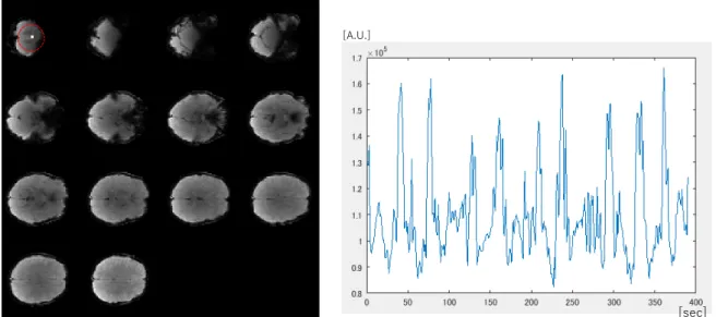
An image voxel containing CSF was found using anatomical information regarding the ventricle location and image contrast(Fig. 1 ). The amplitude of the signal fluctuation was larger than the typical signal variation induced by neuronal activation. Moreover, the signal changes were non-periodic with 20 to 50 seconds intervals. The baseline signal amplitude decreased with the increase of echo time as expected. Typically, if the change of the fMRI signal is due to a different magnetic susceptibility it is proportional to the echo time. However, here we observed the same variations of the signals for all echo times(Fig. 2 ), suggesting that the signal change is induced by the CSF.
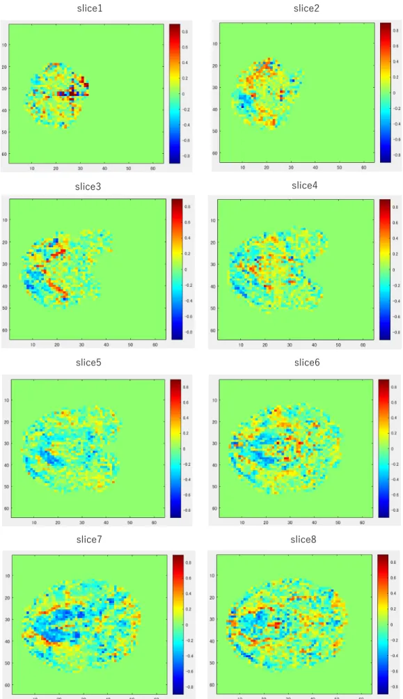
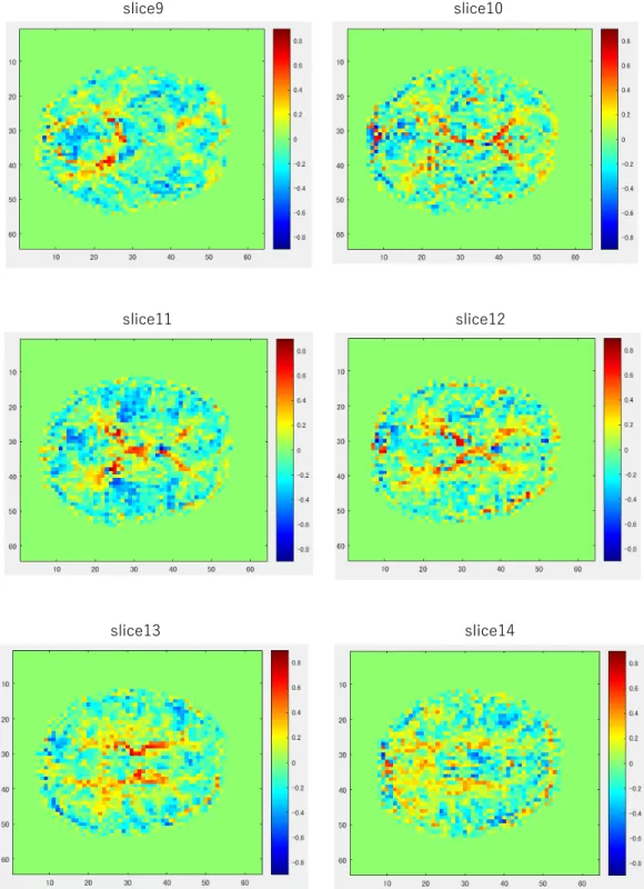
To examine additional image voxels affected by the CSF signal, we performed a correlation analysis of the CSF voxel with all the other image voxels. The generated maps showed high correlation across the brain, in areas occupied by CSF but also in regions containing brain tissue(Fig. 3 ). This indicates that brain voxels contained CSF in addition to tissue suggesting that CSF fluctuation might affect functional signal changes in the brain tissue voxels.
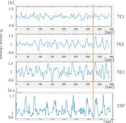
To examine the effect of CSF-induced signal on the neuronal functional signal, we compared the fMRI signal of a voxel from the tissue area with a high correlation value(larger than 0.5)at three- echo times(Fig. 4 ). The signal changes partly aligned with the CSF signal on the temporal axis, indicating a probable contamination of the neuronal function signal with CSF signal. Therefore, fMRI studies by single-echo fMRI might lead to an incorrect evaluation of the neuronal response amplitude or connectivity strength if the contaminant CSF signal is measured as a neuronal-induced signal change. Similarly, the fluctuation might affect the outcome of multi-echo fMRI studies particularly when arithmetic calculations such as the percent signal changes are applied to derive neuronal information
[5].
To eliminate the fluctuation unrelated to the brain neuronal activity a previous study used three-
echo fMRI to separate non-functional signals represented by the proton density(S 0 )from functional
signals represented by transverse relaxivity(R 2 *)[4]. However, the separation might not be perfect
for some processing factors such as the signal amplitude or the statistical power. As described in the
method section, the CSF signal was still found in our data after a series of the preprocessing steps.
Therefore, it is needed to extract signals from local compartments of CSF, identified using anatomical information and empirical knowledge, and to regress out the noise signal from the fMRI signal. To devise a more systematic method to extract those signals will be our future study.
In conclusion, not only the global CSF signal identified in most of previous studies but also non- functional changes in local compartments of CSF may be a physiological noise source and contaminate the functional signal of the brain neuronal tissue. It is therefore necessary to remove the CSF signal at the preprocessing stage to generate more accurate results in fMRI studies.
Figure 2. % signal changes of the time courses of TE1, TE 2 and TE 3 at the CSF voxel.
% sig na l c ha ng e
TE1
TE2
TE3 [%]
[sec]
[sec]
[sec]
Figure 2
Figure 1. MRI images and a signal time course from a CSF voxel. MRI images acquires at echo time TE 2 (left)and a time course from a CSF voxel in the red-dotted circle(right).
Figure1
[A.U.]
[sec]
Figure 3 a. Correlation maps, slice 1 to 8. Color bars stand for correlation value, 08 to -0.8.
slice1 slice2
Figure3 (a) slice1-8, (b) slice9-14
slice3 slice4
slice5 slice6
slice7 slice8
Figure 3 (b)Correlation maps, slice 9 to 14. Color bars stand for correlation value 0.8 to -0.8
slice9 slice10
slice11 slice12
slice13 slice14
Acknowledgement
This study was carried out as a part of the cooperative research project at the Kansei Research Institute of Tohoku Fukushi University, receiving a subsidy of the research facility operation support MEXT, and JSPS KAKENHI Grant Number 19H01111 & 19H00532.
References
1 ) Beissner F, Baudrexel S, Volz S, Deichmann R. Dual-echo EPI for non-equilibrium fMRI – implications of different echo combinations and masking procedures. Neuroimage 2010; 52: 524-31.
2 ) Glover GH, Lemieux SK, Drangova M, Pauly JM. Decomposition of inflow and blood oxygen level- dependent(BOLD)effects with dual-echo spiral gradient-recalled echo(GRE)fMRI. Magn Reson Med 1996; 35: 299-308.
3 ) Speck O, Hennig J. Functional imaging by Io- and T 2 * -parameter mapping using multi-image EPI.
Magn Reson Med 1998; 40: 243-248.
4 ) Kundu P, Inati SJ, Evans JW, Luh W-M, Bandettini PA. Differentiating BOLD signals in fMRI time series using multi-echo EPI. Neuroimage 2012; 60: 1759-70.
5 ) Kang D., Choi U-K., Sung Y-W. Microscopic functional specificity can be predicted from fMRI signals in ventral visual areas. Magnetic Resonance Imaging 32: pp. 1031-1036, 2014
6 ) Sung Y, Kawachi Y, Ogawa S. Evaluating the effects of head motion when processing multi-echo MRI data. Report of Kansei Fukushi Research Institute 21: pp. 65-69.
Figure 4. Time courses of % signal changes from a voxel that has the correlation value of 0.56 with the CSF signal.
Onsets of several signal changes from the baseline of time courses of TE1, TE 2 and TE 3 are aligned with the burst changes of the CSF time course.
1.5 1 -0.6
1.5 1 -0.6
1.5 1 -0.6
% sig na l c ha ng e
TE1
TE2
TE3
CSF [%]
[
A.U.]
1.70.81