Introduction
Chlamydia trachomatis is an obligate intracellular gramnegative bacterium with a unique biphasic growth cycle. After entering host cells, metaboli- cally inert elementary bodies(EBs)rapidly trans- form into metabolically active reticulate bodies
(RBs)that replicate by binary fission within a membranebound vesicle, and this process is known as inclusion. After logarithmic bacterial cell division, the RB reorganizes into infectious EBs, which adapt themselves to survive in the ex- tracellular environment of the host. C. trachomatis
causing disease in humans is classified as a trachoma or sexually transmitted disease(STD).
Although infection with C. trachomatis can be suc- cessfully treated with antibiotics, asymptomatic and persistent infections are common and can re- sult in scarring sequelae such as blinding trachoma and pelvic inflammatory disease. Moreover, chronic or recurrent chlamydial infections have been associated with salpingitis1) or reactive arthri- tis(ReA).2)3)4)
The exposure of in vitro chlamydial infections to cytokines, particularly gamma interferon(IFNγ), produces persistence that can plausibly reflect in vivo events.5)6) IFNγ inhibits intracellular chla-
Correspondence to: Ichiro KONOMI
Mailing address:Hakujinkai Hospital, 22121, Sumiyoshi, Hakataku, Fukuoka, 8120018, Japan Phone:81(92)2720565 Fax:81(92)2721092 Email:[email protected]
Characterization of a Persistent Chlamydial Infection and the Role of Toll like Receptors in the IL 6 Secretion in Chlamydia
Trachomatis Infected Human Synovial Fibroblasts
Ichiro KONOMI, Yurika IKEDADANTSUJI and Ariaki NAGAYAMA
Department of Microbiology and Immunology, Faculty of Medicine, Fukuoka University, Fukuoka, Japan
Abstract:Chlamydia trachomatis a urogenital pathogen is a trigger of reactive arthritis (ReA), which is able to induce interleukin6(IL6)production in human fibroblastlike synovial cells
(HFLS)in vitro, and the persistence of chlamydia might thus play an essential role in stimulating the synthesis of IL6 in HFLS. In addition, a persistent infection has been established in vitro by treatment with gamma interferon(IFNγ)or penicillin and by the deprivation of nutrients. This paper compared the mechanism of chlamydial persistence in C. trachomatisinfected HFLS to that of IFNγ induced persistent infection in HeLa 229 cells. The secretion of IL6 increased only slightly(〜1,000 pg/ml)when HeLa 229 cells were treated with IFNγ regardless of the degree of C. trachomatis infection or tryptophan depletion. On the other hand, pretreatment with an an- tagonistic inhibitor of indoleamine 2,3dioxygenase, 1methyltryptophan, had no inhibitory ef- fect on the production of IL6, and a large amount of IL6 secretion(〜45,000 pg/ml)was observed with the suppression of chlamydial growth in C. trachomatis infected HFLS, i.e., abnor- mal inclusions could not return to their normal shape by the pretreatment with 1 methyltryptophan. These results indicated that IFNγinduced persistence might be different from the persistence of chlamydia in HLFS. The role of Tolllike receptors(TLRs)in IL6 pro- duction in C. trachomatisinfected HFLS was also investigated. The blockade of TLR2 antibody di- minished the infectivity but augmented the IL6 production in C. trachomatisinfected HFLS ; however, TLR4 did not show any correlation with infectivity and IL6 production. These results suggest that TLR2 is involved in both the process of chlamydial infection and the IL6 produc- tion in HFLS.
Key words:Reactive arthritis, Human fibroblastlike synovial cells(HFLS), Persistent chla- mydial infection, TollLike Receptors
mydial growth by the induction of cellular indo- leamine 2,3dioxygenase(IDO)which causes a depletion of intracellular tryptophan.7)8) IDOme- diated tryptophan catabolism can lead to a persis- tent chlamydial infection in cell culture, that is characterized by the production of a few EBs dur- ing the developmental cycle and by the presence of atypical enlarged RBs.9)10) In addition, other mechanisms such as the inducible nitric oxide synthase(iNOS)pathway and iron deprivation could also be attributable to IFNγmediated persistence.11)12) Furthermore, the complexity of the in vivo situation may also be a problem.
ReA can be regarded as a subgroup within infec- tionassociated arthritides. It occurs after an in- fection of the urogenital tract with C. trachomatis or of the gut with such enterobacteria as Yersinia, Sal- monella, Shigella, and Campylobacter jejuni. Chlamydia has been found in the genitourinary tract together with joints of ReA patients, and its involvement in ReA has been further supported by the observation of chlamydial antigens in synovial cells by immu- noelectron microscopy,13) and the identification of chlamydial nucleic acids by the polymerase chain reaction(PCR),14) and ribosomal RNA hy- bridization techniques.15) In addition, abnormal RBs have been found in vivo in the synovial mem- branes of Chlamydiaassociated ReA, where infec- tious EBs can hardly be isolated from the site of infection. These abnormal forms might be in- volved in nonpermissive infections with chronic inflammation. Therefore, a recent study showed that chlamydia can infect fibroblastlike synovial cells in vitro and thereafter multiply to a small de- gree, thus leading to either persistent or latent infection and to the production of interleukin6(IL 6)in human synovial fibroblasts.3) Other studies
have reported that C. trachomatis infection activates the expression of interferon regulatory factor and IDO in human synovial fibroblasts.16)
The Tolllike receptor(TLR)4 is a potentially in- teresting candidate for study in C. trachomatis infections, because signaling through TLR4 is activated by both lipopolysaccharide and heat shock protein 60 of mammalian and microbial origin.17)18)19)20) In addition, recent papers have noted TLR4independent cytokine production from inflammatory cells exposed to live chlamydial
EBs,21)22) and a dominant role for TLR2 versus TLR4 in the recognition process of Chlamydophila pneumoniae.23) In another paper, an antibody to TLR4 inhibited Chlamydiainduced cytokine secre- tion in human dendritic cells.24) These findings suggest that TLR2 and/or TLR4 play important roles in the inflammatory signaling in Chlamydia induced diseases.
The present study studied the characteristics of a persistent chlamydial infection in a human syno- vial fibroblast cell line, to determine whether the mechanism of persistence in human synovial fibro- blasts is similar to that of IFNγ induced persis- tence with IL6 production. In addition, the role of TLR2 and TLR4 in the in vitro response to C. tra- chomatisinduced IL6 production was determined.
Materials and methods
1.Cell cultures and media
Human fibroblastlike synovial cells(HFLS)were obtained from Cell Applications Inc.(San Diego, CA). These primary cells were derived from the inflamed synovial tissue of a rheumatoid arthritis patient. The method used to generate HFLS has been described before.3)16) The HeLa 229 human epithelial cell line(CCL 2.1)and McCoy cell mouse fibroblast cell line(CRL 1696)were used in these experiments. All cell lines were maintained in Dul- becco’s modified Eagle Medium(DMEM;Invitro- gen, Carlsbad, CA)supplemented with 10% heat inactivated fetal calf serum(FCS)and 100μg/ml streptomycin. Proliferating HFLS were grown in a synoviocyte growth medium(Cell Application Inc.).
2.Infection of HFLS or HeLa 229 cells
The chlamydial strain C. trachomatis serovar E
(E/UW5/Cx)was propagated and prepared as previously described.25) HFLS and HeLa 229 cells were seeded into 24well flatbottomed culture plates with or without 13mm glass cover slips.
Stocks of chlamydial strains were diluted with su- crosephosphateglutamate medium(75.0 g sucrose, 0.52 g KH2PO4, 1.22 g Na2HPO4, 0.72 g glutamic acid, 1,000 ml distilled water;pH 7.2)25) and inoculated onto the monolayer cultures of each cell(1×10⁴ cells/well of HFLS and 1×10⁵ cells/well of HeLa
229 cells, respectively). The cells were infected with a multiplicity of infection(MOI)of 1 to HFLS and 0.1 to HeLa 229 cells by centrifugation at 1,000
× g for 60 min. After the inoculum was decanted, the cells were washed in medium to remove the non adsorbed chlamydia and further incubated in 1 ml
DMEM containing 1% FCS(maintenance medium)
without cycloheximide. To examine the effects of IFNγ on either chlamydial growth or IL6 pro- duction, infected cells were cultured with the indi- cated concentration of IFNγ(2.0 and 20 ng/ml).
For UV inactivation, chlamydial suspensions in a 60mm dish were irradiated for 30 min with a ger-
micidal lamp(Westinghouse G30T8)set at a dis- tance of 15cm from the dish. For mock infection, uninfected McCoy cells were frozen and thawed on- ce, and then were used in the same fashion as the infected McCoy cells.
3.Immunofluorescence staining and fluorescence microscopy
HFLS or HeLa 229 cells in a 24well culture plate with glass cover slips were infected by centrifuga- tion, and then were incubated with 1 ml of mainte- nance medium. The infected monolayers were washed with PBS, and the cells were fixed with −20 chilled methanol 72 h after infection. After the specimens had been dried, the inclusion bodies were stained with fluorescein isothiocyanate(FITC)
labeled monoclonal antibody(mAb)against C. tra- chomatis major outer membrane protein(Syva Mi- crotrak, San Jose, CA). The cells were then rinsed with saline and mounted in a 1:1 solution of PBSglycerol. The formation of inclusions and the existence of chlamydial particles were confirmed using a Zeiss Axiophot fluorescence microscope. Infectivity was identified based on the number of inclusionforming units(IFUs).
4.Tissue preparation for electron microscopy The monolayers of infected HFLS or HeLa 229 cells were washed with PBS at 72 h after infection.
The cells were removed from the culture dishes by gentle scraping with a sterile rubber policeman, pelleted by centrifugation, and fixed with 2%
glutaraldehyde in PBS for 2 h at 4. After two washes in PBS, the samples were fixed again for 2 h in 2% osmium tetroxidePBS at 4. They were
then dehydrated in a graded series of ethanol solu- tion and embedded in Epon 812. Thin sections were cut on a Reichert ultramicrotome, placed on 200mesh copper grids, stained with uranyl acetate and lead citrate, and then examined by a Hitachi H 7100 transmission electron microscope at an accel-
erating voltage of 100 kV.
5.Determination of the cytokine levels in culture su- pernatants by ELISA
HFLS or HeLa 229 cells in 24well culture plates without glass cover slips were infected with 0.25 ml of properly diluted stock chlamydial suspension as described above. The plates were centrifuged, and then the cells were incubated in 1 ml of mainte- nance medium. At the indicated time, the super- natants of the infected cultures were collected, and stored at −80 until the cytokine assays could be performed. IL6, IL8, TNFα(tumor necrosis factorα), and IFNα in test supernatants were quantified in duplicate wells by capture enzyme linked immunosorbent assays(ELISAs;Biosource, Ratingen, Germany)according to the manufactur- er’s protocol and calculated from the data using standard cytokines.
6.Tryptophan deleted medium
After infection, the maintenance medium was re- placed with the tryptophan deleted medium, which contained all the constituents of DMEM without tryptophan.
7.1methyltryptophan pretreatment
Before infection, HFLS were pretreated with 12.5 or 25 mM 1methyltryptophan(1MT), the IDO competitive inhibitor, at 37 for 1 h.
8. TLR blocking experiments
HFLS and HeLa 229 cells were preincubated at 37 for 2 h with control human IgG(Southern Bio- tech, Birmingham, AL)and mAb against TLR4
(Abcam, Cambridge, UK)or TLR2(Hycult bio- technology, Uden, Netherlands)at a concentration of 20 μg/ml before C. trachomatis inoculation. At 24 h after infection, the supernatants were col-
lected and stored at −70 until assay. The infec- tivities were determined at 48 h after infection by immunofluorescence staining.
Results
1.Electron microscopic analysis and kinetics of chla- mydial growth
HFLS were less susceptible than HeLa 229 cells to C. trachomatis infection. The number of inclusions in C. trachomatisinfected HeLa 229 cells were 10 fold greater than that in infected HFLS at an inoculum dose of 1×10⁵ IFUs(MOI 1;data not shown). Therefore, in the present experiments,
HFLS and HeLa 229 cells were infected with MOI 1 and 0.1, respectively, in order to adjust the rate of infection. In HFLS showed inclusions that con- tained few EBs and abnormally enlarged atypical RBs by electron microscopy(EM)72 h after infec- tion(Fig. 1A)as reported previously.3) Atypical RBs in HFLS were localized adjacent to the inclu- sion membrane and they seem to have been arrested in the process of multiplication. In addi- tion, the abnormal inclusions of HFLS contained a lot of glycogenlike particles. These particles were confirmed to be glycogen granules by stain- ing with Lugol’s iodine. In contrast, HeLa 229 cells inoculated with C. trachomatis had inclusions that contained a lot of normal EBs and RBs(Fig.
1B).
Further experiments were conducted to obtain the growth curves of C. trachomatis in HFLS and HeLa 229 cells. The infected cells were collected at 10 h and then every 24 h after infection. After two cycles of freezing and thawing, the lysates were di- luted and inoculated into duplicate cultures of McCoy cells in order to estimate the infectivity.
The number of infectious EBs decreased at 10 h in HeLa 229 cells and at 10 h and 24 h after infection in HFLS. However, there was a two thousand fold increase in the number of infective EBs in HeLa 229 cells at 48 h after infection, whereas the number of infective EBs even at 96 h after infection was the same as the initial one in HFLS, i.e., no in- crease in infectivity was observed(Fig. 1C). It is obvious that HFLS were less susceptible than HeLa 229 cells with regard to the growth of chlamydia, and these findings were consistent with the EM ob- servations(Figs. 1A and B).
2.The cytokine levels in culture supernatants in C.
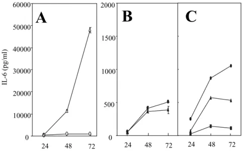
trachomatisinfected HFLS or HeLa 229 cells Cytokine induction was compared in both HFLS and HeLa 229 cells by C. trachomatis infection or mock infection. An infection with C. trachomatis in HFLS induced the production of a large amount of IL6 and IL8 of up to 45,000 and 30,000 pg/ml, re- spectively(Figs. 2A and 3A). On the other hand, HeLa 229 cells chlamydial infection never induced the production of IL6 nor IL8 in HeLa 229 cells
(Figs. 2B and 3B). No TNFα or IFNα secre- tion was detected in supernatants from either in- Fig. 1. Electron microscopic analysis of C. trachomaisin
fected HFLS or HeLa 229 cells and chlamydial growth curves. Micrographs of(A)C. tracho- mais 72 h after infection of HFLS and(B)of HeLa 229 cells. Bars are 5μm, respectively.
Few EBs, abnormal RBs, and a lot of glycogen particles were observed in HFLS. (C)Onestep growth of C. trachomais in HFLS(○)and HeLa 229 cells(●).
fected HFLS or HeLa 229 cells(data not shown).
Previous studies have demonstrated the inhibi- tory effects of IFNγ on C. trachomatis replication, thus resulting in the development of inclusions con- taining enlarged atypical RBs.5)6)9)10) This study examined whether C. trachomatisinfected HeLa 229 cells secreted IL6 during a persistent infection in response to IFNγ. Although IFNγ at 2.0 or 20 ng/ml completely inhibited the chlamydial growth in HeLa 229 cells, the secretion of IL6 was only 1,000 pg/ml regardless of C. trachomatis infection
(Fig. 2C). The IL6 levels of 1,000 pg/ml in IFN γ treated HeLa 229 cells were almost the same as those in uninfected or mock infected HFLS(Figs.
2A and C). IFNγ affects the host cells in vitro by inducing IDO, which catalyzes the initial step in degradation of tryptophan, and correlates with the growth inhibition of Chlamydia.26) This proc- ess is recovered by the addition of an excessive amount of tryptophan, and the depletion of exoge- nous tryptophan induces growth inhibition even further. Therefore, the effect of exogenous trypto- phan on IL6 secretion in infected or mock infected HeLa 229 cells was analyzed. The maintenance me-
dium without tryptophan did not increase IL6 se- cretion in infected HeLa 229 cells(Fig. 2C). Taken together, these observations demonstrated that IFNγ induced persistent infection caused a slight increase of IL6 secretion, which showed no correla- tion with either C. trachomatis infection or the ab- sence of tryptophan. Furthermore, the exogenous IL6(0.01〜100 ng/ml)did not effect the morphol- ogy of inclusions and chlamydial growth in HeLa 229 cells(data not shown).
3.Effect of 1methyltryptophan pretreatment Abnormal inclusions induced by IFNγ could re- turn to a normal shape after the addition of a com- petitive inhibitor of IDO, 1MT, in Chlamydophila pneumoniaeinfected HEp2 cells.27)28) Therefore, C.
trachomatisinduced IL6 production and chlamydial growth were measured in HFLS following expo- sure to 1MT. Pretreatment with 12.5 mM 1MT for 1 h allowed a large amount of IL6 secretion and suppressed chlamydial growth(Figs. 4A and B). In addition, a similar result was observed af- ter the pretreatment with 25 mM 1MT(data not shown).
Fig. 2. Kinetics of IL6 secretion by C. trachomatisinfected HFLS and HeLa 229 cells. In(A)HFLS and(B)HeLa 229 cells, C. tracho- matisinfected cultures(▲ and △), mock infected cultures(● and
○). (C) C. trachomatisinfected HeLa 229 cells were cultured with IFNγ of 2.0 ng/ml (▲) and 20 ng/ml (●), or cultured in de- leted tryptophan medium(■). In(A)and(B), data are mean
±SD(n=3).
4.Effect of neutralizing TLR2 or TLR4 mAb on infec- tivity and IL6 production
TLR blocking experiments were carried out us- ing a TLR neutralizing mAb. The HFLS or HeLa 229 cells were incubated with anti TLR2 or anti TLR4 neutralizing mAb for 2 h before and during
C. trachomatis infection. As shown in Figures 5A and B, the TLR2 mAb blocked approximately 90%
of the infectivity;however, the neutralization of TLR2 mAb produced a fivefold increase in IL6 production in comparison to the control in HFLS. The TLR4 mAb blocked neither the infec- Fig. 3. IL8 production in C. trachomatisinfected HFLS or HeLa 229 cells.
In(A)HFLS and(B)HeLa 229 cells, C. trachomatisinfected cul- tures(▲), and mock infected cultures(●).
Fig. 4. Effect of 1MT on IL6 production and chlamydial growth of C.
trachomatis infected HFLS. In(A)IL6 production and(B)chla- mydial growth, C. trachomatisinfected cultures(●), C. trachomatis infected cultures pretreated with 12.5 mM αMT(▲). In B,
number of infectious EBs were determined on McCoy cells, and shown as number of inclusions. Data are mean±SD(n=3).
tivity nor the IL6 production in C. trachomatisin- fected HFLS. On the other hand, the TLR4 mAb blocked 50% of the infectivity in HeLa 229 cells
(Fig. 5). The TLR2 mAb blocked neither the infec- tivity nor IL6 production in C. trachomatisinfected HeLa 229 cells.
Discussion
A cure for a persistent chlamydial infection in humans is very important;however, it remains difficult to achieve. Because Chlamydiae respond to a variety of environmental stimuli that alter their growth characteristics, Chlamydia thus has the potential to establish a chronic or persistent relationship with the host. If chronic or persis- tent infections are established, then those infec- tions may serve as a reservoir for new infections, and contribute to the immunopathological conse- quences of infection. Therefore, understanding the consequences of persistent chlamydial infection could thus play an important role in the control and prevention of chlamydial disease.
A persistent chlamydial infection causes severe and difficulttotreat diseases such as ReA, which develop in 1 to 3% of patients after a genital infection.29) EM observations of synovial mem- branes from ReA patients show atypical RBs in vivo,30) where infectious EBs cannot be isolated
from the site of infection.31) These morphological observations may correspond to the IFNγ in- duced persistent Chlamydiae seen in vitro.5)6) The characterization of the in vitro persistent phase and of the in vivo evidence suggests that Chlamydiae persist in an altered atypical form during chronic disease. Further study examined whether the mechanism of persistence in HFLS was similar to that of IFNγ induced persistence. The most im- portant mechanism underlying the effect of IFNγ on chlamydial growth is thought to be tryptophan depletion through the activation of the host trypto- phandegrading enzyme IDO.7)8)32) However, C.
trachomatisinfected HeLa 229 cells cultured in IFN γ or the tryptophandeleted medium did not show an increase in the IL6 production(Fig. 2), despite the loss of chlamydial growth. Pretreatment with 1MT could have resulted in a decrease of the IL6 production and chlamydial normal replication if tryptophan depletion occurred in C. trachomatisin- fected HFLS because of the antagonistic effect of α MT on IDO.28) However, the pretreatment with 1 MT could neither restore the abnormal form to a normal shape nor suppress the IL6 production in C. trachomatisinfected HFLS(Fig. 4). Tryptophan depletion may not correlate with persistent infec- tion in synovial fibroblasts. Collectively, the cur- rent results demonstrated that a chlamydial persistent infection in HFLS was different from Fig. 5. Effects on IL6 production and formation of inclusions with pre-
treatment of antiTLR2 or antiTLR4 antibody in C. trachomatis infected HFLS or HeLa 229 cells. IL6 production(□), formation of inclusions(■). In graphs of IL6 production, data are mean
±SD(n=3).
IFNγ induced persistence.
TLRs are innate immune receptors involved in the pattern recognition of bacterial antigens, and it is currently believed that their differential acti- vation helps to discriminate between microbial pathogens.33) It has been suggested that TLR4 is involved in the recognition of gramnegative bacte- ria through their LPS,34) and TLR2 mainly recog- nizes the elements of grampositive bacteria and yeasts,35) although this dichotomy is not always sustained. TLR2 is essential for the early cyto- kine production such as IL1 and IL6 in C. tracho- matis genital infection,17) and an antibody to TLR4 inhibits C. trachomatisinduced IL1β, IL6, and TNFα production by dendritic cells.24) The rec- ognition of C. pneumoniae depends on the TLR2 acti- vation of either dendritic cells or mononuclear cells.21)23) This study investigated the role of TLR2 and TLR4 in the growth of chlamydia and C. tracho- matisinduced IL6 production in both HFLS and HeLa 229 cells. Interestingly, the augmentation of IL6 production inversely correlated with the de- crease in the inclusion formation by TLR2 mAb
(Fig. 5). TLR4 did not contribute to the IL6 pro- duction nor to the formation of inclusions in C.
trachomatisinfected HLFS. On the other hand, al- though the formation of chlamydial inclusions of C. trachomatis in HeLa 229 cells depended on TLR4, it did not imply the stimulation of IL6 production
(Fig. 5). These results suggest that TLR2 might affect both the process of chlamydial infection and the IL6 production in HFLS, and is different from that in HeLa 229 cells.
In conclusion, the present study showed that mechanisms other than IFNγ induced persistence might contribute to the chlamydial persistence on ReA. It is probable that TLR2 and other mecha- nisms might therefore be involved in the IL6 production. Further investigation into the inter- play between chlamydia and its environment is thus needed to both understand the hostmicrobial interactions and provide important insight for the treatment and prevention of ReA.
Acknowledgments
We thank Dr. Ataru Kuroiwa(Faculty of Medi- cine, Fukuoka University, Japan)for his comments
on the manuscript.
References
1)Cates W, Wasserheit JN.:Genital chlamydial infec- tions:epidemiology and reproductive sequelae. Am.
J. Obstet. Gynecol. 164 : 17711781, 1991.
2)Gaston JS : Immunological basis of Chlamydia in- duced reactive arthritis. Sex Transm Infect. 76 : 156 61, 2000.
3)Hanada H, IkedaDantsuji Y, Naito M, Nagayama A:Infection of human fibroblastlike synovial cells with Chlamydia trachomatis results in persis- tent infection and interleukin6 production. Microb.
Pathog. 34 : 5763, 2003.
4)Sieper J, Kingsley G:Recent advances in the patho- genesis of reactive arthritis. Immunol Today. 17 : 160163, 1996.
5)Beatty WL, Byrne GI, Morrison RP:Morphologic and antigenic characterization of interferon gamma mediated persistent Chlamydia trachomatis infection in vitro. Proc. Natl. Acad. Sci. U.S.A. 90:39984002, 1993.
6)Beatty WL, Morrison RP, Byrne GI:Persistent chla- mydiae:from cell culture to a paradigm for chlamy- dial pathogenesis. Microbiol. Rev. 58:686699, 1994.
7)Taylor MW, Feng G:Relationship between inter- ferongamma, indoleamine 2,3dioxygenase, and tryptophan catabolism. FASEB J. 11:25162522, 1991.
8)Wood H, FehlnerGardner C, Berry J, Fischer E, Gra- ham B, Hackstadt T, Roshick C, McClarty G :Re- gulation of tryptophan synthase gene expression in Chlamydia trachomatis. Mol Microbiol. 5:1347 1359, 2003.
9)Beatty WL, Morrison RP, Byrne GI:Immunoelectron microscopic quantitation of differential levels of chlamydial proteins in a cell culture model of per- sistent Chlamydia trachomatis infection. Infect.
Immun. 62:40594062, 1994.
10)Beatty WL, Belanger TA, Desai AA, Morrison RP, Byrne GI:Tryptophan depletion as a mechanism of gamma interferonmediated chlamydial persistence.
Infect. Immun. 62:37053711, 1994.
11)Ding AH, Nathan CF, Stuehr DJ:Release of reactive nitrogen intermediates and reactive oxygen interme- diates from mouse peritoneal macrophages. Compa- rison of activating cytokines and evidence for inde- pendent production. J. Immunol. 141:24072412, 1988.
12)Igietseme JU, Ananaba GA, Candal DH, Lyn D, Black CM:Immune control of Chlamydial growth in the human epithelial cell line RT4 involves multiple mechanisms that include nitric oxide induction, tryp- tophan catabolism and iron deprivation. Microbiol.
Immunol. 42:617625, 1998.
13)Schumacher HR, Magge S, Cherian PV, Sleckman J, Rothfuss S, Clayburne G, Sieck M:Light and elec- tron microscopic studies on the synovial membrane in Reiter’s syndrome:immunocytochemical identifi- cation of chlamydial antigen in patients with early disease. Arthritis Rhum. 31:937946, 1988.
14)TaylorRobinson D, Gilroy CB, Thomas BJ, Keat AC:Detection of Chlamydia trachomatis DNA in joints of reactive arthritis patients by polymerase chain reaction. Lancet. 340:8182, 1992.
15)Rahman MU, Cheema MA, Schumacher HR, Hudson AP:Molecular evidence for the presence of chlamy- dia in the synovium of patients with Reiter’s syn- drome. Arthritis Rheum. 35:521529, 1992.
16)Rdel J, Groh A, Hartmann M, Schmidt KH, Leh- mann M, Lungershausen W, Straube E:Expression of interferon regulatory factors and indoleamine 2,3 dioxygenase in Chlamydia trachomatisinfected synovial fibroblasts. Med. Microbiol. Immunol.
(Berl). 187:205212, 1999.
17)Darville T, O’Neill JM, Andrews CW, Nagarajan UM, Stahl L, Ojcius DM:Tolllike receptor2, but not Tolllike receptor4, is essential for development of oviduct pathology in chlamydial genital tract infection. J. Immunol. 171:61876197, 2003.
18)Bulut Y, Faure E, Thomas L, Karahashi H, Michel- sen KS, O. Equils, Morrison SG, Morrison RP, Arditi M:Chlamydial heat shock protein 60 activates macrophage and endothelial cells through Tolllike receptor 4 and MD2 in a MyD88dependent pathway.
J. Immunol. 168:14351440, 2002.
19)Ohashi K, Burkart V, Flohe S, Kolb H:Cutting edge:heat shock protein 60 is a putative endoge- nous ligand of the tolllike receptor4 complex. J.
Immunol. 164:558561, 2000.
20)Kol A, Lichtman AH, Finberg RW, Libby P, Kurt Jones EA:Cutting edge:heat shock protein(HSP)
60 activates the innate immune response:CD14 is an essential receptor for HSP60 activation of mononu- clear cells. J. Immunol. 164:1317, 2000.
21)Prebeck S, Brade H, Kirschning CJ, da Costa CP, Durr S, Wagner H, Miethke T:The Gramnegative bacterium Chlamydia trachomatis L2 stimulates tu- mor necrosis factor secretion by innate immune cells independently of its endotoxin. Microbes. Infect.
5:463470, 2003.
22)Netea MG, Kullberg BJ, Galama JM, Stalenhoef AF, Dinarello CA, vander Meer JW:NonLPS compo- nents of Chlamydia pneumoniae stimulate cytokine production through Tolllike receptor 2dependent pathways. Eur. J. Immunol. 32:11881195, 2002.
23)Prebeck S, Kirschning C, Durr S, da Costa C, Donath B, Brand K, Redecke V, Wagner H, Miethke T:Pre- dominant role of tolllike receptor 2 versus 4 in
Chlamydia pneumoniaeinduced activation of den- dritic cells. J. Immunol. 167:33163323, 2001.
24)Gervassi A, Alderson MR, Suchland R, Maisonneuve JF, Grabstein KH, Probst P:Differential regulation of inflammatory cytokine secretion by human den- dritic cells upon Chlamydia trachomatis infection.
Infect. Immun. 72:72317239, 2004.
25)Nagayama A, Nakao T, Taen H:In vitro activities of ofloxacin and four other new quinolinecarboxylic ac- ids against Chlamydia trachomatis. Antimicrob.
Agents. Chemother. 32:17351737, 1988.
26)Gupta SL, Carlin JM, Pyati P, Dai W, Pfefferkorn ER, Murphy MJ:Antiparasitic and antiproliferative effects of indoleamine 2,3dioxygenase enzyme ex- pression in human fibroblasts. Infect. Immun. 62:
22772284, 1994.
27)Mehta SJ, Miller RD, Ramirez JA, Summersgill JT:Inhibition of Chlamydia pneumoniae replication in HEp2 cells by interferongamma:role of trypto- phan catabolism. J. Infect. Dis. 177:13261331, 1998.
28)Pantoja LG, Miller RD, Ramirez JA, Molestina RE, Summersgill JT:Characterization of Chlamydia pneumoniae persistence in HEp2 cells treated with gamma interferon. Infect. Immun. 69:79277932, 2001.
29)Hogan RJ, Mathews SA, Mukhopadhyay S, Sum- mersgill JT, Timms P:Chlamydial persistence:be- yond the biphasic paradigm. Infect. Immun. 72:
18431855, 2004.
30)Nanagara R, F Li, Beutler A, Hudson A, Schumacher HR:Alteration of Chlamydia trachomatis biologic behavior in synovial membranes. Suppression of sur- face antigen production in reactive arthritis and Reiter’s syndrome. Arthritis Rheum. 38:14101417, 1995.
31)Keat A, Thomas B, Dixey J, Osborn M, Sonnex C, TaylorRobinson D:Chlamydia trachomatis and re- active arthritis:the missing link. Lancet. 1:7274.
ss, 1987.
32)Sakash JB, Byrne GI, Lichtman A, Libby P: Cytoki- nes induce indoleamine 2,3dioxygenase expression in human atheromaasociated cells:implications for persistent Chlamydophila pneumoniae infection.
Infect. Immun. 70:39593961, 2002.
33)Ulevitch RJ:Toll gates for pathogen selection.
Nature. 401:755756, 1999.
34)Poltorak A, He X, Smirnova I, Liu MY, Van Huffel C, Du X, Birdwell D, Alejos E, Silva M, Galanos C, Freudenberg M, RicciardiCastagnoli P, Layton B, Beutler B:Defective LPS signaling in C3H/HeJ and C57BL/10ScCr mice:mutations in Tlr4 gene. Science.
282:20852088, 1998.
35) Takeuchi O, Hoshino K, Kawai T, Sanjo H, Takada H, Ogawa T, Takeda K, Akira S:Differential roles of TLR2 and TLR4 in recognition of gramnegative and
grampositive bacterial cell wall components.
Immunity. 11 : 443451, 1999.
(Received on June 19, 2009, Accepted on September 9, 2009)