1
Identification of a Major Glucose Transporter in Flavobacterium johnsoniae: Inhibition of F. johnsoniae Colony Spreading by Glucose Uptake
Keigo Imamura1,2, Keiko Sato1, Yuka Narita1,*, Yoshio Kondo2, Daisuke Nakane3, Mariko Naito1, Taku Fujiwara2 and Koji Nakayama1
1Department of Microbiology and Oral Infection and 2Department of Pediatric Dentistry, Graduate School of Biomedical Sciences, Nagasaki University, 1-7-1 Sakamoto, Nagasaki 852-8588 and 3Department of Physics, Gakushuin University, 1-5-1 Mejiro, Toshima-ku,Tokyo 171-8588, Japan
*Present Address: Section of Infection Biology, Department of Functional Bioscience, Fukuoka Dental College, 2-15-1 Tamura, Sawara-ku, Fukuoka 814-0193, Japan
Short Running Title: MFS Glucose Transporter
Correspondence:
Koji Nakayama, Department of Microbiology and Oral Infection, Graduate School of Biomedical Sciences, Nagasaki University, 1-7-1 Sakamoto, Nagasaki 852-8588, Japan.
Tel: +81 95 819 7648; fax: +81 95 819 7650; email address: [email protected]
2
List of Abbreviations: CCCP, carbonyl cyanide m-chlorophenylhydrazone; CYE, casitone yeast extract; DCCD, N,N′-dicyclohexylcarbodiimide; DNP, 2,4-dinitrophenol;
Em, erythromycin; MFS, major facilitator superfamily; PBS, phosphate buffered saline;
RT, room temperature; Sm, streptomycin; 2DG, 2-deoxy-D-glucose
3
ABSTRACT
Many members of the phylum Bacteroidetes such as Flavobacterium johnsoniae can glide over a solid surface: an ability called gliding motility. It can be usually observed on agar plates as thin, flat, spreading colonies with irregular, feathery edges; this phenomenon is called colony spreading. Colony spreading of F. johnsoniae on 1.5%
agar plates containing poor nutrients is dose-dependently inhibited by addition of
D-glucose, as previously reported. Accordingly, here, we created mutants (by transposon mutagenesis) that partially suppressed glucose-mediated inhibition of colony spreading.
Among the isolates, we found that one had a transposon insertion in Fjoh_4565, tentatively named mfsA, which encodes a major facilitator superfamily (MFS) transporter previously shown to be required for growth on glucose, N-acetyl-glucosamine, and chitin. We constructed an mfsA deletion mutant and found that the mutant showed no glucose-mediated acceleration of growth or glucose uptake.
The mfsA gene complemented the phenotype of a glucose-negative Escherichia coli.
These results suggested that the mfsA gene encodes the sole MFS transporter of glucose in F. johnsoniae and that glucose uptake is partially required for the glucose-mediated inhibition of F. johnsoniae colony spreading.
Key words: Bacteroidetes, colony spreading, gliding motility, glucose uptake, major facilitator superfamily transporter
4
INTRODUCTION
Many bacterial species can glide over solid surfaces: an ability that is called gliding motility. This ability can be observed in many members of the phylum Bacteroidetes, Myxococcus xanthus, Mycoplasma mobile, and many cyanobacteria, but these bacteria have their own unique motility machineries (1). Flavobacterium johnsoniae belonging to the phylum Bacteroidetes has been studied for many years to understand the motility mechanism. A large number of F. johnsoniae proteins have been found to be involved in gliding motility, which include Gld, Spr, and Rem (2). Some of these proteins are also components of the type IX protein secretion system (3, 4). We proposed a helical track model, where adhesive SprB filaments are propelled along a left-handed closed helical loop on the cell surface. Attachment of SprB to a substratum results in cell movement (5).
Gliding motility of F. johnsoniae is usually observed on agar plates as thin, flat, spreading colonies with irregular, feathery edges: this phenomenon is called colony spreading (6). This phenomenon requires gliding motility because F. johnsoniae mutants deficient in gld or spr genes show no colony spreading (3, 7-19). Colony spreading takes place on rather nutrient-poor plates, and when nutrients are added, the colonies tend to be raised and smooth-edged (6). Chang & Pate (20) first reported that sugars suppress colony spreading of F. johnsoniae on 1.5% agar plates. In their study, they found that metabolizable sugars including glucose, galactose, fructose, mannose, xylose, and maltose suppress colony spreading, whereas a nonmetabolizable sugar, lactose, does not. More extensive experiments revealed that a nonmetabolizable sugar, sucrose, suppresses colony spreading at a low concentration and minimal inhibitory concentrations for colony spreading vary among metabolizable sugars (21). Gorski et al.
5
(22) found that the inhibitory sugars have a common structural feature regardless of their metabolizable abilities.
In this study, we created F. johnsoniae mutants that showed colony spreading on glucose-containing agar plates using transposon mutagenesis to investigate which genes are involved in the inhibitory effect of glucose on colony spreading of the bacterium.
MATERIALS AND METHODS
Bacterial strains and culture conditions
Bacterial strains and plasmids used in this study are listed in Table 1 (23, 24). F.
johnsoniae cells were grown aerobically in the enriched casitone yeast extract (CYE) medium and on enriched CYE agar. For selection and maintenance of antibiotic-resistant F. johnsoniae strains, antibiotics were added to the medium at the following concentrations: streptomycin (Sm) 100 μg/ml and erythromycin (Em) 100 μg/ml. To observe colony spreading, we grew F. johnsoniae on PY2 agar (7) at 25°C.
Transposon mutagenesis and gene-directed mutagenesis
Transposon mutagenesis in F. johnsoniae strain UW101 by means of Tn4351 was described previously (25). Gene-directed mutagenesis of F. johnsoniae was carried out as follows. After the mating of E. coli S17-1 λpir (carrying a pRR51 derivative) with F.
johnsoniae CJ1827, an Emr transconjugant was obtained to select for integration of the plasmid into the genome by homologous recombination. An erythromycin-resistant clone was grown overnight in CYE, and the loss of the plasmid via a second recombination event was selected by growth on CYE agar containing streptomycin
6
(18).
Construction of plasmids and bacterial strains
For construction of a targeting plasmid vector designed to create an F. johnsoniae mfsA deletion mutant, DNA regions upstream and downstream of mfsA were PCR-amplified from the chromosomal DNA of F. johnsoniae using pairs of primers (F4565-UF-BamHI plus F4565-UR-SalI and F4565-DF-SalI plus F4565-DR-SphI, respectively, where “U”
indicates upstream, “F” indicates forward, “D” indicates downstream, and “R” indicates reverse). Primers used in this study are listed in Table S1. The amplified DNA upstream was double-digested with BamHI plus SalI. The DNA downstream was digested with SalI plus SphI. Both digested products were ligated with pRR51 that had been digested with BamHI and SphI. (Consequently, we obtained pDF1.)
For construction of shuttle vector pNS1 for F. johnsoniae, the multiple cloning site (MCS) region was 1st-PCR-amplified from pFj29 using the primer pair pFj29-1st-F and gfpmut3-R-SphI. Then, the MCS region was 2nd-PCR-amplified from the 1st-amplified DNA using the primer pair pFj29-2nd-F and gfpmut3-R-SphI. The amplified DNA was digested with BglII and SphI and inserted at the BamHI and SphI sites of pFj29, resulting in pNS1.
For construction of a complemented version of the mfsA strain DFJ, the gene encoding Fjoh_4565 was PCR-amplified from F. johnsoniae UW101 chromosomal DNA using the primer pair F4565-F-BamHI and F4565-stop-R-NotI. The amplified DNA was digested with BamHI and NotI and inserted into the corresponding region of pNS1, resulting in plasmid pNS1 containing mfsA (pDF2).
For construction of an F. johnsoniae strain expressing MfsA-Gfp, the gene
7
encoding Fjoh_4565 was PCR-amplified from F. johnsoniae UW101 chromosomal DNA using the primer pair F4565-F-BamHI and F4565-GR-NotI. The amplified DNA was digested with BamHI and NotI and inserted into the corresponding region of pNS1, resulting in plasmid pNS1 containing mfsA-gfp (pDF3).
For construction of a glucose-negative E. coli strain expressing F. johnsoniae MfsA and MfsA-Gfp, the mfsA gene DNA encoding Fjoh_4565 was PCR-amplified from F. johnsoniae UW101 chromosomal DNA and from the pDF2 plasmid DNA using the primer pairs F4565-22bF-NdeI and F4565-22bR-XhoI as well as F4565-22bF-NdeI and Gfpmut3-stopR-XhoI, respectively. The amplified DNAs were digested with NdeI and XhoI and inserted into the corresponding region of pET-22b (Novagen), resulting in plasmids pDF4 and pDF5, respectively. The glucose-negative E. coli strain LJ141 was then transformed with pDF4 and pDF5.
Glucose uptake
F. johnsoniae cells were grown in the CYE medium at 27°C with shaking (165 rpm) overnight to optical density of ~1.0 at 600 nm. The samples were washed two times with 10 mM Tris-HCl (pH 7.5). The cells were exposed to 1 mM 2,4-dinitrophenol (DNP), 10 μM carbonyl cyanide m-chlorophenylhydrazone (CCCP), 50 μM N,N′-dicyclohexylcarbodiimide (DCCD), or 50 mM arsenate for 1 min at room temperature (RT) and incubated in 10 mM Tris-HCl (pH 7.5) supplemented with 2-deoxy-D-glucose (2DG) at RT for 2 h. Glucose uptake was determined by means of the 2DG uptake in an enzymatic photometric assay using the 2DG Uptake Measurement Kit (COSMO BIO Co.) (26, 27).
8
RNA Isolation
Total RNA from cells of the wild-type and the mfsA mutant at different growth conditions (1% PY2 and 1% PYG) from three independent cultures. After 24 h of culture, bacterial cells were collected by cell scraping in RNAlaterR○ solution (Thermo Fisher Scientific) and centrifuged at 8,000 rpm for 10 min. Cell pellets were resuspended with Trizol, and RNA was extracted using an RNeasy Mini Kit (Qiagen) according to the manufacturer’s recommendations. DNA was removed with RNase-free DNase.
Gene Expression Microarrays
According to manufacturers’ instructions, the complementary RNA was amplified and labeled by Low Input Quick Amp Labeling Kit (Agilent Technologies), and hybridized to Agilent-based microarray platform with 4 x 44 K probes per slide (Agilent Technologies). The array contains probe sets to 5,113 open reading frames of F.
johnsoniae UW101. Designing microarray probes was done with the Agilent eArraysystem with the following settings during the microarray probe design: Tm (70°C) matching methodology, 60-mer probe length, 8 probes/gene. All hybridized microarray slides were scanned using an Agilent scanner. Relative hybridization intensities and background hybridization values were calculated using Agilent Feature Extraction Software (ver. 9.5.1.1).
Localization of MfsA
Cells were examined by microscopy to identify MfsA on the cell membrane. Cells of F.
johnsoniae mfsA/mfsA-gfp (200 μl) were placed on a slide glass for 3 min at RT and
9
were washed two times with PBS. DAPI (Invitrogen) and FM4-64 (Invitrogen) were used for detection of DNA and cell membranes, respectively. After two washes with PBS, the cells were incubated with a 1/500 dilution of DAPI (Invitrogen) and FM4-64 (Invitrogen) for 30 min and were washed two times with PBS. The coverslip was mounted on glass and examined under an inverted fluorescence microscope.
Cell growth
The growth curves of the wild-type, mfsA deletion mutant, and mfsA/mfsA+ complemented strain were determined. The cells were incubated in CYE overnight to optical density of ~1.0 at 600 nm. The microorganisms were washed two times with 10 mM Tris-HCl (pH 7.5) and incubated in the mPY2 medium (0.05% peptone, 0.05%
yeast extract) or mPY2 supplemented with D-glucose (15 mM) with shaking at 27°C.
Cell growth was measured by optical density at 600 nm at indicated time points. Error bars show standard deviation.
Statistical analysis
The data of glucose uptake test were analysed using Student’s t-test. Results were considered to be statistically significant with a P value <0.001.
RESULTS
The inhibitory effect of glucose on colony spreading of F. johnsoniae
As previously found (20-22), glucose suppressed colony spreading of F. johnsoniae on PY2 plates in a concentration-dependent manner, and D-glucose with 10 mM completely inhibited colony spreading (Fig. 1A and B). A nonmetabolizable derivative
10
of glucose, 2-deoxy-D-glucose (15 mM), partially suppressed colony spreading (Fig.
1C), suggesting that there were two types of suppression: metabolism-dependent and metabolism-independent.
Construction of a transposon insertion library and screening on the basis of colony spreading
Transposon-containing suicide plasmid R751::Tn4351Ω4 was used for mutagenesis of F.
johnsoniae strain UW101. Cells were grown on agar plates supplemented with 15 mM glucose and 100 mM Em. Seventeen colonies showing higher levels of colony spreading compared to the wild type were found among nearly 48,000 colonies. The transposon insertion sites in all the mutants were determined by DNA sequencing.
Fjoh_4565, which encode a major facilitator superfamily (MFS) transporter, was present at the insertion site of one of the mutants (Fig. 2A). Fjoh_4565 was recently shown to be required for growth on glucose, N-acetyl-glucosamine, and chitin (28). We tentatively named this gene mfsA. We constructed an mfsA deletion mutant (DFJ1), which partially restored colony spreading on the 15 mM glucose-containing plate (Fig.
2B and Fig. S1). E. coli–F. johnsoniae shuttle vector plasmids containing the mfsA+ and mfsA-gfp fusion genes were then introduced into strain DFJ1, resulting in mfsA/mfsA+ and mfsA/mfsA-gfp complemented strains (DFJ1/pDF2 and DFJ1/pDF3).
Complemented strains DFJ1/pDF2 and DFJ1/pDF3 showed no colony spreading on a 15 mM glucose-containing plate, just as the wild type did (Fig. 2B and Fig. S1). The other sixteen mutants had the transposon DNA in different genes and the results will be reported elsewhere.
11
Growth of the mfsA mutant in media with or without glucose
The mfsA mutant, the mfsA/mfsA+ complemented strain, and the wild type were incubated in mPY2 with or without 15 mM glucose, and growth of the strains was determined via optical density at 600 nm. Addition of glucose resulted in increased growth of the wild-type and mfsA/mfsA+ complemented strains, whereas the growth of the mfsA mutant was not changed by addition of glucose (Fig. 2C).
Glucose uptake in the mfsA mutant
Glucose uptake of the mfsA mutant, of the mfsA/mfsA+ complemented strain, and of the wild type was determined. The mfsA/mfsA+ complemented strain and the wild type showed glucose uptake, whereas the mfsA mutant showed no glucose uptake (Fig. 3A).
Glucose uptake of the wild type was decreased by proton motive force inhibitors, CCCP and DNP, but not decreased by ATPase inhibitors, arsenate, and DCCD, indicating that the glucose uptake system in F. johnsoniae depends on proton motive force (Fig. 3B).
Gene expression in the mfsA mutant
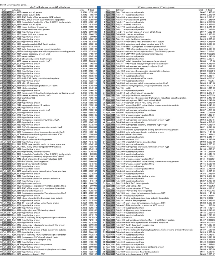
To determine which genes are influenced by mfsA, microarray analysis of the mfsA mutant, which was grown in agar plates supplemented with 15 mM glucose, was performed, and the result was compared to that of the wild type grown in the glucose-supplemented agar plates. The ratio of expression of each gene in the mfsA mutant with glucose versus that in the wild type with glucose was compared with the ratio of expression of each gene in the wild type without glucose versus that in the wild type with glucose (Fig. 4). The result revealed that gene expression in the mfsA mutant correlated with that in the wild type without glucose. The 100 genes most upregulated
12
and downregulated by the disruption of mfsA (mfsA with glucose versus the wild type with glucose) were compared with those under the influence of depletion of glucose in the wild type (wild type without glucose versus wild type with glucose) (Tables S2 and S3). Seventy-seven and 87 of the 100 upregulated and downregulated genes, respectively, were common between the two comparisons. These results suggested that the mfsA mutant experienced glucose starvation even when glucose was added into the medium.
Location of the MfsA protein
To determine intracellular localization of MfsA, we used the mfsA/mfsA-gfp fusion strain. Using fluorescence microscopy, we found that green fluorescence was located around the cell (Fig. 5A), suggesting that MfsA is located in the cell surface membranes.
FM4-64 (red) and DAPI (blue) were used to indicate the areas of lipid layers and cytoplasm, respectively (Fig. 5B, C, and D).
Complementation of the glucose-negative phenotype in E. coli by the mfsA gene We tested whether F. johnsoniae MfsA can complement the glucose-negative phenotype in E. coli. The mfsA and mfsA-gfp genes were placed after the T7 promoter in plasmid pET-22b, resulting in vectors pDF4 and pDF5, respectively. The glucose-negative E.
coli strain LJ141 that lacks detectable glucose transport activity was then transformed with pDF4 and pDF5. The transformed E. coli strains were streaked onto MacConkey agar plates supplemented with 50 mM glucose. Strains LJ141/pDF4 and LJ141/pDF5 formed red colonies because of the fermentation of glucose, whereas strains LJ141 and LJ141 carrying the vector plasmid pET-22b showed non-glucose-fermenting ocher
13
colonies, demonstrating that F. johnsoniae MfsA can function as a glucose transporter in E. coli (Fig. 6). Strains LJ141/pDF4 and LJ141/pDF5 did not form red colonies on MacConkey agar supplemented with 50 mM mannose or mannitol, suggesting that MfsA has no contribution to the uptake of mannose or mannitol (Fig. 6).
DISCUSSION
The results presented here illustrate the role of mfsA in glucose inhibition of colony spreading. A recent study by Larsbrink et al (28) identified a locus containing mfsA and 10 other genes that were involved in F. johnsoniae chitin utilization. mfsA was shown to be required for growth on glucose, N-acetylglucosamine, and chitin. Our results confirm and extend these findings.Genome information on F. johnsoniae reveals that it has no phosphotransferase system but has 8 genes encoding putative major MFS transporters.
The MFS is one of the largest groups of secondary active transporters conserved from bacteria to humans. In this study, we found that (i) the mfsA mutant showed no glucose uptake, (ii) the mfsA mutant did not utilize glucose for its growth, and (iii) the mfsA gene complemented the glucose-negative phenotype of E. coli LJ141. The present findings with the previous one (28) strongly indicate that mfsA (Fjoh_4565, which is one of the 8 above-mentioned genes) encodes the sole glucose transporter in F.
johnsoniae. Comparison with proteins in the MFS family using the IUBMB-approved Transporter Classification Database (www.tcbd.org) revealed that a protein most similar to MfsA is glucose/galactose transporter Ggp (2.A.1.7.2) in Brucella abortus, which belongs to the fucose: H+ symporter (FHS) family (2.A.1.7) and that the top 13 proteins similar to MfsA belong to the FHS family. These 13 proteins including Ggp in B.
abortus have 12 transmembrane segments (TMSs) except for one protein, which has 11
14
TMSs. On the other hand, MfsA appears to have 14 TMSs (Fig. S2). In this study, we found that MfsA requires proton motive force for its glucose uptake; this finding is consistent with the comparison result, which suggested that MfsA may belong to the FHS family.
In 1947, Stanier (6) reported that F. johnsoniae cells form spreading colonies on nutrient-poor plates. The cells formed rather small colonies with smooth edges on a plate with 2.0% tryptone, whereas they formed larger colonies with irregular, feathery edges on a plate containing 0.25% tryptone. Carbohydrates such as glucose, maltose, glucosamine, N-acetylglucosamine, sucrose, and trehalose can suppress colony spreading of F. johnsoniae (21). Most of the carbohydrates are metabolized by F.
johnsoniae, but this bacterium cannot utilize sucrose as an energy source. Nevertheless, sucrose inhibits colony spreading. Similarly, a nonmetabolizable derivative of glucose, 2-deoxy-glucose, also inhibits colony spreading although the inhibitory effect was much weaker than that of D-glucose, suggesting that there may be two types of the carbohydrate-mediated inhibitory effect: metabolism-dependent and metabolism-independent. In this study in F. johnsoniae, we created mutations that suppress the effect of glucose on colony spreading. They included the mutant possessing the transposon DNA in the mfsA gene, which encode an MFS protein. These results suggest that the glucose-mediated inhibitory effect on colony spreading is at least partly attributable to glucose uptake. Further research is needed to find which metabolite(s) in the metabolic pathway inhibits colony spreading.
ACKNOWLEDGMENT
We thank Drs. K. Jahreis and S. Chen for generous gifts of E. coli LJ141 and the shuttle
15
plasmid pFj29, respectively. This work was supported by the Japan Society for the Promotion of Science Kakenhi Grants (Grant IDs 24117006 and 25293375 to KN).
DISCLOSURE
The authors have no conflicting financial interests.
16
REFERENCES
1. Jarrell K.F., McBride M.J. (2008) The surprisingly diverse ways that prokaryotes move. Nat Rev Microbiol 6: 466-76.
2. McBride M.J., Nakane D. (2015) Flavobacterium gliding motility and the type IX secretion system. Curr Opin Microbiol 28: 72-7.
3. Sato K. Naito M., Yukitake H., Hirakawa H., Shoji M., McBride M.J., Rhodes R.G., Nakayama K. (2010) A protein secretion system linked to bacteroidete gliding motility and pathogenesis. Proc Natl Acad Sci U S A 107: 276-81.
4. Nakayama K. (2015) Porphyromonas gingivalis and related bacteria: from colonial pigmentation to the type IX secretion system and gliding motility. J Periodont Res 50: 1-8.
5. Nakane D., Sato K., Wada H., McBride M.J., Nakayama K. (2013) Helical flow of surface protein required for bacterial gliding motility. Proc Natl Acad Sci U S A 110:
11145-50.
6. Stanier R.Y., (1947) Studies on nonfruiting myxobacteria I. Cytophaga johnsoae sp., a chitin-decomposing myxobacterium. J Bacteriol 53: 297-315.
7. Agarwal S., Hunnicutt D.W., McBride M.J. (1997) Cloning and characterization of the Flavobacterium johnsoniae (Cytophaga johnsonae) gliding motility gene, gldA.
Proc Natl Acad Sci U S A 94: 12139-44.
8. Braun T.F., Khubbar M.K., Saffarini D.A., McBride M.J. (2005) Flavobacterium johnsoniae gliding motility genes identified by mariner mutagenesis. J Bacteriol 187: 6943-52.
9. Braun T.F., McBride M.J. (2005) Flavobacterium johnsoniae GldJ is a lipoprotein that is required for gliding motility. J Bacteriol 187: 2628-37.
17
10. Hunnicutt D.W., Kempf M.J., McBride M.J. (2002) Mutations in Flavobacterium johnsoniae gldF and gldG disrupt gliding motility and interfere with membrane localization of GldA. J Bacteriol 184: 2370-8.
11. Hunnicutt D.W., McBride M.J. (2000) Cloning and characterization of the Flavobacterium johnsoniae gliding motility genes gldB and gldC. J Bacteriol 182:911-8.
12. Hunnicutt D.W., McBride M.J. (2001) Cloning and characterization of the Flavobacterium johnsoniae gliding motility genes gldD and gldE. J Bacterial 183:
4167-75.
13. McBride M.J., Braun D.W. (2004) GldI is a lipoprotein that is required for Flavobacterium johnsoniae gliding motility and chitin utilization. J Bacteriol 186:
2295-302.
14. McBride M.J., Braun T.F., Brust J.L. (2003) Flavobacterium johnsoniae GldH is a lipoprotein that is required for gliding motility and chitin utilization. J Bacteriol 185: 6648-57.
15. Rhodes R.G., Samarasam M.N., Shrivastava A. van Baaren J.M., Pochiraju S., Bollampalli S., McBride M.J. (2010) Flavobacterium johnsoniae gldN and gldO are partially redundant genes required for gliding motility and surface localization of SprB. J Bacteriol 192: 1201-11.
16. Shrivastava A., Rhodes R.G., Pochiraju S., Nakane D., McBride M.J. (2012)
Flavobacterium johnsoniae RemA is a mobile cell surface lectin involved in gliding.
J Bacteriol. 194: 3678-88.
18
17. Rhodes R.G., Samarasam M.N., Van Groll E.J., McBride M.J. (2011) Mutations in Flavobacterium johnsoniae sprE result in defects in gliding motility and protein secretion. J Bacteriol. 193: 5322-7.
18. Rhodes R.G., Pucker H.G., McBride M.J. (2011) Development and use of a gene deletion strategy for Flavobacterium johnsoniae to identify the redundant gliding motility genes remF, remG, remH, and remI. J Bacteriol. 193: 2418-28.
19. Rhodes R.G., Nelson S.S., Pochiraju S., McBride M.J. (2011) Flavobacterium johnsoniae sprB is part of an operon spanning the additional gliding motility genes sprC, sprD, and sprF.J Bacteriol 193: 599-610.
20. Chang L.E. Pate J.L. (1981) Nutritional requirements of Cytophaga johnsonae and some of its auxotrophic mutants. Curr Microbiol 5: 235-40.
21. Wolkin R.H., Pate J.L. (1984) Translocation of motile cells of the gliding bacterium Cytophaga johnsonae depends on a surface component that may be modified by sugars. J Gen Microbiol 130: 2651-69.
22. Gorski L., Godchaux III W., Leadbetter E.R. (1993) Structural specificity of sugars that inhibit gliding motility of Cytophaga johnsonae. Arch Microbiol 60: 121-5.
23. Simon R., Priefer U., Puhler A. (1983) A broad host range mobilization system for in vivo genetic engineering: transposon mutagenesis in Gram negative bacteria.
Bio/Technology 2:784-91.
24. Chen S., Bagdasarian M., Kaufman M.G., Bates A.K., Walker E.D. (2007) Mutational analysis of the ompA promoter from Flavobacterium johnsoniae. J Bacteriol 189:5108–18.
19
25. McBride M.J., Baker S.A. (1996) Development of techniques to genetically manipulate members of the genera Cytophaga, Flavobacterium, Flexibacter, and Sporocytophaga. Appl Environ Microbiol 62: 3017-22.
26. Monden M., Koyama H., Otsuka Y., Morioka T., Mori K., Shoji T., Mima Y., Motoyama K., Fukumoto S., Shioi A., Emoto M., Yamamoto Y., Yamamoto H., Nishizawa Y., Kurajoh M., Yamamoto T., Inaba M. (2013) Receptor for Advanced Glycation End Products Regulates Adipocyte Hypertrophy and Insulin Sensitivity in Mice. Diabetes 62: 478-89.
27. Saito K., Lee S., Shiuchi T., Toda C., Kamijo M., Inagaki-Ohara K., Okamoto S., Minokoshi Y. (2011) An enzymatic photometric assay for 2-deoxyglucose uptake in insulin-responsive tissues and 3T3-L1 adipocytes. Anal Biochem 412:9–17
28. Larsbrink J., Zhu Y., Kharade S.S., Kwiatkowski K.J., Eijsink V.G.H., Koropatkin N.M., McBride M.J., Pope P.B. (2016) A polysaccharide utilization locus from Flavobacterium johnsoniae enables conversion of recalcitrant chitin. Biotechnol Biofuels 9: 260.
20
FIGURE LEGENDS
Fig. 1. The inhibitory effect of D-glucose on colony spreading. (A) F. johnsoniae strains wild type (UW101) and gldJ mutant (UW102-55) were grown on PY2 agar with or without 15 mM D-glucose at 25°C for 96 h. (B) A concentration-dependent inhibitory effect of D-glucose on colony spreading. (C) Effects of 2-deoxy-D-glucose on colony spreading.
Fig. 2. Insertion sites of transposon-mediated mutations, and the growth of the mfsA mutant on a PY2 plate with D-glucose and in mPY2 broth with or without
D-glucose. (A) Insertion site of Tn4351 in Fjoh_4565 (mfsA). (B) Colonies of F.
johnsoniae strains: wild type (CJ1827), ΔmfsA (DFJ1), ΔmfsA with a vector plasmid (DFJ1/pNS1), ΔmfsA/pNS1 containing mfsA+ (DFJ1/pDF2), and ΔmfsA/pNS1 containing mfsA-gfp (DFJ1/pDF3) on PY2 agar with 15 mM D-glucose after 5 days incubation at 25oC. (C) Growth of the wild type, ΔmfsA, and ΔmfsA/pNS1 containing mfsA+ (pDF2) in mPY2 broth with (red) or without (blue) 15 mM D-glucose.
Fig. 3. Glucose uptake of F. johnsoniae strains. (A) F. johnsoniae strains—wild type, ΔmfsA/pNS1 containing mfsA+ (pDF2), and ΔmfsA/pNS1 containing mfsA-gfp (pDF3)—were grown in the CYE medium at 27°C to optical density of ~1.0 at 600 nm.
After two washes with 10 mM Tris-HCl buffer (pH 7.5), the cells were incubated in the buffer containing 2-deoxy-D-glucose at RT for 2 h. Glucose uptake was measured by an enzymatic photometric assay. (B) F. johnsoniae wild-type cells were treated with CCCP, DNP, arsenate, or DCCD. *: P<0.001.
21
Fig. 4. Comparison of gene expression between the mfsA mutant with glucose and the wild type without glucose. Ratio of expression of each gene in the mfsA mutant with glucose versus that in the wild type with glucose was compared with ratio of expression of each gene in the wild type without glucose versus that in the wild type with glucose.
Fig. 5. Subcellular localization of MfsA. Cells were examined by microscopy to identify the location of MfsA. To stain DNA and cell membranes, DAPI (Invitrogen) and FM4-64 (Invitrogen) were used, respectively. A, GFP fluorescence; B, FM4-64 fluorescence; C, DAPI fluorescence; D, Merging of A, B, and C. All the images were captured at 100× magnification.
Fig. 6. Complementation of the glucose-negative phenotype of E. coli. E. coli strains LJ141 (glucose-negative), LJ141 harboring pET-22b, LJ141 harboring pET-22b containing mfsA+ (pDF4), and LJ141 harboring pET-22b containing mfsA-gfp (pDF5) were streaked on MacConkey agar plates supplemented with 50 mM glucose, mannose, and mannitol. Red colonies indicate the fermentation of sugars, whereas ocher colonies reflect a deficiency in sugar fermentation.
Supporting Information Table S1. Primers.
Table S2. Upregulated genes. The 100 genes most upregulated by the disruption of
22
mfsA (mfsA with glucose versus the wild type with glucose) were compared with those under the influence of depletion of glucose in the wild type (wild type without glucose versus wild type with glucose).
Table S3. Downregulated genes. The 100 genes most downregulated by the disruption of mfsA (mfsA with glucose versus the wild type with glucose) were compared with those under the influence of depletion of glucose in the wild type (wild type without glucose versus wild type with glucose).
Fig. S1. Colony spreading of F. johnsoniae strains on PY2 agar with or without glucose. F. johnsoniae strains were incubated on PY2 agar with 5 mM glucose (A), with 15 mM glucose (B) and without glucose (C) for 5 days at 25oC. Panel A: 1, wild type (CJ1827); 2, ΔmfsA/pNS1 containing mfsA-gfp (DFJ1/pDF3); 3, ΔmfsA with a vector plasmid (DFJ1/pNS1); 4, ΔmfsA (DFJ1); 5, ΔmfsA/pNS1 containing mfsA+ (DFJ1/pDF2). Panel B: 1, wild type (CJ1827); 2, ΔmfsA with a vector plasmid (DFJ1/pNS1); 3, ΔmfsA/pNS1 containing mfsA-gfp (DFJ1/pDF3); 4, ΔmfsA (DFJ1); 5, ΔmfsA/pNS1 containing mfsA+ (DFJ1/pDF2). Panel C: 1, wild type (CJ1827); 2, ΔmfsA/pNS1 containing mfsA+ (DFJ1/pDF2); 3, ΔmfsA/pNS1 containing mfsA-gfp (DFJ1/pDF3); 4, ΔmfsA with a vector plasmid (DFJ1/pNS1).
Fig. S2. Transmembrane segments of the MfsA protein.
Table 1. Bacterial strains and plasmids used in this study
Strain Description Reference or source
E. coli strain
S17-1 Opir hsdR17 (rK- mK-) recARP4-2-Tc::Mu aph::Tn7Opir lysogen, Smr 23
LJ141 W3110 '[ptsHI crr]::kan galP::cam mgl500::Tn10 K. Jahreis
F. johnsoniae strain
UW101 wild type ATCC
CJ1827 WT (rps1), Background UW101 18
UW102-55 gldJ 9
DFJ1 'mfsA this study
F. johnsoniae plasmid
pFj29 Apr Emr, E. coli-F. johnsoniae shuttle plasmid 24
pNS1 Apr Emr, E. coli-F. johnsoniae shuttle plasmid this study
pDF1 Apr, pRR51 containing mfsA upstream and downstream regions this study
pDF2 Apr Emr, pNS1 containing mfsA+ this study
pDF3 Apr Emr, pNS1 containing mfsA-gfp this study
pRR51 suicide vector 18
E. coli plasmid
pET-22b Apr , expression vector Novagen
pDF4 Apr, pET-22b containing mfsA+ this study
pDF5 Apr, pET-22b containing mfsA-gfp this study
0 10000 20000 30000 40000 50000 60000
WT1 WT2 gldJ WT1 WT2 gldJ WT1 WT2 gldJ WT1 WT2 gldJ WT1 WT2 gldJ
0 0.05 mM 0.1 mM 1 mM 10 mM glucose
15 mM D-glucose
ー
15 mM
2-Deoxy-D-Glucose WT
WT
WT gldJ
ー
WT
gldJ
WT Glucose + WT Glucose -
A B
C
Glucose -
pixel
Fig. 1
Fjoh_4565 (mfsA)
WT ΔmfsA ΔmfsA/pNS1
ΔmfsA/pDF2 ΔmfsA/pDF3
A
B
0.0 0.2 0.4 0.6 0.8 1.0
0 2 4 6 8 10
WT
0.0 0.2 0.4 0.6 0.8 1.0
0 2 4 6 8 10
mfsA
0.0 0.2 0.4 0.6 0.8 1.0
0 2 4 6 8 10
mfsA/mfsA+
C
Tn4351
Time (h)
Time (h)
Time (h)
O.D.600O.D.600O.D.600
Fig. 2
0 0.2 0.4 0.6 0.8 1 1.2
Glucose uptake
B
0 0.2 0.4 0.6 0.8 1 1.2 1.4 1.6 1.8 2
WT Δmft Δmft mft Δmft mft- gfp
Glucose uptake
A
*
* *
Fig. 3
y = 1.2639x
0.0000 1.0000 2.0000 3.0000 4.0000 5.0000 6.0000 7.0000 8.0000 9.0000 10.0000
0.0000 1.0000 2.0000 3.0000 4.0000 5.0000 6.0000 7.0000 8.0000 9.0000 10.0000
WT without glucose / WT with glucose
Δ mfsA with gluc o se / WT with gluc ose
Fig. 4
10 μm
A B
C D
Fig. 5
-
Glucose +
Mannose +
Mannitol +
vector mfsA
+mfsA-gfp LJ141
Fig. 6
Table S1. Primers.
F4565-UF-BamHI: GGATCCTATACGAAAATGCCAAAACATCCC F4565-UR-SalI: GTCGACTACTGGAATCAGGATGTCATTGGC F4565-DF-SalI: GTCGACCTCTTATGTAGTACCACTTATTGG F4565-DR-SphI: GCATGCTCCTCTTGTGGCTTTAGACGTTCG F4565-F-BamHI: GGATCCATGAGTTCAGAAAATGTTCAAACC F4565-stop-R-NotI: GCGGCCGCATTAGTGTCCGCCGCCTTCGCT F4565-GR-NotI: GCGGCCGCAGTGTCCGCCGCCTTCGCTTTC
pFj29-1st-F: GGATCCGGTACCGATATGGCGGCCGCAGTAAAGGAGAAGAAC
pFj29-2nd-F: AGATCTCTTTAAGAAGGAGATATACATATGGGATCCGGTACCGATATG Gfpmut3-R-SphI: GCATGCTTATTTGTATAGTTCATCCATGCC
F4565-22bF-NdeI: CATATGAGTTCAGAAAATGTTCAAACCAAA F4565-22bR-XhoI: CTCGAGGTGTCCGCCGCCTTCGCTTTCAAC Gfpmut3-stopR-XhoI: CTCGAGTTATTTGTATAGTTCATCCATGCC
Table S2. Upregulated genes.
Fjoh definition ratio t-test Fjoh definition ratio t-test
1 Fjoh_3856 Fjoh_3856-hypothetical protein 6436 8.74E-11 1 Fjoh_3856 Fjoh_3856-hypothetical protein 4616 3.91E-08
2 Fjoh_2478 Fjoh_2478-hypothetical protein 2445 1.01E-05 2 Fjoh_2478 Fjoh_2478-hypothetical protein 1589 2.08E-05
3 Fjoh_2477 Fjoh_2477-cytochrome-c peroxidase 1045 6.04E-08 3 Fjoh_4848 Fjoh_4848-hypothetical protein 889 0.002141
4 Fjoh_3855 Fjoh_3855-hypothetical protein 1014 3.37E-07 4 Fjoh_2477 Fjoh_2477-cytochrome-c peroxidase 855 3.06E-07
5 Fjoh_3854 Fjoh_3854-hypothetical protein 867 3.81E-07 5 Fjoh_4849 Fjoh_4849-hypothetical protein 697 1.17E-05
6 Fjoh_3857 Fjoh_3857-TonB-dependent receptor 650 2.17E-05 6 Fjoh_4850 Fjoh_4850-hypothetical protein 610 3.8E-08
7 Fjoh_3853 Fjoh_3853-peptidase S41 586 3.63E-06 7 Fjoh_3855 Fjoh_3855-hypothetical protein 606 8.54E-05
8 Fjoh_0549 Fjoh_0549-hypothetical protein 466 3.44E-08 8 Fjoh_1960 Fjoh_1960-hypothetical protein 605 5.91E-06
9 Fjoh_0852 Fjoh_0852-hypothetical protein 432 7.19E-09 9 Fjoh_4847 Fjoh_4847-hypothetical protein 567 0.006007
10 Fjoh_3858 Fjoh_3858-FecR anti-FecI sigma factor 422 0.000156 10 Fjoh_1959 Fjoh_1959-hypothetical protein 425 0.000585 11 Fjoh_1147 Fjoh_1147-response regulator receiver protein 386 5.69E-05 11 Fjoh_3854 Fjoh_3854-hypothetical protein 402 1.28E-06 12 Fjoh_1148 Fjoh_1148-meta-pathway phenol degradation-like protein 307 0.00022 12 Fjoh_0549 Fjoh_0549-hypothetical protein 397 1.97E-07 13 Fjoh_2894 Fjoh_2894-TonB-dependent receptor, plug 290 1.22E-07 13 Fjoh_1958 Fjoh_1958-hypothetical protein 390 0.0132
14 Fjoh_3852 Fjoh_3852-hypothetical protein 287 7.09E-09 14 Fjoh_1833 Fjoh_1833-hypothetical protein 324 2.45E-22
15 Fjoh_4168 Fjoh_4168-hypothetical protein 277 6.6E-09 15 Fjoh_1147 Fjoh_1147-response regulator receiver protein 289 0.000191 16 Fjoh_1149 Fjoh_1149-hypothetical protein 262 0.004941 16 Fjoh_1957 Fjoh_1957-ATPase central domain-containing protein 257 1.57E-06 17 Fjoh_4169 Fjoh_4169-TonB-dependent receptor 223 6E-06 17 Fjoh_2894 Fjoh_2894-TonB-dependent receptor, plug 240 9.35E-07 18 Fjoh_4889 Fjoh_4889-hypothetical protein 185 5.7E-07 18 Fjoh_2102 Fjoh_2102-amino acid adenylation protein 229 1.63E-07 19 Fjoh_2102 Fjoh_2102-amino acid adenylation protein 171 1.47E-07 19 Fjoh_1148 Fjoh_1148-meta-pathway phenol degradation-like protein 218 0.000551
20 Fjoh_2892 Fjoh_2892-hypothetical protein 163 4.56E-09 20 Fjoh_3857 Fjoh_3857-TonB-dependent receptor 208 4.87E-05
21 Fjoh_2893 Fjoh_2893-hypothetical protein 160 2.07E-09 21 Fjoh_4168 Fjoh_4168-hypothetical protein 207 4.99E-08
22 Fjoh_3414 Fjoh_3414-hypothetical protein 141 3.21E-06 22 Fjoh_2688 Fjoh_2688-SH3 type 3 domain-containing protein 205 7.88E-06
23 Fjoh_2891 Fjoh_2891-hypothetical protein 136 8.48E-09 23 Fjoh_3853 Fjoh_3853-peptidase S41 195 9.36E-06
24 Fjoh_2103 Fjoh_2103-alpha/beta hydrolase fold protein 116 1.13E-06 24 Fjoh_4855 Fjoh_4855-hypothetical protein 194 2.96E-08
25 Fjoh_4177 Fjoh_4177-glycoside hydrolase 101 1.97E-06 25 Fjoh_3858 Fjoh_3858-FecR anti-FecI sigma factor 176 0.001114
26 Fjoh_2098 Fjoh_2098-TonB-dependent receptor 101 6.4E-05 26 Fjoh_0852 Fjoh_0852-hypothetical protein 175 4.49E-08
27 Fjoh_3477 Fjoh_3477-hypothetical protein 99 7.76E-07 27 Fjoh_1149 Fjoh_1149-hypothetical protein 173 0.006355
28 Fjoh_3171 Fjoh_3171-pyridoxal-dependent decarboxylase 98 2.38E-07 28 Fjoh_3227 Fjoh_3227-hydrophobe/amphiphile efflux-1 (HAE1) family protein 152 5.49E-05
29 Fjoh_4723 Fjoh_4723-endonuclease I 95 8.98E-09 29 Fjoh_4169 Fjoh_4169-TonB-dependent receptor 149 2.52E-05
30 Fjoh_3841 Fjoh_3841-hypothetical protein 93 4.58E-05 30 Fjoh_2103 Fjoh_2103-alpha/beta hydrolase fold protein 148 5.92E-07
31 Fjoh_3478 Fjoh_3478-hypothetical protein 93 1.95E-08 31 Fjoh_2892 Fjoh_2892-hypothetical protein 143 3.47E-08
32 Fjoh_3401 Fjoh_3401-hypothetical protein 90 3.43E-05 32 Fjoh_4889 Fjoh_4889-hypothetical protein 140 1.48E-06
33 Fjoh_3170 Fjoh_3170-nitroreductase 88 1.44E-07 33 Fjoh_1832 Fjoh_1832-hypothetical protein 138 8.88E-06
34 Fjoh_3476 Fjoh_3476-OmpA/MotB domain-containing protein 88 0.000179 34 Fjoh_2098 Fjoh_2098-TonB-dependent receptor 137 0.000183
35 Fjoh_1146 Fjoh_1146-hypothetical protein 88 2.27E-07 35 Fjoh_2893 Fjoh_2893-hypothetical protein 137 2.76E-08
36 Fjoh_4175 Fjoh_4175-glycoside hydrolase 85 3.66E-06 36 Fjoh_1423 Fjoh_1423-hypothetical protein 127 0.001784
37 Fjoh_4174 Fjoh_4174-carbohydrate-binding family 6 protein 84 2.75E-08 37 Fjoh_2084 Fjoh_2084-hypothetical protein 125 3.36E-21
38 Fjoh_1423 Fjoh_1423-hypothetical protein 83 0.002033 38 Fjoh_2891 Fjoh_2891-hypothetical protein 122 2.79E-08
39 Fjoh_3349 Fjoh_3349-hypothetical protein 81 7.08E-05 39 Fjoh_3414 Fjoh_3414-hypothetical protein 121 7.56E-06
40 Fjoh_2185 Fjoh_2185-hypothetical protein 80 2.08E-06 40 Fjoh_3239 Fjoh_3239-hydrophobe/amphiphile efflux-1 (HAE1) family protein 120 1.23E-05 41 Fjoh_2097 Fjoh_2097-amino acid adenylation protein 80 1.58E-05 41 Fjoh_2095 Fjoh_2095-amino acid adenylation protein 117 4.8E-06 42 Fjoh_4176 Fjoh_4176-carbohydrate-binding family 6 protein 77 4.51E-05 42 Fjoh_2094 Fjoh_2094-class III aminotransferase 113 0.000261 43 Fjoh_2095 Fjoh_2095-amino acid adenylation protein 72 4.36E-06 43 Fjoh_2097 Fjoh_2097-amino acid adenylation protein 110 1.78E-05 44 Fjoh_4170 Fjoh_4170-FecR anti-FecI sigma factor 71 0.010909 44 Fjoh_3228 Fjoh_3228-RND family efflux transporter MFP subunit 109 1.91E-06
45 Fjoh_2094 Fjoh_2094-class III aminotransferase 70 0.000225 45 Fjoh_4851 Fjoh_4851-hypothetical protein 107 0.000228
46 Fjoh_2084 Fjoh_2084-hypothetical protein 70 2.33E-18 46 Fjoh_3852 Fjoh_3852-hypothetical protein 104 2.61E-08
47 Fjoh_3227 Fjoh_3227-hydrophobe/amphiphile efflux-1 (HAE1) family protein 68 3.56E-05 47 Fjoh_1738 Fjoh_1738-malate synthase 99 4.98E-06
48 Fjoh_2099 Fjoh_2099-thioesterase 67 0.001056 48 Fjoh_4856 Fjoh_4856-hypothetical protein 98 1.08E-15
49 Fjoh_3851 Fjoh_3851-polyphosphate kinase 66 2.1E-05 49 Fjoh_4854 Fjoh_4854-hypothetical protein 96 1.56E-13
50 Fjoh_3350 Fjoh_3350-hypothetical protein 65 0.003495 50 Fjoh_3232 Fjoh_3232-UspA domain-containing protein 93 0.000199
51 Fjoh_4562 Fjoh_4562-TonB-dependent receptor, plug 64 2.44E-05 51 Fjoh_2099 Fjoh_2099-thioesterase 93 0.001601
52 Fjoh_4171 Fjoh_4171-ECF subfamily RNA polymerase sigma-24 factor 63 0.019574 52 Fjoh_2093 Fjoh_2093-amino acid adenylation protein 91 1.99E-07
53 Fjoh_1518 Fjoh_1518-hypothetical protein 62 8.44E-05 53 Fjoh_3477 Fjoh_3477-hypothetical protein 91 3.26E-06
54 Fjoh_3253 Fjoh_3253-ECF subfamily RNA polymerase sigma-24 factor 61 5.25E-05 54 Fjoh_3225 Fjoh_3225-hypothetical protein 89 0.000234
55 Fjoh_3418 Fjoh_3418-hypothetical protein 60 1.5E-16 55 Fjoh_1739 Fjoh_1739-isocitrate lyase 88 0.005507
56 Fjoh_3154 Fjoh_3154-hypothetical protein 60 7.02E-07 56 Fjoh_2100 Fjoh_2100-hypothetical protein 87 1.66E-19
57 Fjoh_2104 Fjoh_2104-hypothetical protein 59 0.013886 57 Fjoh_3841 Fjoh_3841-hypothetical protein 87 8.02E-05
58 Fjoh_4577 Fjoh_4577-metallophosphoesterase 58 0.000581 58 Fjoh_3476 Fjoh_3476-OmpA/MotB domain-containing protein 83 9.57E-05
59 Fjoh_4325 Fjoh_4325-hypothetical protein 57 3.71E-06 59 Fjoh_3692 Fjoh_3692-hypothetical protein 82 7.31E-13
60 Fjoh_2100 Fjoh_2100-hypothetical protein 57 5.94E-18 60 Fjoh_4174 Fjoh_4174-carbohydrate-binding family 6 protein 81 1.27E-07
61 Fjoh_3251 Fjoh_3251-TonB-dependent receptor 57 0.000645 61 Fjoh_2185 Fjoh_2185-hypothetical protein 80 4.86E-06
62 Fjoh_0821 Fjoh_0821-TonB-dependent receptor 56 2.4E-05 62 Fjoh_2096 Fjoh_2096-beta-lactamase domain-containing protein 79 9.86E-05
63 Fjoh_2101 Fjoh_2101-cyclic peptide transporter 55 0.000392 63 Fjoh_4723 Fjoh_4723-endonuclease I 78 2.16E-08
64 Fjoh_3173 Fjoh_3173-L-lysine 6-monooxygenase 55 7.01E-10 64 Fjoh_3478 Fjoh_3478-hypothetical protein 78 4.85E-08
65 Fjoh_1368 Fjoh_1368-TonB-dependent receptor 54 5.33E-06 65 Fjoh_1146 Fjoh_1146-hypothetical protein 77 1.04E-06
66 Fjoh_2093 Fjoh_2093-amino acid adenylation protein 54 2.42E-07 66 Fjoh_2088 Fjoh_2088-amino acid adenylation protein 76 0.000701 67 Fjoh_2096 Fjoh_2096-beta-lactamase domain-containing protein 53 4.13E-05 67 Fjoh_3226 Fjoh_3226-RND efflux system outer membrane lipoprotein 75 9.26E-06 68 Fjoh_3228 Fjoh_3228-RND family efflux transporter MFP subunit 53 2.67E-06 68 Fjoh_4175 Fjoh_4175-glycoside hydrolase 73 1.25E-05
69 Fjoh_2937 Fjoh_2937-hypothetical protein 52 1.13E-07 69 Fjoh_2104 Fjoh_2104-hypothetical protein 73 0.012579
70 Fjoh_0546 Fjoh_0546-hypothetical protein 51 1.1E-08 70 Fjoh_2087 Fjoh_2087-NAD-dependent epimerase/dehydratase 72 2.05E-05 71 Fjoh_2088 Fjoh_2088-amino acid adenylation protein 51 0.001119 71 Fjoh_4177 Fjoh_4177-glycoside hydrolase 72 7.23E-06 72 Fjoh_3232 Fjoh_3232-UspA domain-containing protein 50 0.00018 72 Fjoh_2101 Fjoh_2101-cyclic peptide transporter 71 0.000292 73 Fjoh_2089 Fjoh_2089-amino acid adenylation protein 48 5.25E-05 73 Fjoh_2089 Fjoh_2089-amino acid adenylation protein 71 5.81E-05
74 Fjoh_4857 Fjoh_4857-beta-glucosidase 47 1.23E-05 74 Fjoh_1518 Fjoh_1518-hypothetical protein 68 0.000125
75 Fjoh_2090 Fjoh_2090-amino acid adenylation protein 47 3.46E-06 75 Fjoh_3401 Fjoh_3401-hypothetical protein 68 9.29E-05 76 Fjoh_3239 Fjoh_3239-hydrophobe/amphiphile efflux-1 (HAE1) family protein 46 9.61E-06 76 Fjoh_0821 Fjoh_0821-TonB-dependent receptor 68 2.75E-05 77 Fjoh_2087 Fjoh_2087-NAD-dependent epimerase/dehydratase 46 1.12E-05 77 Fjoh_3238 Fjoh_3238-RND family efflux transporter MFP subunit 68 2.68E-07 78 Fjoh_3153 Fjoh_3153-hypothetical protein 45 1.74E-15 78 Fjoh_2090 Fjoh_2090-amino acid adenylation protein 67 4.18E-06 79 Fjoh_3225 Fjoh_3225-hypothetical protein 45 0.000201 79 Fjoh_2092 Fjoh_2092-AMP-dependent synthetase/ligase 65 9.19E-06
80 Fjoh_2408 Fjoh_2408-hypothetical protein 45 3.22E-13 80 Fjoh_3251 Fjoh_3251-TonB-dependent receptor 65 0.001416
81 Fjoh_2092 Fjoh_2092-AMP-dependent synthetase/ligase 43 1.14E-05 81 Fjoh_3349 Fjoh_3349-hypothetical protein 64 0.00015 82 Fjoh_3175 Fjoh_3175-MATE efflux family protein 42 4.48E-06 82 Fjoh_4176 Fjoh_4176-carbohydrate-binding family 6 protein 63 6.16E-05
83 Fjoh_4561 Fjoh_4561-hypothetical protein 41 0.000353 83 Fjoh_1368 Fjoh_1368-TonB-dependent receptor 61 4.73E-06
84 Fjoh_3155 Fjoh_3155-Rhs element Vgr protein 41 2.85E-05 84 Fjoh_2091 Fjoh_2091-short-chain dehydrogenase/reductase SDR 59 0.000276
85 Fjoh_2502 Fjoh_2502-hypothetical protein 41 6.65E-11 85 Fjoh_4853 Fjoh_4853-hypothetical protein 59 4.04E-15
86 Fjoh_3152 Fjoh_3152-phage tail protein 40 6.08E-07 86 Fjoh_4577 Fjoh_4577-metallophosphoesterase 58 0.000728
87 Fjoh_0820 Fjoh_0820-hypothetical protein 40 4.91E-05 87 Fjoh_3253 Fjoh_3253-ECF subfamily RNA polymerase sigma-24 factor 57 0.000126
88 Fjoh_1142 Fjoh_1142-sulfatase 38 9.1E-07 88 Fjoh_4852 Fjoh_4852-hypothetical protein 56 0.017921
89 Fjoh_3247 Fjoh_3247-leucine-rich repeat-containing protein 37 3.82E-05 89 Fjoh_0546 Fjoh_0546-hypothetical protein 52 1.93E-08
90 Fjoh_2895 Fjoh_2895-FecR anti-FecI sigma factor 37 0.026316 90 Fjoh_2689 Fjoh_2689-hypothetical protein 52 9.89E-08
91 Fjoh_2091 Fjoh_2091-short-chain dehydrogenase/reductase SDR 37 0.000487 91 Fjoh_2408 Fjoh_2408-hypothetical protein 51 6.92E-14
92 Fjoh_2437 Fjoh_2437-hypothetical protein 37 1.54E-06 92 Fjoh_2437 Fjoh_2437-hypothetical protein 51 1.06E-06
93 Fjoh_3157 Fjoh_3157-GPW/gp25 family protein 36 2.06E-07 93 Fjoh_4857 Fjoh_4857-beta-glucosidase 50 1.07E-05
94 Fjoh_2936 Fjoh_2936-PAS/PAC sensor protein 36 4.8E-05 94 Fjoh_0820 Fjoh_0820-hypothetical protein 50 4.89E-05
95 Fjoh_3174 Fjoh_3174-IucA/IucC family protein 36 6.79E-06 95 Fjoh_4170 Fjoh_4170-FecR anti-FecI sigma factor 49 0.017712 96 Fjoh_1346 Fjoh_1346-hypothetical protein 35 1.11E-07 96 Fjoh_2086 Fjoh_2086-4'-phosphopantetheinyl transferase 48 0.000427 97 Fjoh_1715 Fjoh_1715-ADP-heptose--LPS heptosyltransferase-like protein 35 1.13E-05 97 Fjoh_4562 Fjoh_4562-TonB-dependent receptor, plug 48 7.25E-06
98 Fjoh_3140 Fjoh_3140-catalase 35 8.29E-06 98 Fjoh_4561 Fjoh_4561-hypothetical protein 47 0.000646
99 Fjoh_3859 Fjoh_3859-ECF subfamily RNA polymerase sigma-24 factor 34 0.000186 99 Fjoh_3350 Fjoh_3350-hypothetical protein 47 0.003961 100 Fjoh_3226 Fjoh_3226-RND efflux system outer membrane lipoprotein 33 9.55E-06 100 Fjoh_4171 Fjoh_4171-ECF subfamily RNA polymerase sigma-24 factor 46 0.027402
ΔmfsA with glucose versus WT with glucose WT without glucose versus WT with glucose