ORIGINAL ARTICLE
Drug-induced hypersensitivity syndrome/drug reaction with eosinophilia and systemic symptoms due to lamotrigine differs from that due to other drugs
Yasuya TASHIRO,
1Hiroaki AZUKIZAWA,
2Hideo ASADA,
2Hiroyuki NIIHARA,
3Eishin MORITA,
3Teruo YAMAUCHI,
1Yoshiko MIZUKAWA,
4Yoshio KUSAKABE,
5Satoshi NUMAZAWA,
6Miki IZUMI,
7Hirohiko SUEKI,
1Hideaki WATANABE
11
Department of Dermatology, Showa University School of Medicine, Tokyo,
2Department of Dermatology, Nara Medical University School of Medicine, Nara,
3Department of Dermatology, Shimane University Faculty of Medicine, Izumo,
4Department of Dermatology, Kyorin University School of Medicine,
5Faculty of Pharma-Sciences, Teikyo University,
6Division of Toxicology, Department of Pharmacology, Toxicology and Therapeutics, School of Pharmacy, Showa University,
7Department of Medical Education, Showa University School of Medicine, Tokyo, Japan
ABSTRACT
Drug-induced hypersensitivity syndrome (DIHS), also referred to as drug reaction with eosinophilia and systemic symptoms (DRESS), is a multi-organ systemic drug reaction characterized by hematological abnormalities and reactivation of human herpesvirus-6 (HHV-6). DIHS/DRESS is typically associated with a limited number of drugs, such as the anticonvulsants. Our group has treated 12 patients for DIHS/DRESS due to lamotrigine (LTG), but their presentation differed from that of patients with DIHS/DRESS caused by other drugs. The aim of the present study was to identify significant differences between DIHS/DRESS caused by LTG versus other drugs. We retro- spectively reviewed data of 12 patients with DIHS/DRESS caused by LTG and 32 patients with DIHS/DRESS due to other drugs. The increase in alanine aminotransferase level was significantly milder in the LTG group than the DIHS/DRESS group due to other drugs. The percentage of atypical lymphocytes in the blood during DIHS/DRESS was lower in the LTG group. Serum levels of lactate dehydrogenase and thymus and activation-regulated chemo- kine were also lower in the LTG group. There were fewer DIHS/DRESS patients with HHV-6 reactivation in the LTG group than in the group treated with other drugs. Lymphocyte transformation after DIHS/DRESS onset was faster in the LTG group. The two groups did not differ with respect to the interval from first drug intake to rash, white blood cell count, blood eosinophilia or DRESS score. There were no significant histopathological differ- ences between the two groups. The features of LTG-associated DIHS/DRESS and DIHS/DRESS due to other drugs differ.
Key words: drug reaction with eosinophilia and systemic symptoms, drug-induced hypersensitivity syndrome, human herpes virus 6, lamotrigine, thymus and activation-regulated chemokine.
INTRODUCTION
Drug-induced hypersensitivity syndrome (DIHS),1–3 also referred to as drug reaction with eosinophilia and systemic symptoms (DRESS),4–6 is characterized by severe skin erup- tion, fever, lymphadenopathy, hepatitis, hematological abnor- malities with eosinophilia and atypical lymphocytes and, in some cases, the involvement of other organs.1–6 Recent reports have shown that human herpesvirus-6 (HHV-6) reacti- vation contributes to the development of DIHS/DRESS.1,2 Compared with other types of drug eruptions, the onset of
DIHS/DRESS tends be late (2–8 weeks or more after drug exposure).1–7Human HHV-6 DNA is detected in the serum 3– 5 weeks after the onset, followed by a dramatic rise in anti- HHV-6 immunoglobulin (Ig)G titers.3,7 According to published data, among patients with DIHS/DRESS, 75–95% have leuko- cytosis,4,8 18.2–90% show atypical lymphocytes,8,9 52–95%
have eosinophilia5,6 and 75–100% develop hepatic abnormali- ties.5,8
A limited number of drugs cause DIHS/DRESS, namely anti- convulsants, such as carbamazepine, phenytoin, phenobarbital and zonisamide, as well as allopurinol, diaphenylsulfone,
Correspondence: Hideaki Watanabe, M.D., Ph.D., Department of Dermatology, Showa University School of Medicine, 1-5-8 Hatanodai, Shinagawa-ku, Tokyo 142-8666, Japan. Email: [email protected]
Received 31 October 2018; accepted 9 December 2018.
226 © 2019 Japanese Dermatological Association
doi: 10.1111/1346-8138.14776 Journal of Dermatology2019;46: 226–233
273
salazosulfapyridine and mexiletine.1–7 Lamotrigine (LTG) is an antiepileptic drug that is also effective for the treatment of bipolar disorder.10 In Japan, LTG was approved as add-on therapy for patients with recalcitrant epilepsy in 2008. In 2011, approval was granted for its use for suppression of recurrent/
relapsed mood episodes in patients with bipolar disorder, and in 2014 as monotherapy in epileptic patients.11 The primary safety concern with LTG is drug eruption, with ordinary erup- tion occurring in approximately 10% of patients and serious eruption in approximately 0.1%.12To date, our group has trea- ted 12 patients with DIHS/DRESS due to LTG. The presenta- tion in these patients, such as liver dysfunction, differed from that in patients treated with other drugs. Therefore, in this study we investigated differences in DIHS/DRESS between 12 patients treated with LTG and 32 patients receiving other drugs.
METHODS
This study was approved by the ethics committees of Showa University School of Medicine, Nara Medical University School of Medicine, Kyorin University School of Medicine and Shimane University School of Medicine (all Japan), and was conducted according to the Declaration of Helsinki. Informed consent for all diagnostic and research procedures was obtained from all participating patients.
Drug-induced hypersensitivity syndrome was diagnosed according to the criteria established by the Japanese consen- sus group:13high fever, widespread eruption, lymphadenopa- thy, leukocytosis with atypical lymphocytosis and/or eosinophilia, and liver dysfunction. The data of 44 patients seen at our hospital for DIHS/DRESS between 1 April 2000 and 31 August 2018, and who satisfied the full criteria for DIHS, were retrospectively evaluated. There were 12 patients with DIHS/DRESS caused by LTG. There were 32 patients with DIHS/DRESS due to other drugs; data on 20 of these patients were also used in other studies.8
Time from disease onset to the first visit to our hospital, the results of blood examinations, the presence/absence of HHV-6 reactivation and the results of lymphocyte transformation tests (LTT) were evaluated. Biopsy specimens were available for 28 of the 44 patients.
White blood cell (WBC) counts (normal range, 3500–9000/
lL) were determined both at the initial examination and at the time of maximum disease severity. Eosinophils, serum lactate dehydrogenase (LDH; normal range, 105–220 U/L) and serum alanine aminotransferase (ALT; normal range, 5–25 IU/L) levels were determined at the time of maximum disease severity. The serum thymus and activation-regulated chemokine (TARC/
CCL17; normal range,<450 pg/mL) level was measured using a chemiluminescent enzyme immunoassay with the HISCL sys- tem (Sysmex, Hyogo, Japan) with a TARC assay kit (Shionogi, Osaka, Japan). The highest value of TARC during DIHS/DRESS was included in the analysis in this study.
Human herpesvirus-6 infection was evaluated by serological tests of serum samples upon patient admission and at various times thereafter. Titers of IgG and IgM antibodies to HHV-6
were determined in all DIHS/DRESS patients using an indirect immunofluorescence antibody assay. Serum HHV-6 DNA was measured using real-time polymerase chain reaction (PCR), as described previously.1,14 HHV-6 reactivation as evidenced by the increase in HHV-6 IgG titers and HHV-6 DNA levels com- monly occurs 2–3 weeks after onset.13
Lymphocyte transformation tests are commonly performed in Japan because the test is covered by health insurance agencies as a method for diagnosing cutaneous adverse drug reactions (ADR). All patients (12 patients with DIHS/DRESS caused by LTG and 32 patients with DIHS/DRESS due to other drugs) were examined by LTT. LTT were performed as described previously.14 Briefly, peripheral mononuclear cells separated by density-gradient centrifugation were cultured with each possible causative drug for 7 days and the stimulation index (SI), obtained by measuring lymphocyte proliferation, was compared with that of a control. SI of more than 1.8 was considered a positive result.
The RegiSCAR scoring system15 was developed to more clearly define DIHS/DRESS, and patient scores were evaluated in this study.
Histopathological features were investigated by hema- toxylin–eosin staining of skin biopsy samples obtained from the 28 DIHS/DRESS patients for whom biopsy data were available.
The histopathological features of DIHS/DRESS were classified into four patterns, as described by Ortonneet al.:16 interface dermatitis (ID), eczematous, acute generalized exanthematic pustulosis (AGEP)-like and erythema multiforme (EM)-like.
Briefly, the ID pattern was defined as basal lymphocyte exocy- tosis with keratinocyte vacuolization and/or apoptosis; the eczematous pattern as a grade 2 or 3 spongiosis with lympho- cytes exocytosis; AGEP-like as a multilocular subcorneal or intracorneal pustulosis; and EM-like as slight to moderate acanthosis with orthokeratotic hyperkeratosis and perivascular infiltrations in the upper dermis.16 The presence of apoptotic keratinocytes in the epidermis was also examined. The histopathological findings were examined by three experts in dermatopathology (M. I., H. S. and H. W.).
Data analysis
The Mann–WhitneyU-test and Fisher’s exact test were used to identify significant differences between groups. The data are expressed as meansstandard error. P<0.05 was consid- ered to indicate statistical significance in all tests.
RESULTS
Patient dataThe data of 44 patients who satisfied all criteria for DIHS13 were retrospectively evaluated. In 32 patients (21 males and 11 females), DIHS/DRESS occurred due to the usual causative drugs: carbamazepine (n=15), allopurinol (n=4), phenobarbi- tal (n=3), salazosulfapyridine (n=2), mexiletine (n=2), zon- isamide (n=2), and dapsone, febuxostat, phenytoin and trichloroethylene (n=1 each).17 Twelve patients (five males and seven females) developed DIHS/DRESS due to LTG use.
The mean age in the group treated with the usual drugs (UD
© 2019 Japanese Dermatological Association 227
DIHS by lamotrigine versus other drugs
group) and the LTG group was 49.3 and 40.9 years, respec- tively (Table 1). A previous report18showed a greater predomi- nance of women (66.67% female and 33.33% male patients, F : M=2:1) with DIHS/DRESS due to LTG, and 68.42% of patients were over 18 years of age. We did not observe signifi- cant sex differences, but 11 out of 12 DIHS/DRESS cases due to LTG were over 18 years of age. However, only a small num- ber of cases were included in this report, and further studies are required.
Liver dysfunction in DIHS/DRESS is significantly milder in patients treated with LTG than with other drugs
An essential feature in the diagnosis of DIHS/DRESS is liver dysfunction,3,7 which is the most characteristic finding of this drug eruption. In this study, all 44 patients had hepatic abnor- malities, as evidenced by their serum ALT levels being above the normal range (5–25 IU/L). However, liver dysfunction was significantly milder in the LTG group (mean, 110.626.1 IU/L) compared with the UD group (mean, 328.161.4 IU/L;
P<0.01; Fig. 1). In previous reports,18 57.89% of DIHS/
DRESS patients experienced liver dysfunction (ALT,>100 IU/L) due to LTG. In our study, liver dysfunction (ALT, >100 IU/L) was found in 33.3% of patients in the LTG group and 75%
(ALT,>100 IU/L) in the UD group. Therefore, liver dysfunction from DIHS/DRESS due to LTG appears milder compared with that caused by other drugs.
Percentage of atypical lymphocytes, but not white blood or eosinophil counts, are significantly lower in patients treated with LTG than with other drugs Because leukocytosis with atypical lymphocytes of varying amounts is a prominent feature of DIHS/DRESS,3,7we investi- gated whether the two groups differed in their blood examina- tion results. WBC counts exceeding 11 000/lL (normal range, 3500–9000) during the clinical course were found in nine of the 12 patients (75.0%) in the LTG group and in 27 of the 32 patients (84.4%) in the UD group. There was no significant
difference in WBC count between the two groups during the course of the disease. Atypical lymphocytes were found in 10 patients (83.3%) in the LTG group and 30 patients (93.8%) in the UD group. The mean percentage of atypical lymphocytes (maximum value during the disease course) was significantly lower in the LTG group than in the UD group (mean, 3.381.03% vs 9.831.65%, respectively;P<0.05; Fig. 2).
Eosinophilia (≥1500/mm3; normal range: 70–440/lL) was noted in six of 12 patients (50.0%) in the LTG group and in 21 of 32 patients (65.6%) in the UD group during the clinical course of DIHS/DRESS. There was no significant difference between the LTG and UD groups in the incidence of eosinophilia or the mean eosinophil count in WBC (2391.4574.3 vs 3448.6569.4 mm3, respectively) during the disease course.
DIHS/DRESS-related serum LDH levels are significantly lower in patients treated with LTG versus other drugs
Mean serum LDH levels were significantly lower in cases with DIHS/DRESS caused by LTG (453.154.2 U/L) than in those caused by other drugs (639.678.2 U/L,P<0.05; Fig. 3a).
DIHS/DRESS-related serum TARC/CCL17 levels are significantly lower in patients treated with LTG versus other drugs
A previous report demonstrated a correlation between serum TARC levels of patients in the acute stage of DIHS/DRESS and disease activity.19,20In our patients, mean serum TARC levels were significantly lower in the LTG group than in the UD group (4442.01027.8 vs 14 736.33334.6 pg/mL, P<0.05;
Fig. 3b).
Table 1.Characteristics of the patients
Other drugs Lamotrigine
Numbers of patients 32 12
Sex (male/female) 21/11 5/7
Age (years, meanSE) 49.32.81 40.94.37 Causative drug
(numbers of patients)
Carbamazepine (15)
Lamotrigine (12) Allopurinol (4)
Phenobarbital (3) Mexiletine (2) Salazosulfapyridine
(2)
Zonisamide (2) Dapsone (1) Febuxostat (1) Phenytoin (1) Trichloroethylene (1)
Figure 1. Serum alanine aminotransferase (ALT) levels in drug- induced hypersensitivity syndrome/drug reaction with eosino- philia and systemic symptoms (DIHS/DRESS) caused by lamot- rigine (LTG group) and the usual drugs (UD group). Patients in both groups had hepatic abnormalities, based on serum ALT levels that were above the normal range (5–25 IU/L). However, mean liver dysfunction was significantly milder in the LTG group than in the UD group (110.626.1 vs 328.161.4 IU/
L,**P<0.01).
228 © 2019 Japanese Dermatological Association
Y. Tashiroet al.
275
HHV-6 reactivation
Drug-induced hypersensitivity syndrome/DRESS is a multi- organ systemic reaction closely associated with the reactiva- tion of herpes virus, especially HHV-6.1–3 Among the 44 patients in this study, HHV-6 reactivation was detected in one of the 12 LTG patients and 23 of the 32 UD patients with DIHS/DRESS; there were fewer DIHS/DRESS patients with HHV-6 reactivation in the LTG group than in the UD group (P<0.01, Fisher’s exact test). DIHS/DRESS patients with HHV-6 reactivation also had significantly higher levels of serum LDH and TARC (bothP<0.01).
Onset of a positive LTT
Drug-specific T-cell responses are often diagnosed using LTT.
In DIHS/DRESS patients, a high rate of positive LTT results 4 weeks after disease onset (after the disappearance of erup- tions) has been reported.21 We examined all patients (12
patients with DIHS/DRESS caused by LTG and 32 patients with DIHS/DRESS due to other drugs) and observed positive results in eight of 12 patients caused by LTG and 23 of 32 patients due to other drugs. In the present study, the mean time from disease onset to a positive LTT result was shorter in the LTG group than in the UD group (12.03.89 vs 69.319.9 days,P<0.05; Fig. 4).
Histopathological features are not associated with HHV-6 reactivation
The histopathological features of DIHS/DRESS were investi- gated in the 28 patients for whom skin biopsy samples were available (Table 2). The histopathological findings were classi- fied as described in a previous study:16 eczematous, ID, AGEP-like or EM-like. The most common histological pattern on biopsy was ID (n=8, Fig. 5a), followed by an EM-like pat- tern (n=7, Fig. 5b) and an AGEP-like pattern (n=1, Fig. 5c).
While an eczematous pattern alone was not seen in any of the specimens, it did occur together with other patterns. In addi- tion to the four patterns listed above, a lichenoid-tissue reac- tion was seen alone in a single biopsy specimen (n=2, Fig. 5d) but co-occurred with other findings in other samples.
The co-occurrence of two or more patterns in a single skin specimen was common (10/28 patients, 35.7%), similar to pre- vious reports.16,22,23A report from Taiwan showed that patients with both histological patterns tended to have a higher rate of HHV-6 reactivation.23 However, none of the histological pat- terns (including the coexistence of two or more patterns) was statistically associated with HHV-6 reactivation. HHV-6 reacti- vation was noted in six of seven patients with an EM-like pat- tern alone, but there was no significant difference in prevalence between an EM-like pattern and other patterns (P=0.0604, Fisher’s exact test). Moderately apoptotic keratinocytes were observed on the biopsies of 11 of the 28 patients (Fig. 5d), but did not correlate with HHV-6 reactivation.
Other findings
There was no significant difference in the interval from first drug intake to skin rash, or in skin manifestations such as a purpuric erythematous rash and/or periorbital and facial Table 2. Histological features of patients with DIHS/DRESS
Histological pattern Lamotrigine Other drugs Total Apoptotic cells DRESS score (mean) HHV-6 reactivation
EM 0 7 7 2 6.29 6
ID 3 5 8 3 5.38 3
Eczema 0 0 0 – – –
AGEP 0 1 1 – 8 1
LTR 1 1 2 1 5 1
EM+ID 0 3 3 2 6.67 1
EM+Eczema+AGEP 0 1 1 – 5 –
EM+AGEP+ID 1 1 2 – 7.5 1
EM+AGEP+LTR 0 1 1 1 7 1
ID+AGEP 0 1 1 1 7 –
ID+LTR 0 2 2 1 6.5 1
Data are numbers of patients unless otherwise stated. AGEP, acute generalized exanthematic pustulosis; EM, erythema multiforme; ID, interface der- matitis; LTR, lichenoid-tissue reaction.
Figure 2. Atypical lymphocytes in the two groups. Atypical lymphocytes were detected in 83.3% of the patients in the lamotrigine (LTG) group and 93.8% of those in the usual drugs (UD) group. The mean percentage of atypical lymphocytes (maximum value during the disease) was significantly lower in the LTG than in the UD group (3.381.03% vs 9.831.65%,*P<0.05).
© 2019 Japanese Dermatological Association 229
DIHS by lamotrigine versus other drugs
edema, which are characteristic of DIHS/DRESS, between the LTG and UD groups. In addition, there was no difference in DRESS score. The DRESS score is used for classification of DIHS/DRESS; neither DIHS caused by LTG nor DIHS caused by other drugs affected the diagnosis of DIHS/DRESS. In the UD group, three of 32 patients showed reactivations of both HHV-6 and Epstein–Barr virus (EBV) and five of 32 patients showed reactivation of both HHV-6 and cytomegalovirus (CMV). There were no patients who showed reactivation of HHV-6, EBV and CMV. In the LTG group, one patient showed only CMV reactivation and another showed only EBV
reactivation. There were no differences in the DIHS/DRESS relapse rate between LTG and other drugs.
DISCUSSION
Lamotrigine is one of the causative drugs of DIHS/DRESS, and it can also cause other types of severe drug eruptions, includ- ing Stevens–Johnson syndrome (SJS) and toxic epidermal necrolysis (TEN).10,24,25In Japan, there is a system managed by the Pharmaceuticals and Medical Devices Agency (PMDA) designed to aid those suffering from ADR. Saeki et al.11 reported that 92 out of 309 patients (29.8%) with LTG-related ADR reported to the PMDA had DIHS/DRESS. However, whether DIHS/DRESS due to LTG differs from DIHS/DRESS due to other drugs is unclear and was investigated in this study.
The main feature of DIHS/DRESS is a cutaneous rash that develops after exposure to the causative drug, and is associ- ated with fever and organ involvement.3,7 Hepatic failure, including elevation of serum transaminases, is a common find- ing.5,8In this study, all 44 patients had liver dysfunction, but it was milder in the LTG group than in the UD group. The reason for this difference in DIHS/DRESS due to LTG versus other drugs, including anticonvulsants such as carbamazepine and phenytoin, remains unclear. Carbamazepine and phenytoin are typical cytochrome P450 (CYP) substrates. Whereas LTG is mainly metabolized by uridine 50-diphospho-glucuronosyltrans- ferase (UGT), carbamazepine is metabolized to the toxic metabolite carbamazepine-10, 11-epoxide, by the enzyme CYP3A4,26while phenytoin is mainly metabolized to 40-hydro- xylated phenytoin by CYP2C9, and to a minor extent by CYP2C19.27Generally, unstable reactive metabolites metaboli- cally activated by CYP enzymes induce hepatotoxicity. LTG contains a triazine ring that is metabolized at the 2-position by UGT to form a quaternary ammonium glucuronide.28A signifi- cant pharmacokinetic interaction exists between valproate and LTG that increases the risk of LTG-related drug rash due to the inhibition of UGT by valproate.29 It has therefore been
(a) (b)
Figure 3. Serum lactate dehydrogenase (LDH) and thymus and activation-regulated chemokine (TARC) levels in drug-induced hypersensitivity syndrome/drug reaction with eosinophilia and systemic symptoms (DIHS/DRESS). (a) The mean serum LDH levels were significantly lower in patients with DIHS/DRESS caused by lamotrigine (LTG; 453.154.2 U/L) than in those with DIHS/
DRESS caused by other drugs (639.678.2 U/L,*P<0.05). (b) Mean serum TARC levels were also significantly lower in the LTG than in the usual drugs group (4442.01027.8 vs 14 736.33334.6 pg/mL,P<0.05). DIHS/DRESS patients with HHV-6 reactiva- tion had significantly higher serum LDH (P<0.01) and TARC (**P<0.01) levels.
Figure 4. Lymphocyte transformation test (LTT) results. We performed LTT in all patients and obtained positive results for eight of 12 patients with drug-induced hypersensitivity syn- drome/drug reaction with eosinophilia and systemic symptoms (DIHS/DRESS) caused by lamotrigine (LTG), and for 23 of 32 patients with DIHS/DRESS due to other drugs. The mean time from disease onset to a positive LTT result was shorter in the LTG group than in the usual drugs group (12.03.89 vs 69.2619.9 days,*P<0.05).
230 © 2019 Japanese Dermatological Association
Y. Tashiroet al.
277
hypothesized that unmetabolized LTG is the cause of the ADR.
A direct interaction between LTG and macromolecules, such as human leukocyte antigen, also triggers ADR. However, this does not explain the difference between DIHS/DRESS due to LTG versus other anticonvulsants; therefore, further studies are required.
Thymus and activation-regulated chemokine/CCL17, a member of the CC family of chemokines,30is a ligand for CC chemokine receptor (CCR)4, expressed on type 2 helper T (Th2) lymphocytes.31–33 TARC plays important roles in Th2- type immune responses, by selectively incorporating CCR4+ Th2-polarized memory/effector T cells into inflamed tissues, such as those seen in atopic dermatitis.34 Ogawa et al.19 determined a correlation between serum TARC levels and dis- ease activity in patients in the acute stage of DIHS/DRESS, consistent with our finding of significantly lower serum TARC levels in DIHS/DRESS due to LTG than that due to other drugs. Moreover, both previous investigations19,20 and our own suggest that elevated serum TARC levels during the early stage of disease is a useful marker for early recognition of HHV-6 reactivation. Our results also showed higher serum LDH levels in patients with DIHS/DRESS than in those without HHV-6 reactivation, in agreement with a previous study.8 Thus, both serum TARC and serum LDH levels in patients with DIHS/DRESS may be biomarkers of HHV-6 reactivation.
Moreover, serum TARC levels may be an indicator of DIHS/
DRESS severity.
Lymphocyte transformation test positivity after disease onset occurred significantly earlier in the LTG group than in the UD group in this study. Previous reports noted that positive LTT reactions during the acute, but not the recovery, stage of maculopapular drug eruptions and SJS/TEN, while the oppo- site situation characterized DIHS/DRESS.21 In this study, a positive LTT was also observed in patients during the recovery stage of DIHS/DRESS. The time to LTT was faster in the LTG group than in the UD group, although patients in both groups suffered from the same syndrome, DIHS/DRESS. Thus, it may be possible to identify causative drugs by performing LTT at an early stage when DIHS/DRESS is suspected due to LTG.
Hanafusaet al.35detected drug-specific CD8+cytotoxic T lym- phocytes in the acute stages of DIHS/DRESS and SJS, whereas CD4+ T-cell proliferation predominated in most patients in the recovery stage of DIHS/DRESS, and in those with maculopapular-type drug eruption or EM. Moreover, dur- ing the course of DIHS/DRESS, there was a dramatic switch in the predominant drug-specific proliferating T-cell population, in which first CD8+ cytotoxic T cells, but later CD4+T cells, pre- dominated, followed by proliferation of drug-specific CD4+ CD25+ Foxp3+ regulatory T cells during the recovery stage of DIHS/DRESS.35 These findings are suggestive of a
(a) (b)
(c)
(e)
(d) Figure 5. Histopathological features of
drug-induced hypersensitivity syndrome/
drug reaction with eosinophilia and sys- temic symptoms (DIHS/DRESS). (a) Inter- face dermatitis. Spongiosis and ballooning degeneration are seen in the epidermis together with perivascular inflammation in the upper dermis (hematoxylin–eosin [HE], bar=200lm). (b) An erythema multi- forme-like pattern featuring moderate acanthosis with orthokeratotic hyperker- atosis and perivascular infiltrations in the upper dermis (HE, bar=200lm). (c) Acute generalized exanthematic pustulo- sis, characterized by subcorneal pustules, papillary dermal edema and infiltration by lymphocytes, eosinophils and neutrophils, is seen in the upper dermis (HE, bar=200lm). (d) Lichenoid tissue reac- tion, characterized by hyperkeratosis, focal acanthosis and a dense infiltration, is seen in the upper dermis, together with liquefaction degeneration between the epidermis and dermis. The rete ridges are irregularly elongated (HE, bar=200lm).
(e) In some specimens, apoptotic ker- atinocytes are scattered within the epider- mis. Apoptotic cells (arrows) in the epidermis were seen to some extent in samples from 11 of the 28 DIHS/DRESS patients for whom biopsy tissue was available (HE, bar=200lm).
© 2019 Japanese Dermatological Association 231
DIHS by lamotrigine versus other drugs
predominant drug-specific proliferating T-cell population in the acute stage of LTG-related DIHS/DRESS. LTT are also used to diagnose drug-induced liver injury; therefore, we examined the relationship between LTT positivity and liver dysfunction. There were no significant differences in LTT results between patients with ALT of 100 IU/L or more and those with ALT of less than 100 IU/L. In addition, there was no significant difference in ALT value between the LTT-positive and LTT-negative groups.
Among the four histopathological patterns of DIHS/DRESS identified by Ortonne et al.,16namely eczematous, ID, AGEP- like and EM-like patterns,~54% of our patients had ID or an EM-like pattern. Only one patient had an AGEP-like pattern. In addition, the eczematous pattern also occurred together with one or more of the other types of pattern. We further identified lichenoid tissue reaction as a characteristic feature of DIHS/
DRESS, occurring alone and with other histopathological pat- terns. A recent study demonstrated that patients with certain histological patterns tended to have a higher rate of HHV-6 reactivation.23 However, none of the histological patterns (in- cluding cases with coexistence of two or more patterns) was statistically associated with HHV-6 reactivation. Interestingly, among the seven biopsy specimens with only an EM-like pat- tern, six were obtained from patients with HHV-6 reactivation, but the incidence did not differ between study groups. Two studies reported a correlation between apoptotic keratinocytes in skin biopsies and severe DIHS/DRESS,16,22whereas in our study scattered apoptotic keratinocytes were seen in 39.3% of the DIHS/DRESS samples. However, there was no correlation between the presence of these cells in the epidermis and DIHS/DRESS severity. Histological differences between the LTG and UD groups were not observed.
In conclusion, DIHS/DRESS due to LTG seems to be char- acterized by symptoms that are milder than those occurring in DIHS/DRESS due to other drugs, including liver dysfunction and the percentage of atypical lymphocytes, but there was no difference in the DRESS score between our UD and LTG groups. Fewer patients in the LTG group had HHV-6 reactiva- tion than was the case in the UD group, with both TARC and LDH levels correlating with HHV-6 reactivation. Moreover, the time to LTT positivity after DIHS/DRESS onset was significantly faster in the LTG group. However, histological differences between the two groups were not observed.
ACKNOWLEDGMENTS:
This study was supported by a Grant-in-Aid for Scientific Research from The Ministry of Education, Culture, Sports, Science and Technology Japan (no. 18K08277), and, in part, by Health and Labor Sciences Research Grants from The Ministry of Health, Labor and Welfare of Japan (no. H26-nanchi(nan)-ippan-081).CONFLICT OF INTEREST:
None declared.REFERENCES
1 Tohyama M, Yahata Y, Yasukawa Met al.Severe hypersensitivity syndrome due to sulfasalazine associated with reactivation of human herpesvirus 6.Arch Dermatol1998;134: 1113–1117.
2 Suzuki Y, Inagi R, Aono T, Yamanishi K, Shiohara T. Human herpesvirus 6 infection as a risk factor for the development of severe drug-induced hypersensitivity syndrome. Arch Dermatol 1998;134: 1108–1112.
3 Shiohara T, Inaoka M, Kano Y. Drug-induced hypersensitivity syn- drome (DIHS): a reaction induced by a complex interplay among herpesvirus and antiviral and antidrug immune responses. Allergol Int2006;55: 1–8.
4 Bocquet H, Bagot M, Roujeau JC. Drug-induced pseudolymphoma and drug hypersensitivity syndrome (Drug Rash with Eosinophilia and Systemic Symptoms: DRESS). Semin Cutan Med Surg 1996;
15: 250–257.
5 Kardaun SH, Sekula P, Valeyrie-Allanore Let al.Drug reaction with eosinophilia and systemic symptoms (DRESS): an original multisys- tem adverse drug reaction. Results from the prospective RegiSCAR study.Br J Dermatol2013;169: 1071–1080.
6 Wei CH, Chung-Yee Hui R, Chang CJet al. Identifying prognostic factors for drug rash with eosinophilia and systemic symptoms (DRESS).Eur J Dermatol2011;21: 930–937.
7 Watanabe H. Recent advances in drug-induced hypersensitivity syndrome/drug reaction with eosinophilia and systemic symptoms.
J Immunol Res2018;2018: 5163129.
8 Uno H, Kabashima K, Tohyama Met al.TNF-aas a useful predictor of human herpesvirus-6 reactivation and indicator of the disease process in drug-induced hypersensitivity syndrome (DIHS)/drug reaction with eosinophilia and systemic symptoms (DRESS).J Der- matol Sci2014;74: 177–179.
9 Ang CC, Wang YS, Yoosuff EL, Tay YK. Retrospective analysis of drug-induced hypersensitivity syndrome: a study of 27 patients.J Am Acad Dermatol2010;63: 219–227.
10 Mockenhaupt M, Viboud C, Dunant Aet al.Stevens-Johnson syn- drome and toxic epidermal necrolysis: assessment of medication risks with emphasis on recently marketed drugs. The EuroSCAR- study.J Invest Dermatol2008;128: 35–44.
11 Saeki H, Yamada K, Morikawa N, Asahina A, Ochiai T, Iijima M.
Severe drug eruptions due to lamotrigine in Japan based on data from the relief system of the Pharmaceuticals and Medical Devices Agency.Allergol Int2017;66: 156–158.
12 Dunner DL. Safety and tolerability of emerging pharmacological treatments for bipolar disorder.Bipolar Disord2005;7: 307–325.
13 Shiohara T, Iijima M, Ikezawa Z, Hashimoto K. The diagnosis of a DRESS syndrome has been sufficiently established on the basis of typical clinical features and viral infections. Br J Dermatol 2007;
156: 1083–1084.
14 Kato K, Kawase A, Azukizawa Het al.Novel interferon-cenzyme- linked immuneSpot assay using activated cells for identifying hyper- sensitivity-inducing drug culprits.J Dermatol Sci2017;86: 222–229.
15 Kardaun SH, Sidoroff A, Valeyrie-Allanore Let al. Variability in the clinical pattern of cutaneous side-effects of drugs with systemic symptoms: does a DRESS syndrome really exist? Br J Dermatol 2007;156: 609–611.
16 Ortonne N, Valeyrie-Allanore L, Bastuji-Garin Set al.Histopathology of drug rash with eosinophilia and systemic symptoms syndrome: a morphological and phenotypical study. Br J Dermatol 2015; 173:
50–58.
17 Watanabe H, Tohyama M, Kamijima Met al.Occupational trichlor- oethylene hypersensitivity syndrome with human herpesvirus-6 and cytomegalovirus reactivation.Dermatology2010;221: 17–22.
18 Wang XQ, Lv B, Wang HFet al.Lamotrigine induced DIHS/DRESS:
manifestations, treatment, and outcome in 57 patients.Clin Neurol Neurosurg2015;138: 1–7.
19 Ogawa K, Morito H, Hasegawa Aet al.Identification of thymus and activation-regulated chemokine (TARC/CCL17) as a potential marker for early indication of disease and prediction of disease activity in drug-induced hypersensitivity syndrome (DIHS)/drug rash with eosi- nophilia and systemic symptoms (DRESS).J Dermatol Sci2013;69:
38–43.
20 Ogawa K, Morito H, Hasegawa Aet al.Elevated serum thymus and activation-regulated chemokine (TARC/CCL17) relates to
232 © 2019 Japanese Dermatological Association
Y. Tashiroet al.
279
reactivation of human herpesvirus 6 in drug reaction with eosinophi- lia and systemic symptoms (DRESS)/drug-induced hypersensitivity syndrome (DIHS).Br J Dermatol2014;171: 425–427.
21 Kano Y, Hirahara K, Mitsuyama Y, Takahashi R, Shiohara T. Utility of the lymphocyte transformation test in the diagnosis of drug sensi- tivity: dependence on its timing and the type of drug eruption.
Allergy2007;62: 1439–1444.
22 Skowron F, Bensaid B, Balme Bet al.Drug reaction with eosinophi- lia and systemic symptoms (DRESS): clinicopathological study of 45 cases.J Eur Acad Dermatol Venereol2015;29: 2199–2205.
23 Cho YT, Liau JY, Chang CYet al.Co-existence of histopathological features is characteristic in drug reaction with eosinophilia and sys- temic symptoms and correlates with high grades of cutaneous abnormalities.J Eur Acad Dermatol Venereol2016;30: 2077–2084.
24 Han SH, Hur MS, Youn HJet al.Drug reaction with eosinophilia and systemic symptom syndrome induced by lamotrigine.Ann Dermatol 2017;29: 206–209.
25 Iriki H, Ouchi T, Ito H et al. Case of lamotrigine-induced drug adverse reaction under tocilizumab treatment with clinical and viro- logical features of drug-induced hypersensitivity syndrome.J Der- matol2018;45: 738–741.
26 Shi L, Dang XL, Liu XY, Wei HM, Yang MM, Zhang Y. Effect of Sophora flavescens on the pharmacokinetics of carbamazepine in rats.Arch Pharm Res2014;37: 1617–1623.
27 Komatsu T, Yamazaki H, Asahi Set al.Formation of a dihydroxy metabolite of phenytoin in human liver microsomes/cytosol: roles of cytochromes P450 2C9, 2C19, and 3A4.Drug Metab Dispos2000;
28: 1361–1368.
28 Gidal BE, Sheth R, Parnell J, Maloney K, Sale M. Evaluation of VPA dose and concentration effects on lamotrigine pharmacokinetics:
implications for conversion to lamotrigine monotherapy. Epilepsy Res2003;57: 85–93.
29 Magdalou J, Herber R, Bidault R, Siest G.In vitroN-glucuronidation of a novel antiepileptic drug, lamotrigine, by human liver micro- somes.J Pharmacol Exp Ther1992;260: 1166–1173.
30 Imai T, Yoshida T, Baba M, Nishimura M, Kakizaki M, Yoshie O.
Molecular cloning of a novel T cell-directed CC chemokine expressed in thymus by signal sequence trap using Epstein-Barr virus vector.J Biol Chem1996;271: 21514–21521.
31 Imai T, Baba M, Nishimura M, Kakizaki M, Takagi S, Yoshie O. The T cell- directed CC chemokine TARC is a highly specific biological ligand for CC chemokine receptor 4. J Biol Chem 1997; 272:
15036–15042.
32 Sallusto F, Lanzavecchia A, Mackay C. Chemokines and chemokinereceptors in T cell priming and Th1/Th2 mediated responses.Immunol Today1998;19: 568–574.
33 D’Ambrosio D, Iellem A, Bonecchi Ret al.Selective up-regulation of chemokine receptors CCR4 and CCR8 upon activation of polarized human type 2 Th cells.J Immunol1998;161: 5111–5115.
34 Hamid Q, Naseer T, Minshall EM, Song YL, Boguniewicz M, Leung DYM.In vivoexpression of IL-12 and IL-13 in atopic dermatitis.J Allergy Clin Immunol1996;98: 225–231.
35 Hanafusa T, Azukizawa H, Matsumura S, Katayama I. The predomi- nant drug-specific T-cell population may switch from cytotoxic T cells to regulatory T cells during the course of anticonvulsant- induced hypersensitivity.J Dermatol Sci2012;65: 213–219.
© 2019 Japanese Dermatological Association 233
DIHS by lamotrigine versus other drugs
CONCISE COMMUNICATION
Case of lamotrigine-induced drug adverse reaction under tocilizumab treatment with clinical and virological features of drug-induced hypersensitivity syndrome
Hisato IRIKI, Takeshi OUCHI, Hiromi ITO, Miho SAWADA, Miho MUKAI,
Hisashi NOMURA, Yuko BABA, Takeya ADACHI, Takeru FUNAKOSHI, Masayuki AMAGAI, Hayato TAKAHASHI
Department of Dermatology, Keio University School of Medicine, Tokyo, Japan ABSTRACT
The pathological mechanisms and immunological kinetics of drug-induced hypersensitivity syndrome (DIHS), including the relevance of interleukin (IL)-6, remain unclear. We report a case of drug adverse reaction that does not fulfill the diagnostic criteria of DIHS but mimics its characteristic features. Because the patient was under anti-IL-6 therapy at the onset, some symptoms typically seen in DIHS were absent, such as fever and leukocyte count abnormalities. However, the characteristic features of DIHS were clearly observed in the subse- quent course, including the repeated recurrence of skin rash, prolonged liver dysfunction and reactivation of herpes viruses. This case suggested that IL-6 role at the onset is not a main factor to determine the subse- quent pathomechanism of DIHS and attention should be paid to the preceding therapy for achieving accurate diagnosis.
Key words: drug adverse reaction, drug-induced hypersensitivity syndrome, interleukin-6, lamotrigine, tocilizumab.
INTRODUCTION
Drug-induced hypersensitivity syndrome (DIHS) is a life-threa- tening adverse drug reaction. It is characterized by a skin rash that usually develops several weeks to months after starting one of a limited number of causative drugs including anticonvulsants, allopurinol and minocycline.1 Typically, the patient develops a fever, lymphadenopathy and hepatic or renal dysfunction in addition to a rash. The delayed onset after starting the causative drug and prolonged relapsing clinical manifestations after discontinuing the causative drug are unique features of DIHS that differ from other cutaneous adverse drug reactions. Some clinical manifestations are considered to be related to reactivation of herpes viruses.
Although DIHS is distinct because of such features, its pathological mechanisms and immunological kinetics remain unclear. Here, we report a case of lamotrigine-induced drug adverse reaction coincidentally under anti-interleukin (IL)-6 biologic therapy. This case did not fulfill diagnostic criteria of DIHS but followed its characteristic clinical features including relapsing rash and herpes virus reactivation. Based on this case, we discuss the role of IL-6 at the onset in DIHS pathogenesis.
CASE REPORT
A 29-year-old Japanese woman had been treated with tocilizu- mab for rheumatoid arthritis (RA). Because she developed epi- lepsy in addition to RA, she was started on oral lamotrigine.
Three weeks later, she visited our outpatient clinic after devel- oping a fever of 37.7°C, diarrhea, cervical lymphadenopathy, a maculopapular rash on her entire body and pustules around her mouth (Fig. 1a,b). Blood tests on the first visit (day 0) showed leukopenia (1100/lL) and a slight increase in lactate dehydrogenase (246 IU/L). Serum levels of thymus and activa- tion-regulated chemokine (TARC) were not elevated (380 pg/
mL). Histopathologically, a skin biopsy specimen on day 0 revealed mild infiltration of lymphocytes and eosinophils and liquefaction degeneration in the basal keratinocytes. Her clini- cal findings suggested a diagnosis of DIHS caused by lamot- rigine. Before developing rash, tocilizumab had been administrated 360 mg/month for four times, and lamotrigine was administrated 50 mg/day for 18 days and subsequently 100 mg/day for 9 days.
Oral prednisone (1 mg/kg) was started immediately, and all of her other drugs were either discontinued or replaced with others (Fig. 2). The skin rash disappeared temporarily.
Correspondence: Hayato Takahashi, M.D., Ph.D., Department of Dermatology, Keio University School of Medicine, 35 Shinanomachi, Shinjuku- ku, Tokyo 160-8582, Japan. Email: [email protected]
Received 15 May 2017; accepted 6 February 2018.
738 © 2018 Japanese Dermatological Association
doi: 10.1111/1346-8138.14288 Journal of Dermatology2018;45: 738–741
281
However, three recurrences of the rash were observed despite the treatment with prednisone, including erythema in a some- what annular form at day 10 (Fig. 1c), a maculopapular erup- tion over her entire body on day 20 (Fig. 1d) and slight erythema on the back on day 75 (not shown). These rashes disappeared spontaneously without additional treatment. The dose of prednisone was gradually reduced from day 29 and was discontinued 1 year after onset.
Blood tests showed that serum levels of alanine transami- nase and aspartate transferase were slightly elevated during each recurrence (Fig. 2). The lymphocyte transformation test (LTT) for lamotrigine was negative on day 0 but then turned positive and increased in value thereafter (stimulation index 1.7 on day 23, 2.3 on day 44 and 3.1 on day 119). Furthermore, real-time polymerase chain reaction was used to profile the peripheral lymphocytes for DNA derived from various human herpes viruses (HHV). This allowed detection of Epstein–Barr virus (EBV; 4700 copies/106 peripheral blood mononuclear
cells [PBMC]) and HHV-7 (820 copies/106PBMC) on days 35 and 43, respectively (Table 1), whereas HHV-6 DNA was not detected and anti-HHV-6 immunoglobulin G was not elevated.
C-reactive protein (CRP) was also measured sequentially and the levels remained low until the skin rash recurred on day 75 (Fig. 2). Until day 40, CRP was undetectable due to tocilizumab treatment but it was subsequently detected intermittently when the rash recurred.
DISCUSSION
Drug-induced hypersensitivity syndrome has a variety of char- acteristic features including clinical symptoms, virological find- ings, immunologicalin vitrotests and so on, some of which are represented as diagnostic criteria.1 In addition to the criteria, Kanoet al.2reported as one of the characteristic DIHS features that positive LTT reactions were frequently confirmed at 5– 8 weeks after the onset but not at the onset in DIHS. Another
(a) (b)
(c) (d)
Figure 1. Skin rashes observed in this case. (a,b) Maculopapular rash on the entire body on day 0. (c) Erythema, partly arranged in an annular form, on the knee on day 10. (d) Maculopapular eruption on the entire body on day 20.
© 2018 Japanese Dermatological Association 739
Drug rash with DIHS features on IL-6 Tx
characteristic of DIHS is that the reactivation of multiple herpes viruses including not only HHV-6 but others can occur during the course.3In this case, only four criteria were confirmed dur- ing the course that are “prolonged clinical symptoms after dis- continuation of the causative drug”, “maculopapular rash developed more than 3 weeks after starting with a limited num- ber of drugs”, “liver abnormalities” and “lymphadenopathy”, while two criteria necessary for making diagnosis of DIHS,
“fever” and “leukocyte abnormalities”, were not observed probably due to tocilizumab effect at the onset. Although this case met only four of the seven diagnostic criteria, repeated recurrence of skin rash, prolonged liver dysfunction, the sequential reactivation of EBV and HHV-7 and the negative to positive conversion of the LTT results observed in our case are well consistent with the characteristic clinical course of DIHS.
These clinical circumstances supported that it was reasonable to consider that this case was essentially DIHS but clinically modified by anti-IL-6 therapy at the onset.
Interleukin-6 is a key cytokine in CRP synthesis as a hepatic response to inflammatory conditions.4Serum levels of CRP are
depressed when the serum tocilizumab concentration is 1lg/
mL or more.5In our study, the change in CRP levels suggested that tocilizumab had acted until approximately day 40. There- fore, we were able to evaluate the role of IL-6 at the onset of DIHS pathogenesis. Actually, our patient lacked a fever of more than 38°C as well as the leukocyte abnormalities that are usually seen in a typical case of DIHS including leukocytosis, atypical lymphocytosis or eosinophilia. In terms of fever, IL-6 is an endogenous pyrogen,6so it is reasonable to postulate that anti-IL-6 biologics suppressed her fever. In addition, because neutropenia is a reported side-effect of anti-IL-6 biologics,7,8 we postulated that the use of tocilizumab might have masked leukocyte abnormality. Moreover, this case does not show ele- vation of serum TARC level, which is typically elevated at the onset of DIHS. Lack of elevation in serum TARC level is also considered the effect of anti-IL-6 biologics because Rachel et al.reported that IL-6 KO mice showed lower serum TARC level than wild-type mice in T-cell-mediated acute inflamma- tion.9In addition, liver dysfunction was mild at the onset, even though it was prolonged. Considering this evidence, anti-IL-6 biologics may modify and moderate the symptoms of DIHS at the onset.
However, more importantly, the characteristic features of DIHS that usually happen several weeks after onset, including recurrent skin rash, prolonged liver dysfunction and successive reactivation of herpes viruses, were clearly observed, when the effect of tocilizumab had worn off. These features, particularly herpes virus reactivation, are important factors associated with the severity and prognosis of DIHS,10,11 and anti-IL-6 therapy only at the onset was not able to obscure these features, at least in this case, even if it could modify some symptoms at the onset. Because the importance of IL-6 may depend on the disease phases of DIHS, anti-IL-6 therapy throughout the Figure 2. Schematic summary of the clinical course. The important drug history and results of blood tests are shown along the time course. ALT, alanine aminotransferase; AST, aspartate aminotransferase; CRP, C-reactive protein;c-GT,c-glutamyltransferase.
Table 1.Quantitative polymerase chain reaction results for human herpes viruses (copies/106 peripheral blood mononuclear cells)
Days after onset 0 7 29 35 44 94
EBV ND ND ND 4700 ND ND
CMV ND ND ND ND ND ND
HHV-6 ND ND ND ND ND ND
HHV-7 ND ND ND ND 820 ND
CMV, cytomegalovirus; EBV, Epstein–Barr virus; HHV, human herpe- svirus; ND, not detected.
740 © 2018 Japanese Dermatological Association
H. Irikiet al.
283
course is needed to more precisely understand IL-6 roles in herpes virus reactivation and prolonged symptoms in DIHS.
This case suggests that physicians’ attention should be carefully paid to the management of potential DIHS patients under biologic treatment that masks key symptoms at the onset.
ACKNOWLEDGMENTS:
This work was partly supported by Health and Labor Sciences Research Grants (Research on Intractable Diseases) from the Ministry of Health, Labour and Welfare of Japan.CONFLICT OF INTEREST:
None declared.REFERENCES
1 Avancini J, Maragno L, Santi CG, Criado PR. Drug reaction with eosinophilia and systemic symptoms/drug-induced hypersensitivity syndrome: clinical features of 27 patients.Clin Exp Dermatol2015;
40: 851–859.
2 Kano Y, Hirahara K, Mitsuyama Y, Takahashi R, Shiohara T. Utility of the lymphocyte transformation test in the diagnosis of drug sen- sitivity: dependence on its timing and the type of drug eruption.
Allergy2007;62: 1439–1444.
3 Shiohara T, Inaoka M, Kano Y. Drug-induced hypersensitivity syn- drome (DIHS): a reaction induced by a complex interplay among herpesviruses and antiviral and antidrug immune responses.Allergol Int2006;55: 1–8.
4 Heinrich PC, Castell JV, Andus T. Interleukin-6 and the acute phase response.Biochem J1990;265: 621–636.
5 Nishimoto N, Terao K, Mima T, Nakahara H, Takagi N, Kakehi T.
Mechanisms and pathologic significances in increase in serum interleukin-6 (IL-6) and soluble IL-6 receptor after administration of an anti-IL-6 receptor antibody, tocilizumab, in patients with rheuma- toid arthritis and Castleman disease.Blood2008;112: 3959–3964.
6 Dinarello CA, Cannon JG, Mancilla J, Bishai I, Lees J, Coceani F.
Interleukin-6 as an endogenous pyrogen: induction of prostaglandin E2 in brain but not in peripheral blood mononuclear cells.Brain Res 1991;562: 199–206.
7 Kremer JM, Blanco R, Brzosko M,et al.Tocilizumab Inhibits Struc- tural Joint Damage in Rheumatoid Arthritis Patients With Inadequate Responses to Methotrexate: Results From the Double-Blind Treat- ment Phase of a Randomized Placebo-Controlled Trial of Tocilizu- mab Safety and Prevention of Structural Joint Damage at One Year.
Arthritis Rheum2011;63: 609–621.
8 Jones G, Sebba A, Gu J, et al. Comparison of tocilizumab monotherapy versus methotrexate monotherapy in patients with moderate to severe rheumatoid arthritis: the AMBITION study.Ann Rheum Dis2010;69: 88–96.
9 McLoughlin RM, Jenkins BJ, Grail D,et al.IL-6 trans-signaling via STAT3 directs T cell infiltration in acute inflammation. Proc Natl Acad Sci U S A2005;5(102): 9589–9594.
10 Asano Y, Kagawa H, Kano Y, Shiohara T. Cytomegalovirus disease during severe drug eruptions: report of 2 cases and retrospective study of 18 patients with drug-induced hypersensitivity syndrome.
Arch Dermatol2009;145: 1030–1036.
11 Tohyama M, Hashimoto K, Yasukawa M, Kimura H, Horikawa T, Nakajima K,et al.Association of human herpesvirus 6 reactivation with the flaring and severity of drug-induced hypersensitivity syn- drome.Br J Dermatol2007;157: 934–940.
© 2018 Japanese Dermatological Association 741
Drug rash with DIHS features on IL-6 Tx
Vancomycin Mediates IgA Autoreactivity in Drug-Induced Linear IgA
Bullous Dermatosis
Jun Yamagami
1,10, Yoshio Nakamura
1,10, Keisuke Nagao
1,2, Takeru Funakoshi
1, Hayato Takahashi
1, Akiko Tanikawa
1, Takahisa Hachiya
3, Toshiyuki Yamamoto
4, Akemi Ishida-Yamamoto
5,
Toshihiro Tanaka
6, Noriki Fujimoto
6, Chikako Nishigori
7, Tetsuya Yoshida
8, Norito Ishii
9, Takashi Hashimoto
9and Masayuki Amagai
1Vancomycin (VCM) is known to induce linear IgA bullous dermatosis (LAD). However, in contrast to con- ventional LAD, in which circulating IgA autoantibodies against basement membrane proteins are commonly detected, patient sera from VCM-induced LAD yields negative results in indirect immunofluorescence mi- croscopy, and the targeted autoantigen remains undetermined. By using sera from a typical patient with VCM- induced LAD, we identified that co-incubation of sera with VCM resulted in linear IgA deposition at the basement membrane zone by indirect immunofluorescence. Patient sera reacted with the dermal side of 1 mol/L NaCl-split skin and with the recombinant noncollagenous (i.e., NC1) domain of type VII collagen by both immunoblot and ELISA in the presence of VCM. The investigation of an additional 13 patients with VCM- induced LAD showed that 10 out of the 14 sera (71.4%) reacted with the NC1 domain of type VII collagen by ELISA when spiked with VCM, whereas only 4 (28.6%) tested positive without it. The enhancement of reactivity to NC1 by VCM, as determined by optical density via ELISA, was observed in 10 out of the 14 sera (71.4%). These findings indicate that type VII collagen is a target autoantigen in VCM-induced LAD and that VCM mediates IgA autoreactivity against type VII collagen, providing an insight into mechanisms involved in drug-induced autoimmune disease.
Journal of Investigative Dermatology(2018)138,1473e1480;doi:10.1016/j.jid.2017.12.035
INTRODUCTION
Linear IgA bullous dermatosis (LAD) is an autoimmune blis- tering disease characterized by subepidermal blisters with linear deposits of IgA along the basement membrane zone (BMZ) on direct immunofluorescence (DIF). Clinical mani- festations of LAD vary, from patients with vesicular lesions, which often appear in a herpetiform arrangement on erythemas, to those with tense blisters indistinguishable from
bullous pemphigoid (Gottlieb et al., 2016). Although in most patients LAD is unassociated with an apparent triggering event, there exists a subset of patients (2.3%) in which the onset of LAD is attributed to drug administration (Horiguchi et al., 2008). LAD has been reported to be induced by a wide range of drugs (e.g., captopril, diclofenac, etc.) in more than 100 patients. Vancomycin (VCM) is the most frequent culprit, accounting for 46.2% of cases (Chanal et al., 2013;
Fortuna et al., 2012; Gabrielsen et al., 1981; Kuechle et al., 1994). Drug-induced LAD is often triggered within a shorter timeframe relative to other drug hypersensitivities and drug-induced autoimmune diseases, appearing as early as 1 day after VCM administration in cases of VCM-induced LAD (vLAD) (Neughebauer et al., 2002; Richards et al., 1995;
Whitworth et al., 1996; Zenke et al., 2014). Given the time required for the establishment of adaptive immune responses (Brugat et al., 2017), these observations suggest that the onset of drug-induced LAD may not involve de novo adaptive immune responses that are generated against the causative drugs. We hypothesized that the causative drugs might modify or enhance the reactivity of a preexisting immuno- logical repertoire that is otherwise nonreactive or weakly reactive to an LAD antigen.
With the currently available technology, it is not possible to reliably distinguish between conventional and drug- induced LAD. The diagnosis of drug-induced LAD largely relies on the timing of drug administration, the onset of mucocutaneous lesions, and spontaneous resolution of the
1Department of Dermatology, Keio University School of Medicine, Tokyo, Japan;2Dermatology Branch, National Institute of Arthritis and
Musculoskeletal and Skin Diseases, Bethesda, Maryland, USA;3Medical and Biological Laboratories Company, Nagoya, Japan;4Department of Dermatology, Fukushima Medical University, Fukushima, Japan;
5Department of Dermatology, Asahikawa Medical University, Asahikawa, Japan;6Department of Dermatology, Shiga University of Medical Science, Otsu, Japan;7Division of Dermatology, Department of Internal Related, Graduate School of Medicine, Kobe University, Kobe, Japan;8Department of Dermatology, Tokyo Medical Center, Tokyo, Japan; and9Department of Dermatology, Kurume University School of Medicine, Fukuoka, Japan
10These authors contributed equally to this work.
Correspondence: Jun Yamagami, Department of Dermatology, Keio University School of Medicine, 35 Shinanomachi, Shinjuku-ku, Tokyo 160-8582, Japan. E-mail:[email protected]
Abbreviations: BMZ, basement membrane zone; COL7, type VII collagen;
DIF, direct immunofluorescence; IIF, indirect immunofluorescence; LAD, linear IgA bullous dermatosis; TCR, T-cell receptor; VCM, vancomycin;
vLAD, vancomycin-induced linear IgA bullous dermatosis
Received 26 July 2017; revised 13 December 2017; accepted 20 December 2017; accepted manuscript published online 2 February 2018; corrected proof published online 14 March 2018
ORIGINAL ARTICLE
ª2018 The Authors. Published by Elsevier, Inc. on behalf of the Society for Investigative Dermatology.
285
www.jidonline.org 1473lesions after drug withdrawal. Only a few cases of vLAD have been reported with supportive evidence for the cause-effect relationship, such as positive lymphocyte transformation tests and patch tests (Fortuna et al., 2012).
However, such results need to be carefully interpreted, because they reflect immune responses limited to T cells, whereas IgA-mediated humoral immunity is the relevant mechanism involved in LAD.
Several basement membrane proteins, such as type XVII collagen (BP180) and its fragments, including LAD-1 and LABD97 (antigen with molecular weight of 97 kDa), lamin- 332, and type VII collagen (COL7), have been reported as the target antigens of IgA autoantibodies in conventional LAD (Ishiko et al., 1998; Schumann et al., 2000; Tsuchisaka et al., 2015; Zenke et al., 2014). Autoantigens in conven- tional LAD can be determined by studying the autoanti- bodies that circulate in peripheral blood in approximately 70% of patients (Zone et al., 1990). Paradoxically, although DIF from vLAD patients shows linear IgA deposition, indirect immunofluorescence (IIF) results are usually negative, suggesting that circulating antibodies in LAD patients are either low in level or incapable of binding BMZ
antigens in their native form. As such, targeted auto- antigens, such as type XVII collagen and LAD285 (antigens with molecular weight of 285 kDa), have been identified in only a few cases (Palmer et al., 2001; Tashima et al., 2014).
Thus, the lack of BMZ reactivity of patient sera and better definitions of targeted antigens are major issues that need to be explored in vLAD.
In this study, we discovered that co-incubation of VCM renders IgA from most vLAD patients reactive to COL7, as confirmed by IIF against normal human skin, ELISA, and immunoblotting analysis using recombinant COL7. Thus, COL7 is the major targeted autoantigen in vLAD, and the observation that the antigen-antibody reaction required VCM provides insight into the mechanisms involved in drug- induced autoimmune diseases.
RESULTS
Serum IgA in a typical case of vLAD acquires reactivity to the BMZ in the presence of VCM
A 73-year-old woman with mixed connective tissue disease undergoing treatment with 5 mg/day of prednisolone presented with a chronic ulcer on her left lower limb that was
Figure 1. Clinical, histopathological, and immunological features of a typical patient with vancomycin- induced linear IgA bullous
dermatosis.(a, b) Annular edematous erythemas with tense blisters and erosions appeared on the abdomen, buttocks, and groin. (c) Blisters were arranged along the edge of erythema in a ring shape. (d) Blisters and erosions were also seen on the oral mucosa and vulva.
(e) Histopathological findings of the skin lesion showed a subepidermal blister filled with numerous neutrophils. (f) Direct immunofluorescence showed linear IgA deposition along the basement membrane zone. Scale bars¼50mm.
J Yamagami et al.
Vancomycin in Linear IgA Bullous Dermatosis
associated with subcutaneous calcinosis due to her under- lying disease. Routine culture showed superinfection with methicillin-resistant Staphylococcus aureus, and the patient underwent treatment with intravenous VCM at a dose of 1.5 g twice daily. Six days into VCM treatment, she developed rapidly expanding annular, edematous erythemas with peripherally arranged tense blisters and erosions on the abdomen, buttocks, and groin (Figure 1aec). Blisters and erosions were also seen on the oral mucosa and vulva (Figure 1d). Skin biopsy showed a subepidermal blister that was filled with numerous neutrophils (Figure 1e). DIF showed linear IgA deposition along the BMZ, and neither IgG nor complement component 3 deposition was observed (Figure 1f). Lymphocyte transformation test for VCM yielded a stimulation index of 3,980% (positive 180%). These find- ings collectively led to the diagnosis of vLAD, and VCM was immediately discontinued. The woman was further treated with systemic administration of prednisolone (1 mg/kg/day) and dapsone (75 mg/day), which led to complete resolution of skin lesions. No relapse was seen after the discontinuation of dapsone and reduction of corticosteroid to a baseline dose.
Despite positive DIF findings, IIF with normal human skin as a substrate did not show IgA binding to the skin in this case (Figure 2a). This suggested that IgA antibodies in circulation either did not exist at sufficient concentrations or were not reactive to BMZ antigens in their native form.
Recent studies on the mechanisms of T cell-mediated drug hypersensitivities proposed that causative drugs are capable of directly binding to class I MHC or to the T-cell receptors (TCRs), thereby resulting in altered specificity of the preexisting T-cell repertoire (Chung et al., 2016; Illing et al., 2012; Watkins and Pichler, 2013). We therefore hypothesized that VCM might bind to vLAD patients’ IgA, modifying it to BMZ antigens. Indeed, VCM spiked into patient serum at a concentration of 2 m g/ml rendered serum
reactivity against BMZ via IIF, showing IgA binding along the BMZ, a pattern identical to that obtained via DIF (Figure 2b). Serial dilution of VCM resulted in a loss of reactivity against BMZ at concentrations below 0.004 m g/ml, indicating that IgA reactivity is dependent on VCM concentration (Figure 2cee). VCM levels peak as high as 25e50 m g/ml at 1e2 hours after administration, and trough levels are commonly maintained at 10e20 m g/ml.
Thus, the concentration of VCM used for IIF reflects the in vivo condition.
COL7 as the target antigen of IgA autoantibodies in this case of vLAD
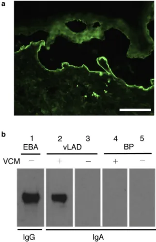
To further characterize the target of IgA autoantibodies in vLAD, we performed IIF with 1 mol/L NaCl-split skin (salt- split skin) as the substrate. A serum with 0.5 m g/ml of VCM showed IgA binding to the dermal side of the split skin (Figure 3a), indicating that the autoantibodies reacted with an autoantigen below the lamina lucida, such as COL7, which form the anchoring fibrils. We then performed immunoblot analysis using the recombinant noncollagenous (i.e., NC1) domain of COL7 to determine whether the serum bound to COL7 with or without 0.5 m g/ml of VCM. The serum showed a positive band of 150 kDa corresponding to the NC1 domain of COL7 only in the presence of VCM (Figure 3b). Furthermore, we examined serum reactivity against COL7 containing the mixture of the recombinant NC1 and NC2 domains by ELISA. VCM enhanced ELISA reactivity (optical density at 280 nm) of IgA against COL7 from 0.243 to 0.636 (2.617-fold increase) at 0.5 m g/ml concentration (Table 1, Case 1). The binding activity of circulating IgA to BMZ via IIF was lost in sera that were obtained after vLAD remission (Table 1, patient 1). However, ELISA detected the enhancement of IgA reactivity against COL7 mediated by VCM, from 0.031 to 0.125 (4-fold
Figure 2. Serum from a patient with vancomycin (VCM)-induced linear IgA bullous dermatosis (vLAD) showed IgA deposition along the basement membrane zone (BMZ) only in the presence of VCM.(a) The serum of a patient with vLAD did not show IgA binding to the skin on indirect immunofluorescence with healthy human skin as a substrate.
(b) Linear IgA deposition
along the BMZ was observed when VCM was added to the serum.
Scale bars¼50mm. (cee) IgA reactivity to the BMZ was dependent on the concentration of VCM.
VCM concentrations: (c) 0.1mg/ml, (d) 0.02mg/ml, and (e) 0.004mg/ml.
Scale bars¼50mm.
J Yamagami et al.
Vancomycin in Linear IgA Bullous Dermatosis
www.jidonline.org 1475
287
increase) (Table 1), suggesting that a particular repertoire of IgA that had transiently expanded during acute methicillin- resistant Staphylococcus aureus infection was modified by VCM. Taken together, IgA antibodies in this case of vLAD reacted with the NC1 domain of COL7, and autoreactivity was significantly enhanced by VCM.
COL7 is a major autoimmune target in vLAD
To extend these findings, we investigated sera from an addi- tional 13 patients with vLAD (Table 1). With the initial patient included, 12 out of the 14 patients were men, and their ages ranged from 38 to 84 (average ¼ 67) years. The onset of vLAD since the administration of VCM ranged from 4 to 16 days.
The diagnosis of vLAD in all patients was based on skin manifestations (erythema, blister, and erosion on the body), subepidermal blister formation with neutrophilic infiltration by histology, and linear IgA deposition along the BMZ by DIF.
Most patients were treated with systemic corticosteroid and/or dapsone, but in two patients vLAD resolved without any treatment other than the discontinuation of VCM. The disease severities varied from mild to severe, and the duration of treatment ranged from one to several weeks.
Although IIF using normal human skin as a substrate did not show IgA binding to the skin in any of the samples tested in the absence of VCM, 3 (21.4%) out of 14 serum samples displayed IgA reactivity to BMZ with spiked VCM concen- trations of 0.5 m g/ml. IIF with sodium-split skin showed that all three sera reacted with the dermal side of the skin in the presence of VCM (Table 1, patients 1, 7, and 12). We then tested the reactivity of vLAD sera to COL7 by ELISA. Only 4 of the 14 sera (28.6%) showed positive reactivity (optical density at 280 nm > 0.06) in the absence of VCM. In contrast, the addition of VCM resulted in positivity in 10 of the 14 sera (71.4%) (Table 1). Furthermore, the ratios of optical density values in the absence or presence of VCM increased in 10 of the 14 sera (71.4%) tested (P ¼ 0.009) (Table 1 and Figure 4). The higher rate of positivity by ELISA than by IIF likely reflects differences in the sensitivities of the two assays.
It is possible that VCM binds to and modifies the anti- genicity of COL7, rather than binding to IgA antibodies. To address this, we pretreated frozen human skin sections with 0.5 m g/ml of VCM or vehicle and then used these sections as substrates for IIF. However, incubation of VCM with the substrate did not result in IgA reactivity to BMZ with any of the three positive sera (data not shown). We also pretreated COL7-coated ELISA plates with 0.5 m g/ml of VCM, but IgA reactivity to COL7 was not observed (see Supplementary Table S1 online). These results suggest that VCM mediates IgA autoreactivity by modifying IgA rather than the antigen, COL7.
Several drugs other than VCM have been reported to cause drug-induced LAD, such as sulbactam/ampicillin and tazobactam/piperacillin, as well as teicoplanin, which belongs to the same class of glycopeptide antibiotics that share similar structures to VCM. To determine whether such drugs were also capable of mediating IgA reactivity to COL7, we co-incubated these drugs with vLAD sera and performed IIF and ELISA. However, IgA binding to the BMZ was not observed upon the addition of any of these drugs in IIF and ELISA, indicating that the acquired or enhanced reactivity of IgA to COL7 was mediated specifically by VCM in the tested cases (see Supplementary Table S2 online).
In aggregate, these findings identify COL7 as a major autoimmune target in vLAD and show that IgA antibody autoreactivity to COL7 was mediated by VCM.
DISCUSSION
Drug-induced autoimmunity is a well-recognized condition manifesting in a wide range of autoimmune diseases, but evidence for mechanisms by which drugs induce autoim- munity is scarce. Certain drugs such as hydralazine, mino- cycline, procainamide, and others have been associated with drug-induced lupus erythematosus. These drugs induce the production of autoantibodies, such as antinuclear anti- bodies, anti-single strand DNA antibody, and anti-histone
Figure 3. Identifying the target antigen of IgA autoantibody in vLAD.(a) The vLAD serum with 0.5mg/ml VCM showed binding of IgA to the dermal side in indirect immunofluorescence using 1 mol/L NaCl-split skin. Scale bar¼50mm. (b) Immunoblot analysis using the recombinant noncollagenous (NC1) domain of COL7. Lane 1: IgG reacting with the NC1 domain of COL7 was detected in serum from a patient with EBA (positive control). Lane 2: IgA reacting with the NC1 domain of COL7 in a vLAD serum was detected when VCM was added. Lane 3: The vLAD serum did not react with the NC1 domain of COL7 without VCM. Lanes 4 and 5: Serum from a patient with BP did not react with the NC1 domain of COL7 either with or without VCM (negative control). BP, bullous pemphigoid; COL7, collagen type VII; EBA, epidermolysis bullosa acquisita VCM, vancomycin;
vLAD, vancomycin-induced linear IgA bullous dermatosis.
J Yamagami et al.
Vancomycin in Linear IgA Bullous Dermatosis