A Study on Biomechanical Simulation of Human
Respiratory System with Finite Element Method
Sept. 2016
28
9
Zhang Guangzhi
CONTENTS
List of figure .. i
Acknowledgement v
Abstract vii
1 Introduction 1
1.1 Anatomy and biomechanical phenomena in human respiratory system ..1 1.2 Previous studies on modeling human respiratory system .. ....4 1.3 Approach and outline of this study . ....9
2 Action of intercostal muscle contraction on rib movement ..11
2.1 Method ...14
2.1.1 Finite element model of chest ..14
2.1.2 Transversely isotropic hyperelastic material
model for respiratory muscles . 16
2.1.3 Simulation condition 22
2.1.4 Simulation procedure ... 23
2.2 Results . 25
2.2.1 Rib movement generated by intercostal muscles in a single
interspace . 25
2.2.2 Deflection of ribs during muscle contraction .. 25 2.2.3 Rib movement generated by individual intercostal muscle . 26 2.2.4 Rib movement generated by entire inspiratory and expiratory
Intercostal muscles 38
2.3 Discussion 41
2.3.1 Reproduction of the theory of Hamberger ... 41 2.3.2 Relationship between rigid rotation and deformation of ribs ... ... 41 2.3.3 Action of intercostal muscle contraction on rib . 42 2.3.4 The net effect during entire intercostal muscle contraction .48
2.4 Conclusions ... ..51
3 Simulation of thorax deformation by activating intercostal muscles
and diaphragm .. . 53 3.1 Method .54 3.1.1 Simulation conditions ..54 3.1.2 Simulation procedure ... 55 3.2 Results . 57 3.2.1 Rib motion ... 57 3.2.2 Diaphragm motion . 64
3.2.3 Thorax deformation during normal quiet breathing and comparison
with 4D-CT images .. 69
3.3 Discussion 77
3.3.1 Rib motion ... 78
3.3.2 Diaphragm motion ... 80
3.4 Conclusions . 85
4 Simulation of lung deformation and ventilation .. 86
4.1 Method . 86
4.1.1 Porous hyperelastic material model . 86
4.1.2 Simulation conditions ..94
4.2 Results and discussion . 94
4.2.1 Lung deformation . 94
4.2.2 Ventilatory volume, alveolar and pleural pressures during
normal quiet breathing .. 96
4.4 Conclusions . 98
5 Influences of ventilation on blood circulation during cardiopulmonary
resuscitation ... 99
5.1 Method . 100
5.1.1 Connection of the circulatory system .. 100
5.1.2 Simulation procedure . 101
5.2 Results and discussion 102
5.2.1 Deformation of lung and heart 102 5.2.2 Open airway and occluded airway cardiopulmonary resuscitation 103 5.2.3 Open-occluded airway and occluded-open airway
cardiopulmonary resuscitation . 106
5.2.4 Occluded-open-occluded-open airway cardiopulmonary
resuscitation . 108
5.3 Conclusions ... 111
6 Conclusions ... 113
List of figure
Fig. 1-1 Locations of the lungs, ribs, intercostal muscles, diaphragm and pleural
space in human chest .. . 2
Fig. 1-2 Rib rotation axis and representative rib motion ... 2
Fig. 1-3 Locations of central tendon and muscles in the diaphragm . 3
Fig. 2-1 FEM model of the rib cage an 15
Fig. 2-2 FEM model of lungs and heart 15
Fig. 2-3 FEM model of diaphragm 15
Fig. 2-4 Fiber orientation of intercostal muscles measured by Loring .. 16
Fig. 2-5 Fiber orientation of diaphragm 16
Fig. 2-6 -element muscle model 18
Fig. 2-7 Active force-length relationship of the diaphragm and intercostal muscles
of an adult baboon 20
Fig. 2-8 The rib movements due to the contraction of the external and internal
interosseous intercostal muscles in one interspace locating between the
rib 5 and 6 26
Fig. 2-9 Different motions and structures of the upper and lower ribs . .. 27
Fig. 2-10 Portion 1, 2 and 3 divided according to the distribution of moment along
curved ribs studied by De Troyer et al. .. 28
Fig. 2-11 The fragments of the internal intercostal muscles 31
Fig. 2-12 The fragments of the external intercostal muscles 31
Fig. 2-13 The normalized displacement of the sternum generated by separate
fragments of the internal intercostal muscles 32
Fig. 2-14 The normalized displacement at lateral extremes of the ribs from rib 1 to 5
generated by separate fragments of the internal intercostal muscles 32
Fig. 2-15 The normalized displacement at lateral extremes of the ribs from rib 6 to 10
generated by separate fragments of the internal intercostal muscles 33
Fig. 2-16 The normalized displacement of the sternum generated by separate
fragments of the external intercostal muscles 33
Fig. 2-17 The normalized displacement at lateral extremes of the ribs from rib 1 to 5
Fig. 2-18 The normalized displacement at lateral extremes of the ribs from rib 6 to 10
generated by separate fragments of the external intercostal muscles 34
Fig. 2-19 Rib motion caused by fragment 4 of external intercostal muscles
in portion 1 35
Fig. 2-20 Rib motion caused by fragment 3 of internal interosseous intercostal
muscles in portion 1 35
Fig. 2-21 Rib motion caused by fragment 7 of external intercostal muscles
in portion 2 36
Fig. 2-22 Rib motion caused by fragment 7 of internal intercostal muscles
in portion 2 36
Fig. 2-23 Rib motion caused by fragment 8 of internal intercostal muscles
in portion 3 37
Fig. 2-24 The front view of the inspiratory chest movement generated by
the inspiratory 39
Fig. 2-25 The lateral view of the inspiratory chest movement generated by
the inspiratory 39
Fig. 2-26 The front view of the inspiratory chest movement generated by
the expiratory 40
Fig. 2-27 The lateral view of the inspiratory chest movement generated by
the expiratory 40
Fig. 2-28 Mechanical actions of the intercostal muscles in portion 2 and 3
and simulated deformation results 47
Fig. 2-29 Deformation shift direction due to component going along the ribs
of the muscle force and net effect direction due to the component
normal 48
Fig. 3-1 Time dependence of activation of the diaphragm and
intercostal muscles 55
Fig. 3-2 Front view of inspiratory rib motion generated by the
intercostal muscles 58
Fig. 3-3 Rear view of inspiratory rib motion generated by the
intercostal muscles 58
Fig. 3-4 Lateral view of inspiratory rib motion generated by the
intercostal muscles 59
Fig. 3-5 Front view of expiratory rib motion generated by the
intercostal muscles 59
intercostal muscles 60
Fig. 3-7 Lateral view of expiratory rib motion generated by the
intercostal muscles 60
Fig. 3-8 Comparison of simulated and experimental bucket-handle angle
during inspiratory chest movement . 62
Fig. 3-9 Comparison of simulated and experimental pump-handle angle
during inspiratory chest movement 62
Fig. 3-10 Simulated bucket-handle angle during expiratory chest movement 63
Fig. 3-11 Simulated pump-handle angle during expiratory chest movement 63
Fig. 3-12 Clinical observation of the diaphragm motion and chest movement
on the sagittal plane in MRI scans 65
Fig. 3-13 Simulation results of the right hemidiaphragm for comparison
with the clinical observation . 66
Fig. 3-14 Front view of the diaphragm deformation 66
Fig. 3-15 Rear view of the diaphragm deformation 67
Fig. 3-16 Motion of the rib cage caused by isolated contraction of the diaphragm 67
Fig. 3-17 Distribution of muscle forces based on the radial fibre orientation
in the diaphragm ..68
Fig. 3-18 Simulation results of the right hemidiaphragm from simultaneous
activation of the diaphragm and inspiratory intercostal muscles for
comparison with the clinical observation 68
Fig. 3-19 The front view of obtained thorax deformation 71
Fig. 3-20 The lateral view of obtained thorax deformation 71
Fig. 3-21 The oblique view of obtained thorax deformation 72
Fig. 3-22 Location of planes X and Z in the chest 72
Fig. 3-23 CT images on plane X 73
Fig. 3-24 Extracted motion of plane X from CT images 73
Fig. 3-25 The simulation results on plane X 74
Fig. 3-26 CT images on plane Z 74
Fig. 3-27 Extracted motion of plane Z from CT images 75
Fig. 3-28 The simulation results on plane Z 75
Fig. 3-29 Movement at location L1 on plane Z compared with 4D-CT images 76
Fig. 3-31 Movement at location L3 on plane Z compared with 4D-CT images 77
Fig. 4-1 Front view of lung deformation during normal quiet breathing 95
Fig. 4-2 Lateral view of lung deformation during normal quiet breathing 96
Fig. 4-3 Variations of alveolar, pleural pressures and lung volume change
during normal quiet breathing respiration 96
Fig. 5-1 The main and lung circulation, and preload and afterload in human body .101 Fig. 5-2 Circuit of the preload and afterload connected to the FEM model
of the heart 101
Fig. 5-3 Deformation results of the thorax 102
Fig. 5-4 Deformation results of the lung 103
Fig. 5-5 Deformation results of the heart .103
Fig. 5-6 Heart volume change during open and occluded airway CPR .. 104
Fig. 5-7 Pleural pressure during open and occluded airway CPR 105
Fig. 5-8 Pressure in the heart during open and occluded airway CPR 105
Fig. 5-9 Heart volume change during occluded-open and open-occluded
airway CPR 106
Fig. 5-10 Pleural pressure during occluded-open and open-occluded
airway CPR 107
Fig. 5-11 Pressure in the heart during occluded-open and open-occluded
airway CPR 107
Fig. 5-12 The displacement of the sternum in dorsal direction during
occluded-open-occluded-open airway CPR ...109
Fig. 5-13 Heart volume change during occluded-open-occluded-open airway CPR 110 Fig. 5-14 Pleural pressure during occluded-open-occluded-open airway CPR 110
Acknowledgement
I would like to personally thank the following people who supported me during writing this thesis.
First I would like to express my deepest appreciation to my supervisor, Professor Xian CHEN. Thank you for your time and patience for the discussion of my research, careful explanation of mechanical problems, and critical review of my work. Thank you for all the care for me all the time. I also thank the examiner committee, Associate Professor Junji OHGI, Professor Takashi SAITO, Associate Professor Koji MORI, Professor Shoji KIDO, Associate Professor Yasushi HIRANO for their suggestions and reviews during the examination.
Moreover, I would like to thank to Professor Toshiaki HISADA for his valuable advice and support. And I also appreciate the Doctor Seiryo SUGIURA for his helpful advice of my research, Doctor Toshiro MIURA, Radiological Technologist Akira NAKAMOTO and Radiological Technologist Chikanori MATSUMURA for the supply of medical data and review comments.
I really appreciate all of members in Medical Mechanical engineering laboratory Yamaguchi University Japan, thank you for giving me a very pleasant researching and study environment.
I extend special thanks to Dr. S. H. Loring, (Harvard Medical School, USA) for the experimental data on intercostal muscle orientation under NIH grant HL33009. This research was supported by the Japan Society for the Promotion of Science (JSPS) through the Funding Program for World-Leading Innovative R&D on Science and Technology (FIRST
Program) initiated by the Council for Science and Technology Policy (CSTP).
Finally, I really want to appreciate my father Juming Zhang and mother Fengxia Wang for bringing me up and 10 years support during study abroad in Japan.
Guangzhi ZHANG
Abstract
The human respiratory system has a complex structure that stretches from the trachea to the alveoli and involves many physiological phenomena such as the airflow, lung deformation, oxygen exchange, pleural pressure distribution, alveolar pressure distribution, respiratory muscle activation, and chest movement. Unfortunately, many respiratory diseases such as chronic obstructive pulmonary disease (COPD), pneumothorax, diaphragmatic paralysis, and lung cancer adversely affect this system. The biomechanical simulation of human respiratory system is expected to be a useful tool for diagnosis and treatment of respiratory diseases. This work aims to establish a computational model in order to reproduce the biomechanical phenomena happened during human breathing. In order to reproduce a reasonable mechanical environment inside the chest during breathing, we focus on activating the respiratory muscles to generate respiratory chest movements in terms of the actual physiological activity during breathing from a biomechanical point of view. According to pathological needs on diagnosis and treatment of respiratory diseases, the basic physiological phenomena during breathing are firstly essential to be reproduced by the computational model, such as the rib motion, diaphragm motion, lung deformation, ventilation, alveolar pressure and plural pressure inside the chest.
During human breathing, the biomechanical phenomena inside the thorax are determined by the thorax deformation, therefore at first we simulated the thorax deformation. The thorax deformation consists of rib motion driven by the contraction of the intercostal muscles and diaphragm motion.
Hence, in order to provide a reasonable mechanical environment for the organs inside the chest, the mechanical action of the intercostal muscles on the rib cage was firstly investigated, that what rib motion can be generated by the contraction of the intercostal muscles and why these motions are happened. After that the thorax deformation was reproduced by considering the diaphragm motion additionally. To reproduce the muscle contractions, we introduced the Hill-type transversely isotropic hyperelastic continuum skeletal muscle model, which allows the intercostal muscles and diaphragm to contract along the muscle fiber directions with clinically measurable muscle activation and active force-length relationship. The anatomical fiber orientations of the intercostal muscles and diaphragm were introduced. We first reproduced the representative motions of the rib and diaphragm in physiology. Afterwards, by activating the diaphragm and intercostal muscles simultaneously, the thorax deformation was simulated. Furthermore, the deformation results were compared with four-dimensional computed tomography (4D-CT) images for validation.
After reproduced the thorax deformation, in order to simulate the lung deformation, ventilation and pressures inside the chest during breathing, we modeled the lungs as porous hyperelastic material and established the respiratory model. The porous hyperelastic material can macroscopically reproduce the lung deformation and the airflow during ventilation simultaneously by considering the permeability between the lung parenchyma and air flow. Consequently, the variations of lung volume change, alveolar and pleural pressures during respiration were also reproduced and have a good consistence with the clinical observation.
The ventilation can affect the mechanical environment in the chest. With this established respiratory model, as an application, we additionally investigated influences of the ventilation on blood circulation during cardiopulmonary resuscitation (CPR). In order to reproduce the heart volume change due to inflow and outflow of blood inside the heart, we considered the blood inside the heart as the porous hyperelastic material and connected preload and afterload circuits representing the blood circulatory system. From the simulation results of the pleural and the cardiac chamber pressure and the volume change of the heart, we found the occluded airway system could promote the blood flow in the heart during CPR, as obtained in the experiments performed on domestic swine. Based on this, we also investigated that an occluded-open-occluded-open airway process could better increase flow rate of the blood to promote the efficiency of CPR.
Introduction
1.1 Anatomy and biomechanical phenomena in human
respiratory system[1], [2]
During human breathing, the lungs are passive tissues and expanded by the thorax deformation. The thorax deformation is provided by the contraction of respiratory muscles. The main respiratory muscles are the intercostal muscles and the diaphragm. The intercostal muscles are inserted between the ribs consist of two muscle layers, which are the external and internal intercostal muscles. The diaphragm is located between the thoracic and abdominal cavity, as shown in Fig. 1-1. During normal breathing, the external intercostal muscles and diaphragm contract simultaneously to provide an increase of chest circumference anteroposteriorly and transversally and descend the bottom of the lungs inferiorly. Meanwhile, negative pleural pressure is generated in the pleural space, which is filled with incompressible fluid to provide lubrication for the sliding between the lungs and the thorax. Therefore, the lungs could be expanded and slide against the chest wall during breathing.
The rib motion and the diaphragm deformation determine the lung deformation during breathing. The rib motion is generated by the contraction of the intercostal muscles, and the direction of rib rotation is determined by the rib rotation axis consisting of two vertebral joints (costovertebral and costotransverse joints). Fig. 1-2 A and B illustrate the
location of the rib rotation axis for the upper and lower ribs. The directions of rib rotation axis are in the lateral directions for the upper ribs, and in the dorsal-ventral directions for the lower ribs. Therefore, when the intercostal
Fig. 1-1 Locations of the lungs, ribs, intercostal muscles, diaphragm and pleural space in human chest.
muscles contract, the upper and lower ribs perform pump-handle and bucket-handle motions, respectively, as show in Fig. 1-2 A and B. The pump-handle and bucket-handle motions make the upper lungs be expanded anteroposteriorly and lower lungs be expanded transversally, respectively.
Rib rotation axis
A
B
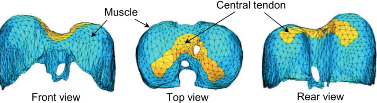
Central tendon Muscle
Front view Top view Rear view
Fig. 1-3 Locations of central tendon and muscles in the diaphragm
On the other hand, the diaphragm expands the lungs in supero-inferior direction. It consists of two domes and the right dome is located more superior than the left dome. As Fig. 1-3 illustrates, the diaphragm is constituted of a central tendon and muscle fibers [3]. The muscle fibers are connected with the lower rib cage. Therefore, the contraction of the muscle fibers lowers the central tendon during breathing as well as pulls the lower ribs along the muscle fiber directions of the diaphragm. Because the intercostal muscles also act on the ribs, hence there is an interaction between diaphragm and intercostal muscles during breathing. Thereby, it is significant that the action of intercostal muscle contraction on rib movement, the diaphragm deformation and the interaction between the intercostal muscles and the diaphragm dominate the thorax deformation as well as the lung deformation. Consequently, the respiratory muscle contraction is fundamental biomechanical phenomenon which leads to a series of physiological and biomechanical phenomena during breathing exercise, such as the rib motion, diaphragm motion, lung deformation, ventilation, alveolar pressure and plural pressure inside the chest.
1.2 Previous studies on modeling human respiratory system
A computational model of the human respiratory system would be a valuable aid to the diagnosis and treatment of the respiratory diseases and elucidation of physiological problems of the respiratory system. For example, chronic obstructive pulmonary disease (COPD) is characterized by the presence of airflow limitation, which is assessed by spirometry test or medical images such as multi detector-row computed tomography (MDCT) [4] in the diagnosis processes. However, the former does not provide information inside of the lung and the later only represents the morphologic feature without the airflow details. Computational models have been proposed to investigate the relationship between imaging measurements and disease severity for developing improved diagnostic methods [5]. On the other hand, computational fluid dynamic analyses have been performed to simulate pharmaceutical aerosol delivery by predicting the site of pharmaceutical aerosol deposition within the airways in the inhaled medicine for treating respiratory diseases [6]. Furthermore, lung deformation simulations have been applied to the radiation therapy for accurate prediction of the lung tumour motion due to breathing [7]. Moreover, the patients with pectus excavatum easily become short of breath and tire during exercise, and the cardiorespiratory function could be significantly improved after surgery [8]. However, the postoperative pain and other effects after surgery also puzzle the doctors in clinical. For that reason, the finite element analyses of rib cage were performed to elucidate stress distribution on the rib cage [9] and dynamic effects on the spine with asymmetric thoraces [10] after Nuss procedure.
The extreme goal of this research is to build a biomechanical computational model of respiratory system coupling the airflow in the bronchial system with the lung deformation for the diagnosis and treatment of the respiratory disease. To that end, it is first essential to develop an approach to simulating thorax deformation produced by respiratory muscle contraction as well as reproducing the ventilation and lung deformation in terms of the actual physiological activity during breathing from a biomechanical point of view.
Regarding the previous studies of modeling human respiratory system with biomechanical computational approach, because of the breathing exercise consists of the biomechanical phenomena: respiratory muscle contraction, thorax deformation, lung deformation and ventilation, so that some studies concentrated on revealing the respiratory muscles action [11] [15], and some studies focused on reproducing the thorax deformation during breathing[16] [23], furthermore some studies investigated the approaches to simulate the lung deformation [24] [26], the air flow and ventilation [27] [34].
The intercostal muscles and diaphragm are the main muscle during human breathing. Loring investigated the action of the intercostal muscles by modeling the intercostal muscles and rib cage with beam elements. Base on the anatomic and mechanical measurements in dogs and human cadavers, the canine [12] and human [11] FEM model were constructed. The muscles forces were applied along the muscle fiber directions in each interspace of ribs. Loring demonstrated the two dimensional geometric model of Hamberger, that the external and internal intercostal muscles can cause inspiratory and expiratory rib movements. In addition, with this three
dimensional FEM model, Loring found the intercostal muscles at the side of the rib cage generate the anteroposterior chest motion and the muscles at the front and back of the rib cage produce the transverse chest motion during human breathing. The diaphragm action is directly linked to the respiration. Because the complex geometric structure of the diaphragm, the diaphragm was modeled using three dimensional continuum approach with tetrahedral [17] and shell [13], [14] finite elements. Ladjal et al. considered the diaphragm as elastic and compressible tissues, and the Young's modulus and Poisson's ratio were applied in this study [17]. For the muscle contraction force, external forces were applied on the surfaces of the tetrahedral meshes. Simulated deformations of cross-sections of the diaphragm were compared with the clinical data observed in CT images for validation. Although obtained diaphragm movement was consistent with the observation in CT images with a small diaphragm movement, the linear elastic constitutive law is just suitable for small displacement problem, and the diaphragm always perform large movement during breathing exercise. Pato et al. simulated the diaphragm motion by using non-linear constitutive law and considering the muscle fibers as incompressible transverse isotropy hyperelastic tissues [13], [14]. These studies also introduced
three-element skeletal muscle model into the constitutive law to make the muscle contract along the fiber directions measured from a female cadaver. Unfortunately, obtained diaphragm motion was not confronted with clinical data for validation. More importantly, not only the diaphragm, the intercostal muscles also participate the breathing exercise and generate rib movement, furthermore the rib movement provide a boundary condition for simulating the diaphragm motion. Regrettably, the influence from the rib
cage was not been considered in the studies mentioned above [13], [14],[17].
Indeed, DiMarco et al. have demonstrated that there is a synergism between the contraction of the diaphragm and intercostal muscles [35]. The intercostal muscle could decrease the shortening of the diaphragm muscle fibers via the rib movements to make the diaphragm generate greater muscle force to resist the abdominal and pleural pressures during breathing. Therefore, the diaphragm and rib motion should be considered and reproduced simultaneously, thereby the thorax deformation is obtained. Michel et al. simulated the thorax deformation using a hexahedral finite element model of the upper human body, which developed for rad safety [16]. The rib cage, lung heart, diaphragm and abdominal tissues were included in this FEM model and meshed independently. The muscle force was applied with beam element along the muscle fiber direction for the intercostal muscles and diaphragm. They simulated the thorax deformation during a respiratory cycle from functional residual capacity to total lung capacity. Unfortunately, they did not used a continuum model for the muscle contractions especially for the diaphragm, and they did not give details of the lung deformation and discuss the mechanical environment during the lung deformation. With a series of investigations, Ladjal et al. proposed an approach to simulating the thorax deformation including the rib movement and the diaphragm motion [17] [21]. They first developed an approach to simulate the rib movements by considering the ribs as rigid bodies which doing rigid rotation around the around the spinal joints. In addition, the displacement caused by the rib rigid rotation was applied as the displacement boundary condition in order to simulate the thorax
deformation. The diaphragm deformation was simulated by applying pressures on the surface of diaphragm meshes. With experiments [36] and simulations [11], [12], it is demonstrated that the ribs could be easily bent by the contraction of the intercostal muscles and the rib cage could store the strain energy during inhaling process to assist the expiration, therefore it is not appropriate to consider the ribs as pure rigid bodies in order to simulate the thorax deformation.
Reproducing the thorax deformation could provide a boundary condition for the lung simulation, and the organ playing the most important role during respiration is the lung. Hence, lots of studies directly focused on constructing a computational mechanical model for the lung. Some studies simulated pulmonary air flow with computational fluid dynamics (CFD) method in order to understand the etiology of lung pathology to improve drug delivery methods [6], [31]. However, they did not consider the influence from the lung deformation. On the other hands, the lung deformation simulating approaches also have been investigated to predict the lung tumour movement during radiation therapy [21], [24] [26]. In these studies, the lung was considered as an isotropic elastic or hyperelastic tissue with a displacement or pressure boundary condition instead of the respiratory muscle contraction. Therefore, the simulation results were mainly concentrated on the lung deformation without any discussion of the mechanical environment inside or outside the lung. Conversely, revealing the relationship between the mechanical environment, airflow inside the lung and the lung deformation has a wider range of applications in clinical, such as contributions to the improvement of mechanical ventilation method or development of advanced diagnostic and treating methods of COPD [27].
In order to consider the lung deformation and the air conduction simultaneously, some studies concentrated on simulating the coupling between the air conducting part (the trachea and bronchi) and the respiratory part (the parenchymal lung tissue) of the respiratory system [27] [29]. This method requires very fine meshes and therefore need large-scale high-performance computing platforms and scalable parallel solvers to calculate the coupled system. Detailed information such as the pressure at the end of the airway tree and the deformation of surrounding tissues during breathing could be obtained. During breathing exercise, the airflow inside the lung is mainly influenced by the lung deformation, in addition the lung deformation is determined by the thorax deformation due to the respiratory muscle contraction. Therefore, in order to reproduce reasonable biomechanical environment for the lung behavior during human breathing, the respiratory muscle contraction and thorax deformation cannot be ignored. Unfortunately, these studies did not take account of the respiratory muscles and the thorax deformation, moreover there has no study simulated the human breathing by activating the respiratory muscles to reproduce a series of physiological and biomechanical phenomena during breathing exercise, range from the rib motion and diaphragm motion to the mechanical environment inside the chest, in terms of the actual physiological activity during breathing from a biomechanical point of view.
1.2 Approach and outline of this study
In order to establish a computational mechanics model of the human respiratory system, this work attaches importance to the respiratory muscle contraction that is relationship between the respiratory muscle contraction
and the thorax deformation, and an important foundation for the simulation of the lung behaviour. Due to the complicated structure of the rib cage, we first investigates the detailed action of the intercostal muscles on the rib movements in chapter 2 and the diaphragm motion in chapter 3, thereby the thorax deformation is reproduced. After that, in chapter 4, the lung deformation and the ventilation is simulated by introducing porous hyperelastic material model. This approach let us obtained the alveolar pressure and pleural pressure generating the mechanical environment inside and outside the lungs during breathing. By the way, concerning the influence from the bronchial tree on the lung deformation, it is demonstrated that the stiffness property of the bronchial tree has no significant effect on simulating the lung deformation with finite element method [26] and the resistance happened in the bronchial tree is relatively small in the normal cases, therefore at current stage we do not introduce the bronchial tree provisionally. During inspiration, decreasing of the pleural pressure expands the lung to perform ventilation, and conversely the ventilation can also affect the mechanical environment in the chest. In chapter 5, established respiratory model is further used to investigate influence of the ventilation on the blood circulation during cardiopulmonary resuscitation (CPR) by additionally connecting blood circulatory system to the mesh of heart. According to the simulation results, we also propose a more efficient way to perform CPR, which could make more blood flow out of the heart during CPR.
Action of intercostal muscle
contraction on rib movement
The intercostal muscles are the predominant muscle generating the chest movement during human breathing. In order to model the human breathing, it is essential to understand the action of the intercostal muscles on generating the rib motion. The action of the intercostal muscles has been a subject in physiology for a long time. Unfortunately, there are also some controversies about the action of the intercostal muscle. Some studies described the intercostal muscles have respiratory actions [37] [39], [36], [40], and some investigations agreed with the intercostal muscles mainly contribute to the trunk motion [41] [43]. Recently, investigating the action of the intercostal muscles has been divided into two steps, (1) is to find out the mechanical action of the inspiratory muscles that is the relationship between the muscle contraction and the rib displacement, and (2) is to confirm the connections between the rib displacement and the respiratory effect [37], [40]. Step (2) could be obtained by applying external forces or displacements to each rib and measuring the airway pressure [37], [40]. However, because it is almost impossible to selectively activate the individual muscle groups as confessed by Wilson and De Troyer [39], step (1) has not been able to be directly obtained. Therefore, in order to understand the function of the intercostal muscles, the problem is to investigate the mechanical action of the inspiratory muscles.
The rib motions generated by the intercostal muscle contractions have just been roughly considered as doing rigid rotation around the spinal joints by far. It had been first explained by the theory of Hamberger over two centuries [44] that attributed the rib motion to the moment about spinal joints resulted from the contraction of intercostal muscles. Furthermore, this 2-dimensional theory was extended to describing 3-dimensional curved ribs by De Troyer [37]. This extended Hamberger theory involves the moment distribution on curved ribs for rib rigid rotation around the spinal joints. However, the experiments also illustrated that the ribs and especially the costal cartilages could not only rigidly rotate but also deform to restore the strain energy for expiration during breathing [45], and two adjacent ribs could also be bent toward to each other by the muscle forces of the intercostal muscles [36]. It implied that the cartilage and the ribs could be easily deformed under the intercostal muscle forces. Hence, in order to find out the relationship between the muscle contraction and the rib displacement, it is necessary to consider not only the rigid rotation of the rib cage but also the issue deformation in the rib cage happened during intercostal muscle contraction.
As described by Wilson and De Troyer, the complicated structure of the rib cage causes complex relation between the muscle force and the bone displacement throughout the rib cage [39]. For such a complicated structure of the human rib cage, finite element analysis (FEA) is a useful tool to qualitatively study the rib displacement and investigate the mechanism during intercostal muscle contraction. By using FEA, Loring inferred the rib movement generated by the internal intercostal muscles, external intercostal muscles, parasternal intercostal muscles and levator costae
muscles in the rib cages of human [12] and dogs [11], and explained the obtained rib motion with the original 2-dimensional Hamberger theory. Unfortunately, Loring had not considered the influence from the tissue deformation and distribution of the moment during the intercostal muscles contraction.
On the other hands, by applying external force on the ribs, the experiments resulted a hypothesis that the compliances of the ribs could determine the rib displacement generated by the intercostal muscle contraction, that in the end-expiratory lung volume the ribs could be displaced cranially more easily than caudally whether by the external or internal intercostal muscles [36]. It also means that the mechanical action of the intercostal muscles could also be covered and influenced by the compliances of the ribs. Unfortunately, there has been no study that selectively activates the intercostal muscles and simultaneously eliminates the compliance of the costovertebral and interchondral joints during experiments.
This study arms to investigate the mechanical action of the intercostal muscles that what rib motion can be generated by the contraction of the intercostal muscles and why these motions are happened. By using FEA, we consider not only the rigid rib rotation but also the rib and costal cartilage deformation and simultaneously eliminate the influence from the compliance of the costovertebral and interchondral joints during the intercostal muscle contraction. Considering the tissue deformation additionally lets us understand why the soft costal cartilages gradually run obliquely in the caudal direction and keep a right angle with the ribs in human trunk.
2.1 Methods
2.1.1 Finite element model of chest
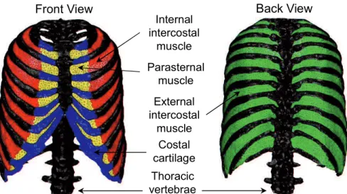
Based on a voxel data set of the chest which was segmented from CT slices of a male volunteer to be used in the simulation of the electrophysiological activity of the heart [46], a 3D reconstruction of the sternum, rib, vertebrae, intercostal muscles, diaphragm, heart and lungs was performed and rendered to form STL format data. Since it is difficult to segment the intercostal muscles from CT slices, the 3D models of the intercostal muscles and diaphragm were constructed by referencing an anatomy textbook [47]. The finite element meshes shown in Fig. 2-1, Fig. 2-2, and Fig. 2-3, consisting of 424,304 tetrahedra elements and 92,841 nodes, were then generated from the 3D models using mesh generation software ANSYS ICEM CFD (ANSYS, Inc.).
The fiber direction of the intercostal muscles was assigned based on human cadaver data obtained from Loring (personal communications, 2012), as shown in Fig. 2-4. For the diaphragm, we referenced an anatomy textbook [47] to determine the fiber direction, as shown in Fig 2-5. We assumed that the fiber distribution had a radial pattern from the central tendon to the bottom edge.
External intercostal muscle Internal intercostal muscle Parasternal muscle Thoracic vertebrae Costal cartilage
Front View Back View
Fig. 2-1 FEM model of the rib cage and intercostal muscles.
A
B
Fig. 2-2 FEM model of lungs and heart A: lungs. B: heart.
Central tendon Muscle
Front view Top view Rear view
Internal intercostal External intercostal Parasternal
Fig. 2-4 Fiber orientation of intercostal muscles measured by Loring [11].
Fig. 2-5 Fiber orientation of diaphragm
2.1.2 Transversely isotropic hyperelastic material model for
respiratory muscles
The intercostal muscles and diaphragm were considered to be incompressible hyperelastic materials. The function UV detC 1 represents the incompressible constraint and is enforced by the Lagrange multiplier , where detC is the determinant of the right Cauchy-Green
tensor C . To exclude the effect of the volume-changing deformation, the strain energy U was assumed to depend on the first reduced invariant of R
C . This was defined as 2/3
1 tr
C
I J C, where J detF denotes the
determinant of the deformation gradient F. By denoting t as the external conservative force on the Neumann boundary, the total potential energy of the muscles can be formulated as a function of the displacement u and Lagrange multiplier , as shown in equation (2.1):
, URd UVd dS
u t u . (2.1)
Given the principle of stationary potential energy, the variation in is equal to zero: d d dS 0 R V V U U U E E t u E E , (2.2)
where E is the Green-Lagrange strain tensor. Thus, the weak form of the governing equation is as follows:
d dS 0 d 0 R V V U U U E t u E E . (2.3)
The Lagrange multiplier corresponds to the hydrostatic pressure p by 2
p [48]. In this study, the strain energy function U proposed by R
-element muscle model to three dimensions. As shown in Fig. 2-6, the parallel element (PE) and series element (SE) are nonlinear springs which represent passive behaviour. The contractile element (CE) produces a contractile force when the muscle is excited.
The muscle force F can be assumed to be the sum of the forces in the PE and CE or SE:
SE CE
PE
F F
Fig. 2-6 -element muscle model
PE SE PE CE
F F F F F . (2.4)
Then, the nominal stress along the fibre direction T can be written as:
PE SE PE CE
T T T T T . (2.5)
The stress T is defined as follows: PE
PE M
0 PE f
2 1 PE 2 1 ,for 1 0 ,otherwise f a f f f aA e f (2.7)
where f is the stretch ratio in the fibre direction given by
1/2 2/3 :
f J C N N . N is the initial direction of the muscle fibre,
M 0
T denotes the maximum muscle nominal stress determining the
maximum muscle contraction force, a and A are material constants [50], and fPE f is the passive force-length relationship of the parallel
element PE [50]. The stress T is given by CE
CE M
0 CE f t
T T f , (2.8)
where m 0 to 1,
represents the time dependence of the activation and for which the maximum value can be 1, and fCE f is the active force-length
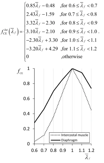
relationship of the contractile element CE. In this study, this was considered to be the active force-length relationship for the muscle tissue. Fig 3 shows the experimental data for the active force-length relationship of the intercostal muscles and diaphragm as measured from an adult baboon [51].
According to the experimental data, fCE f of the intercostal muscles
CE 0.85 0.48 ,for 0.6 0.7 2.43 1.59 ,for 0.7 0.8 3.32 2.30 ,for 0.8 0.9 3.10 2.10 ,for 0.9 1.0 2.30 3.30 ,for 1.0 1.1 3.20 4.29 ,for 1.1 1.2 0 f f f f f f Int f f f f f f f f ,otherwise . (2.9) f CE
f
Fig. 2-7 Active force-length relationship fCE f of the diaphragm and
intercostal muscles of an adult baboon
CE 1.80 0.80 ,for 0.6 0.7 2.37 1.20 ,for 0.7 0.8 1.61 0.59 ,for 0.8 0.9 1.42 0.42 ,for 0.9 1.0 0.55 1.55 ,for 1.0 1.1 2.11 3.27 ,for 1.1 1.2 0 f f f f f f Dia f f f f f f f f ,otherwise . (2.10)
The muscles were modelled as incompressible transversely isotropic hyperelastic materials for which the strain energy function can be written as follows: 1 , , C f R I f U U I U (2.11) 1C 3 1 b I I U ce (2.12) PE CE , , , , f f f f U U U (2.13) M PE 0 1 PE d f f U T f (2.14) M CE , , 0 1 CE t d f f U T f , (2.15)
where UI and U are the strain energies stored in the isotropic f
hyperelastic matrix and muscle fibre, respectively, and c and b are material constants. For an incompressible hyperelastic material, by
denoting p as the hydrostatic pressure, the second Piola-Kirchhoff stress tensor S can be derived as follows:
1 1 1 1 1 3 1 1 1 1 M 1 0 PE CE + + 2 + 2 3 1 t 3 C C f f R I C f b I C f f f f U U U I p p I p bce I T f f S C C E E E C I C N N C . (2.16) The material parameters used for the intercostal muscles and the diaphragm were M
0
T = 0.3 MPa [51], c = 0.009348 MPa, and b = 1.4939 MPa
[52]. The Lamé parameters and of the isotropic elastic model were 569.23 and 853.85 MPa, respectively for bone [11]; 65.38 and 98.08 MPa, respectively, for cartilage [11]; and 5.18 and 10.05 MPa, respectively, for tendons [13].
2.1.3 Simulation condition
The muscles activation were set at 0.5 ( 1 0 . 0 5. ) to make the
muscle forces be equivalent to approximately half of the maximal absolute force at optimal muscle length. The fiber direction of the intercostal muscles was assigned based on the human cadaver data from Loring [11], as shown in Fig. 2. All the degrees of freedom at the bottom of vertebra were fixed as displacement boundary condition, and the simulations were performed with a nonlinear finite element analysis program developed in-house.
In order to investigate how the intercostal muscle contraction moves the rib cage, we did not consider the influence from the other muscles in the
human trunk. This is because all of the respiratory muscles are inserted and acts on the rib cage, therefore the entire effect could be considered as the summation of those individual muscles. Moreover, because the compliance of ribs depends on the lung volume during breathing and influences the rib displacement generated by the intercostal muscle contraction [36], the compliance of the costovertebral joint was set at relatively small in order to eliminate the influence from the compliance of the costovertebral and interchondral joints and model a neutral position of the rib cage during breathing.
2.1.4 Simulation procedure
The theory of Hamberger has been used to explained rib motion for over two centuries and generally accepted. Recently, because this theory, however, had been questioned due to the experiments of measuring rib compliance with external forces [36] which indicated the ribs could move cephalad more easily than caudad at end-expiratory lung volume and hypothesized that different rib moving directions are mainly caused by the distribution of the rib compliance in cranial and caudal direction throughout the rib cage, and not mainly relate to the torques around the spinal joints generated by the different fiber orientation of internal and external intercostal muscles as described in the theory of Hamberger. Therefore, we activated the intercostal muscles in a single interspace located in the middle of the rib cage in order to obtain the rigid rib rotation as described in the 2-dementional model by Hamberger.
Obtained rib rigid rotation is generated by the moment around the spinal joints due to the different fiber directions of the internal and external
intercostal muscles. Therefore, we virtually changed the fiber direction of the intercostal muscles to along the craniocaudal direction that the moment for rib rotation becomes zero to obtain the relationship between the rigid rotation and deformation of the ribs under the contracting forces of the intercostal muscles.
As De Troyer et al. [37] pointed out, the moment for rib rotation distributes along curved adjacent two ribs from dorsal to ventral portion of the rib cage and its direction also revised around the costochondral articulations. Therefore, from obtained relationship of between the rigid rotation and deformation of the ribs, the rib deformations caused by different intercostal muscles along the curved ribs also could be critical. For that reason, we individually activated the intercostal muscle groups from dorsal to ventral portion of the rib cage, and explained the mechanisms when the ribs were simultaneously rotated and deformed by different intercostal muscles throughout the rib cage.
After confirmed the partial mechanical action of the intercostal muscles, in order to investigate the net effect for entire intercostal muscles, we activated entire inspiratory and expiratory intercostal muscles and reproduced inspiratory and expiratory chest movements as observed clinically.
2.2 Results
2.2.1 Rib movement generated by intercostal muscles in a
single interspace
The external and internal interosseous intercostal muscles in the interspace between rib 5 and 6 were activated. As the simulation results shown in Fig. 2-8-A, three points a, b and c in the rib cage were plotted for the comparison of the rib movement. Point a is at the middle of the sternum, point b and c locates at the lateral extremes of ribs 5 and 6. If the cranial direction is positive, the cranial displacement of point a was 0.2mm and points b and c were -0.3mm and 1.0mm, respectively, for the contraction of external intercostal muscles. Fig. 2-8-B shows the deformation for the contraction of internal interosseous intercostal muscles, the cranial displacement of point a was -0.3mm and points b and c were -0.8mm and 0.3mm, respectively. The results shows the external and internal intercostal muscles could rotate the sternum and ribs cranially and caudally, respectively.
2.2.2 Deflection of ribs during muscle contraction
When the moment for rib rotation become zero, as expected, we obtained nearly the same two deformation results for the external and internal interosseous intercostal muscles, and one of them is shown in Fig. 2-8-C. The cranial displacement of point a was 0.05mm and points b and c were -1.0mm and 2.0mm, respectively. The adjacent two ribs 5 and 6 were prominent bent and moved more close to each other than during the contraction of external and internal intercostal muscles.
A
B
C
a
: Plotted points
b
c
Fig. 2-8 The rib movements due to the contraction of the external and
internal interosseous intercostal muscles in one interspace locating between the rib 5 and 6 (Amplification factor 3.0; before muscle contraction: blue; after muscle contraction: orange). A: The lateral view of the rib movement caused by the external intercostal muscles. B: The lateral view of the rib movement caused by the internal interosseous intercostal muscles. C: The lateral view of the rib movement when the muscle fibers were artificially oriented in craniocaudal direction.
2.2.3 Rib movement generated by individual intercostal
muscle
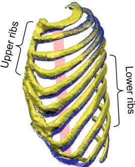
The rib rotation axes determine the rib rotation directions that upper ribs do the pump-handle motion (Fig. 2-9-A) and the lower ribs perform the bucket-handle motion (Fig. 2-9-B) [45]. In addition, the direction of the costal cartilage gradually shifts to oblique and nearly perpendicular to the
ribs, and our result illustrates this right angle in the costochondral articulations, as shown in Fig. 2-9-B, has a predominant function on the rib displacement during breathing. Hence, in order to explain the detailed mechanism later, first we distinguished the upper and lower ribs from anatomical structures.
Rib rotation axis
A
B
Oblique direction of the coastal
cartilage
Fig. 2-9 Different motions and structures of the upper and lower ribs. A:
The upper ribs. B: The lower ribs.
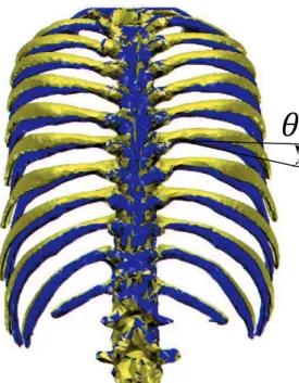
The ribs was further differentiated into 3 portions as shown in Fig. 2-10-A, according to the distribution of moment along curved ribs around the spinal joints for rib rotation as shown in Fig. 2-10-B. Portion 1 has large moment and portion 2 has relative small moment. Regarding the separation for portion 3, because the direction of the moment is reversed around the costochondral articulations, therefore the mechanical action is also different between portion 2 and 3, as pointed out by De Troyer et al [37], [40]. This theory is the extended version of the Hamberger 2-demontional model in order to consider the influence from 3-demontional curved ribs on variation
of moment difference about two adjacent ribs, and concretely explained by De Troyer et al. [37], [40]. 3 Costal cartilage 1 2
Rib rotation axis
External intercostal Internal intercostal 1 2 3
A
B
aFig. 2-10 Portion 1, 2 and 3 divided according to the distribution of
moment along curved ribs studied by De Troyer et al. [37]. A: Location of portion 1, 2 and 3. B: Distribution of moment.
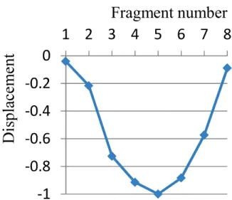
In order to activate the intercostal muscles from dorsal to ventral portion, as Fig. 2-11 and Fig. 2-12 shows, we properly separated both the internal intercostal (Fig. 2-11) and external intercostal (Fig. 2-12) muscles to 8 fragments, and the angle of each fragment is approximately 20 degrees from the center of the body. For visualizations, different colors were adopted for each muscle fragment, and fragments 1 to 8 were assigned from dorsal extremes to ventral extremes of the intercostal muscles.
For discussing the rib motion caused by these 8 fragments of internal and external intercostal muscles, as shown in Fig. 2-11 and Fig. 2-12, we selected the points at the middle of the sternum and the lateral extremes of
ribs from rib 1 to rib 10 as landmarks for representing rib motion. In addition, the displacements at the landmarks were normalized by the magnitude of the plotted sternum displacement for easier to assess, therefore the plotted sternum displacement reached -1 and 1 for the internal intercostal and external intercostal muscles, respectively.
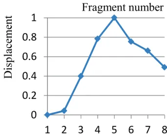
During activating the internal intercostal muscles from fragments 1 to 8, the caudal displacement of the sternum gradually increased from fragments 1 to 5 and decreased from fragments 5 to 8, as shown in Fig. 2-13. For the upper rib 1 to 5, as shown in Fig. 2-14, the caudal displacement of rib 1 varied similarly to the displacement of the sternum in Fig. 2-13. The caudal displacement of ribs from 2 to 5 reached the maximal value between fragments 3 and 4, after that the caudal displacement gradually became smaller and reversed to cranial displacement between fragments 5 and 6, reached largest cranial displacement at fragment 7. For the lower rib 6 to 10, as Fig. 2-15 shows, the caudal displacement reached the maximal value between fragments 2 and 3, after that the caudal displacement gradually became smaller and reversed to cranial displacement between fragments 3 and 6, then decreased to fragment 8. These results illustrate that large downward rib and sternum displacements were happened during the contraction of the internal intercostal muscles in portion 1 of the rib cage. Some reversed rib displacements were generated by the internal intercostal muscles in portion 2 and 3 of the upper and lower rib cage, especially around the costochondral articulations. Therefore, it implies the mechanism is different between the portion 1, 2 and 3 of the rib cage.
Regarding the movements generated by the fragments of the external intercostal muscles, the cranial displacement of the sternum reached -1 at
fragment 5 and gradually decreased to 0.5 at fragment 8, as shown in Fig. 2-16. For the upper rib 1 to 5, as shown in Fig. 2-17, the cranial displacement of rib 1 varied similarly to the displacement of the sternum in Fig. 2-16. The cranial displacement of ribs from 2 to 5 reached the maximal value at fragment 4, after that the cranial displacement gradually became smaller and reversed to caudal displacement between fragments 5 and 6, reached largest caudal displacement at fragment 8. For the lower rib 6 to 10, as Fig. 2-18 shows, the displacement retain almost cranial, and the displacement of each rib first increased along with the fragment changed and then gradually decreased, after that the cranial displacement turned to increase again and then decreased from around fragment 6. These results illustrate that upward rib and sternum displacements were happened during the contraction of the external intercostal muscles not only in portion 1 but also in portion 2 of the rib cage. However, revised rib displacements occurred in the upper rib cage, and the two times increases of cranial displacement in the lower rib cage implies the mechanisms are different between the intercostal muscles in portion 1 and 2.
1 2 3 4 5 6 7 8 : Plotted points
Fig. 2-11 The fragments of the internal intercostal muscles.
1 2 3 4 5 6 7 8 : Plotted points
Fragment number
Fig. 2-13 The normalized displacement of the sternum generated by
separate fragments of the internal intercostal muscles (The horizontal axis shows number of the separate fragments, and the vertical axis shows normalized cranial displacement).
Fragment number
Fig. 2-14 The normalized displacement at lateral extremes of the ribs from
rib 1 to 5 generated by separate fragments of the internal intercostal muscles (The horizontal axis shows number of the separate fragments, and the vertical axis shows normalized cranial displacement).
Fragment number
Fig. 2-15 The normalized displacement at lateral extremes of the ribs from
rib 6 to 10 generated by separate fragments of the internal intercostal muscles (The horizontal axis shows number of the separate fragments, and the vertical axis shows normalized cranial displacement).
Fragment number
Fig. 2-16 The normalized displacement of the sternum generated by
separate fragments of the external intercostal muscles (The horizontal axis shows number of the separate fragments, and the vertical axis shows normalized cranial displacement).
Fragment number
Fig. 2-17 The normalized displacement at lateral extremes of the ribs from
rib 1 to 5 generated by separate fragments of the external intercostal muscles (The horizontal axis shows number of the separate fragments, and the vertical axis shows normalized cranial displacement).
Fragment number
Fig. 2-18 The normalized displacement at lateral extremes of the ribs from
rib 6 to 10 generated by separate fragments of the external intercostal muscles (The horizontal axis shows number of the separate fragments, and the vertical axis shows normalized cranial displacement).
A
B
Fig. 2-19 Rib motion caused by fragment 4 of external intercostal muscles
in portion 1 (Amplification factor 4.0; blue: before muscle contraction; orange: after muscle contraction). A: Lateral view. B: Rear view.
A
B
Fig. 2-20 Rib motion caused by fragment 3 of internal interosseous
intercostal muscles in portion 1 (Amplification factor 4.0; blue: before muscle contraction; orange: after muscle contraction). A: Lateral view. B: Rear view.
Fig. 2-21 Rib motion caused by fragment 7 of external intercostal muscles
in portion 2 (Amplification factor 4.0; blue: before muscle contraction; orange: after muscle contraction).
Fig. 2-22 Rib motion caused by fragment 7 of internal intercostal muscles
in portion 2 (Amplification factor 4.0; blue: before muscle contraction; orange: after muscle contraction).
Fig. 2-23 Rib motion caused by fragment 8 of internal intercostal muscles
in portion 3 (Amplification factor 4.0; blue: before muscle contraction; orange: after muscle contraction).
For visualization, simulated rib motions caused by the intercostal muscles in portion 1, 2 and 3 were shown from Fig. 2-19 to Fig. 2-23. The red areas are the locations of the contracted muscle fragments. The external (fragment 4) and internal (fragment 3) intercostal muscles in portion 1 mainly caused inspiratory and expiratory rib rigid rotations, respectively, as shown in the lateral views of Fig. 2-19 and Fig. 2-20. The arrows in the rear views of Fig. 2-19 and Fig. 2-20, which illustrate the rib moving directions for visualization, show the rib rotation gradually shift from pump-handle to bucket-handle motion in caudal direction due to the rotation axes in the spinal as mentioned above, as observed clinically [45]. Fig. 2-21 shows the rib motion generated by the external intercostal muscles in portion 2 (fragment 7). We could observe the sternum was elevated and somewhat
caudal displacement was generated in the lateral portion of the upper ribs. Regarding rib motions caused by the internal intercostal muscles in portion 2 (fragment 7), Fig. 2-22 the sternum was lowered and the lateral portions of the ribs were elevated. The internal intercostal muscles in potion 3 (fragment 8) lowered the sternum and elevated the lateral portions of the ribs, as shown in Fig. 2-23.
2.2.4 Rib movement generated by entire inspiratory and
expiratory intercostal muscles
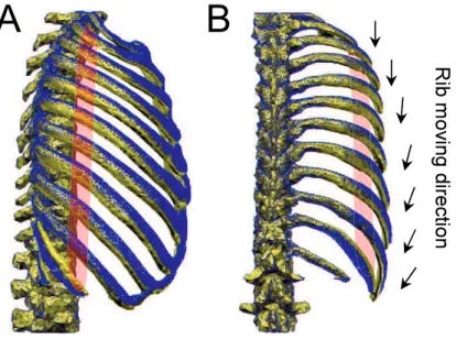
After confirmed the partial mechanical action of the intercostal muscles, by activating entire inspiratory and expiratory intercostal muscles, we reproduced inspiratory and expiratory chest movements as observed clinically. Fig. 2-24 and Fig. 2-25 illustrate the inspiratory chest movement caused by contraction of the inspiratory muscles (external and parasternal intercostal muscles). The arrows show the rib rotation directions. We could observe the bucket-handle motion that the transverse diameter of the rib cage became larger from the front view in Fig. 2-24, and the pump-handle motion that the rise of the sternum was accompanied by the extending of the anteroposterior diameter of the rib cage from the lateral view in Fig. 2-25. The expiratory chest movement generated by activating the expiratory muscles (internal interosseous intercostal muscles) was shown in Fig. 2-26 and Fig. 2-27. The front view in Fig. 2-26 illustrates the bucket-handle motion that the rib cage was narrowed medially, and the lateral view in Fig. 2-27 shows the pump-handle motion of the ribs, shortening of the anteroposterior diameter of the rib cage.
Fig. 2-24 The front view of the inspiratory chest movement generated by
the inspiratory intercostal muscles.
Fig. 2-25 The lateral view of the inspiratory chest movement generated by
Fig. 2-26 The front view of the inspiratory chest movement generated by
the expiratory intercostal muscles.
Fig. 2-27 The lateral view of the inspiratory chest movement generated by
2.3 Discussion
2.3.1 Reproduction of the theory of Hamberger
When the intercostal muscles contracted in a single interspace, for both the external and internal layer muscles, we obtained the ribs close to each other as obtained by De Troyer et al. [36] and Loring [12]. In addition, external intercostal muscles caused an inspiratory rib rotation due to the cranial displacement of sternum and ribs in the ventral region, and internal interosseous intercostal muscles generated an expiratory rib rotation due to caudal displacement of sternum and ribs in the ventral region. This cranial and caudal movement is consistent with the describing in the theory of Hamberger. Although some opposite displacement occurred in the dorsal-lateral region, the respective effect could be explained by the experiment results which concluded the displacement in the ventral region has a larger respiratory effect than in the dorsal region [37]. Therefore, although the rib compliance could determine the rib displacement, without considering the influence from the rib compliance, we concluded that the fiber orientation have a large significance on generating inspiratory and expiratory rib motions as the described by Hamberger.
2.3.2 Relationship between rigid rotation and deformation
of ribs
The theory of Hamberger had not considered the deformation of the ribs. The ribs are acted on the contraction of the muscle forces. After the muscle forces and moment are balanced, because the ribs are not pure rigid body as Fig. 2-8-C and also the experiment [36] and FEM simulation [12] show, the
ribs could be bent and deformed especially. Although the fiber orientation of the external and internal interosseous intercostal muscles caused upward and downward rib rotations, respectively, when we arterially changed the fiber orientation of the intercostal muscles, the craniocaudal fiber orientation did not generated rib rotation and just strongly bent the adjacent two ribs 5 and 6. Absolutely, the respiratory effect also could be ignored. That is when the rib rotation was not clearly generated, the rib deformed predominantly. Conversely, when the rib rotations were obviously happened, the rib deflection and deformation became inconspicuous.
It is easy to consider that, during the intercostal muscle contraction, if the torque generated by the muscle force is small for rib rotation, remained potential could bend and deform the bones. Also, if the deformation of the bones is not predominant, the contracting energy is utilized to resist the reaction force generated by the passive muscles and other tissues during rib rotation. Consequently, by considering the deformation of the rib cage during intercostal muscle contraction, the mechanical action of the intercostal muscles contraction on the rib cage is equivalent to the total action from the rib rotation and deformation. It is worth mentioning that this simple conclusion implies that the mechanical action of the intercostal muscle could shift between the rigid rotation and deformation. Notably, as shown in Fig. 2-10, the moment for the rigid rib rotation distributes along curved ribs and reaches zero around the costochondral articulations.
2.3.3 Action of intercostal muscle contraction on ribs
In portion 1 of the rib cage where the muscles are close to the spinal joints and far from the cartilage, large moment caused dominant rib rigid rotation