Japanese Joumal of Tropical Medicine and Hygiene
第14巻一第2号 昭和61年6月15日
内 容一 原 著
人寄生海洋性裂頭条虫の未知種2例(英文)
一加茂 甫,矢崎 誠一,福本 宗嗣,
前嶋 條士,坂口 祐二 79−86 アフリカ大陸で感染した良性三日熱型マラリア(ハoσσ」8とP.吻砿)(英支)
一海老沢 功 87−90 第27回日本熱帯医学会総会講演抄録(1)
目 次………一……・ 91−92
特別講演……・…・………・ 93−94 シンポジウム礫灘1欝齢1!ll』ll:』ll.1.lll』lll!』』 ll二ll9
皿 熱帯地における旅行者感染症
一その現状と対策を中心に一…一……一・ 109−114 英文抄録一 115−150 会 報
昭和61年度第1回幹事会記録・………・………・・…・……… 151−153
投稿規定
日熱医会誌
JapanJ.T.M.H. 日 本熱帯医学会
TWO UNKNOWN MARlNE
DI PH YLL OB O THRI UM SPECIES OF THE GENUS
FROM HUMAN CASES
HAIIME KAMOl, SEIICHI YAZAKll, SOJI FUKUMOTO1, JOJI MAEJIMA1 AND YUJI SAJ AGUCH12
Received March 22 1986/Accepted May 15 1986
Abstract : The morphological characteristics of two unknown marine species of the genus Dlphyllohothrium were recorded. A cestode spontaneously expelled from a 27‑year‑old seaman resembled D. cameroni Rausch, 1969 from a Japanese seaman by Kamo et al. (198D in the external morphology, but was different in the relative position of the uterine pore. It was related to D. hians (Diesing, 1850) in the position and shape of the cirrus sac, and in the relative position of the cirrus sac to the seminal vesicle, but was different in the shape and size of the scolex. Another cestode expelled after treatment with kamala from a 50‑year‑old seaman re‑
sembled D. hians in the shape of proglottids, the development of inner longitudinal muscle layer, the position of genital atrium, the uterine loops, and egg‑sizes. This was rather related to D. elegans (Krabbe, 1865) in the shape and position of the cirrus sac and the relative position of the cirrus sac to the serninal vesicle, and was different from D. hians in these characteristics.
The definite identification should be established in the future on the basis of more detailed materials and methods.
INTRODUCTION
Sakaguchi et al. (1971) have reported five human cases irLfected with diphynobothriid ces‑
todes found from the inhabitants of fishing viuages in Nagasaki Prefecture, Japan. Three of those cestodes had been identified as Dlplogonopurus grandis, while the remaining two had been taxonomically indeterminable. Later Kamo et al. (1979) observed the densely dlstributed deep pits of their eggshell surfaces by the use of scarming electron microscope, suggesting their association with marine environments.
Since Kamo et al. (1977) suggested the occurrence of human infection with a certain species of Dlphyllobothrium of marine origin, a few marine species of the genus Dlphyllobothrium have been recorded from human cases in Japan such as D. yonagloense by Yamane et al. (1981), D. cameroni by Kamo et al. (1981), and D. pactficum by Kamo et al. (1982). The morphological characteristics of both specimens under consideration were dlfferent from those known species of marine origin in Japan. Moreover, they were not identical with descriptions of any other known species from marine mammals.
Then their morphological characteristics were recorded here for further comparison and identification.
1
2
Department of Medical Zoology, Tottori University School of Medicine, Yonago 683, Japan Departrnent of Parasitic Diseases, Kumamoto University Medical School, Kumamoto 860, Japan
MATERIALS AND METHODS
Case No, I : On 10th October, 1969 a strobila with the scolex has been expelled spontane‑
ously from K. Y. , a 27‑year‑old seaman, who resided at Kanai‑Goto‑Machi, Nagasaki Prefecture.
The yellowish‑brown stout worm measured 460 mm in length, 12.5mm in maxirnum width, and 1.8‑2.0 mm thick.
Case No. 2 : On 26th November, 1969 a cestode without the scolex has been expelled aiter treatment with kamala from I. Y. , a 50‑year‑old seaman, who resided at Ko‑Yagi=Machi, Nagasaki Prefecture. The milkish‑white, thin worm was undulated, measuring 1,020 mm in length and 7. O mrn in maximum width.
The worm8 have been preserved in formalin solution. Whole mount preparations of various maturity‑levels of proglottids were stained with Semichon's acetic carmine. Serial sections of mature proglottids were prepared in horizontal, sagittai and transversal planes, being stained with modified trichrome stain solution. The eggshell surfaces were observed by the scaming electron microscopy.
MORPHOLOGICAL DEscRIPTroNS (All measurments given are in miWmeters)
Cestode from Case No. I : Contracted specimen with scolex, body length 460, lacking termirral segments, maximum width 12. 5, maxirnum thickness about 2.0 (Figure 1).
Strobila muscular, slightly arched dorsad ; margins slightly serrate. Strobila composed of as many as 500 segments ; maximum width attained near terminal one fourth of strobila. Segments
Figure 1 Whole body: specimen No. I (left), No. 2 (right).
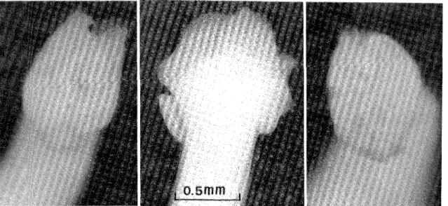
Figure 2 Scolex of specimen No. I : dorsoventral view (left), Iateral view (middle), and apic'al view (right).
i; ;{/(i )i':!i:/i ;i:: ;;:i;,:1;::{n ;ij'iij{ .
.;=<' * =
=;:"i{ : ;*" " '>'"'1"' ':'* ' ̲ '* *>*;=' ="*; ' ' '*' = =
***' "'=
Figure 3 Uterine field of mature segment : specirnen No. I (1eft), and No. 2 (right).
wider than long, with length increasing posteriad. Length/width ratio of pregravid segments about I : 15; of temtinal gravid segments about I : 10. Iunermost layer of longitudinai muscle fibres weu developed, as much as O.288 thick; adjacent layer of circular fibres fairly developed (Figure 4). Calcareous corpuscle abundant. Polygonal scolex, with deep bothria extending full length, 1.08 Iong x 1. 13 wide in lateral view. Bothria with folded margins diversing at apex (Figure 2). Neck absent ; posterior edge of seolex slightly overlapping frst segrnent. Genital pore visible within 70 mm posterior to scolex, situated ventrally on midline at anterior margin of segment, covered by velum of preceeding segment (Figure 3). Genital atrium lined by rounded papmae . Cirrus sac elongate, with margins somewhat undulating in sagittal sections, 1.123‑
"+;
(= = ‑ *"+ "'=' "* ‑'* * ="* *i * ‑' : *'* ' i**‑ **
** ‑ ** ' '; * '* '+' '^= * i*t **'**>*' ' = +* * +*' ! ; * ' =' ** ' ; ' I !! i'f {::;;;::::;i;:1;:;; ::'j : ;/:; "I"";" :# i;:# / :
.... ̲ , ,̲ """ "̲ '̲ ' ̲ ^' ̲' ' ' ::̲ " { f i: ;;; ii, ' /'" " :;::; 1 ' {t / / '!;/;i(1' )"' i ; " "̲' '̲ ' < ̲='" ‑‑ ‑i{ : ;:. :
. > ‑ . ; ='# '< '" *+** = s i ; ̲
=‑' ' X ̲'i=: = '= <t$: l‑ ='
. .. :
* ! ' ; ;s{ ̲ ,., ̲ ̲ ' " '>'='>" "' ' "‑ =‑ ' '=' "= ' "=' ' :;;;;::::..,= ‑ =' ,.=# ' ;"" ' <'=' ' "= ' =‑>"';;:{;;:i' = :/ j* "'* " ̲ ^ ̲ ^ ' ' ' i '‑ ** ' '; ' " :;"' ̲= *' fi ̲ ' ' '"
' " " < ' L
= ' ";i " : i ';i:i ' '< = ' = ii';i ii' ;'==:i :" = f ' ; : =' ' I ^ i ; i{'=' ' '̲ '^ ̲̲̲ S‑ ' '
** ' + *" ‑ ' " " ' ""' '* '>= "'* : * '3s '*' ';=^ + ' ' ‑‑ ' **' "*' =‑*‑ ' ' ; : ( : "' ‑ ‑ ‑^ '>' > ' "‑'*' ' ‑ i**+ .
* := 9 'i'* ; :: * '; " = "'= ' : 't i " * :' "' s'̲;i . . ̲
= ‑ '‑* ‑ ̲" ‑‑ ' ' ' ‑'‑' '^= ^ ̲ 'E* '* ‑ ' " "<' " '' * s "'= * ' ' **"'+ "* * "" ; * $"* = { ' "' '* ==' ' ;='s‑ s " 'i' * ‑ ' . '^'> ‑ .. < ^" ‑ ‑+'* ‑ " ' ‑ ‑ "= ' ‑ ' ‑ =‑ ‑ ! '= ‑ 'i' ‑' ' "*=* '='; ';"' {i' ̲ =" === :‑' ' ' "' = iS ̲ , .* .̲ .
: =̲ ; ' ' ̲"̲ ' ' ^ <" ' ' '
' = '*' '̲ ̲ " ' ' :̲ ' *'
‑'>'= S ‑ ‑ ' ' ‑ = ' ‑ ' ' ' 'ii, ' ' ** '‑ ‑ : "*‑" ‑ * ' *‑' *' '> ' ^̲ , . ^‑ : ̲̲ ^ ̲' ' *=* ****'= = "‑="‑' ' == ; ‑ ' t‑ '#<' lti" '*"* ' '< ' : ‑‑ '* i* i"'* '*=+* ** * ‑* : ' *..' " : *' " >'#' '"' ""i ' ' ::' := ;" =' * '* ‑ '=' ' ' '>x :;1 ; (:;!=; ' r ; :'; ‑< +
#" ; ' ' n "++*.+
+ '== :*;i' ' ;
;;i: li ' :;i :'t':;:1 :;::;i:; ; ‑ ;'i;{';{'i 'i {';="'+'*'=* +* =':1 *: * **" *: i..!,* {
'; "('t'"'; ; ;:'* / ! := " '*'# s /!/(* : *'<;:*; i :;;; :i :i
{' {" !'i' ;:*== = '!' <
Figure 4 sagrttal (A) and cross (B) sect ons specunen No I (left) and No 2 (right)'
l.164 IongXO.098‑0.133 in diameter. Seminal vesicle elongate, O.412‑0.525 in dorsoventral dimension by O. 124‑0.139 in diameter, situated adjacent and parauel to cirrus sac posteriorly, and connected with latter by short duct (Figure 4). Subspherical testes numerous, 0.103‑0. 175 in greatest diameter, arranged in single layer in lateral fields. Terminal portion of vagina running anteriad, then turning ventrad into genital atrium and opening posterior to opening of cirrus sac. Bilobed ovary situated transversely near posterior margin of segment. Viteuine follicles (viteuaria) abundant, forming two lateral fields, O.052‑0.072 in greatest diameter.
Gravid uterus fomting compact loops extending through length of segment from posterior margin to level of anterior edge of genital atrium (Figure 3). Uterus opening through uterine pore posterior to genital pore, usually to right or left of midline. Eggs ellipsoidal to subcpherical with
apical knob, 0.046‑0.056X0.033‑0.044 (avg. O.049 0.02XO.036̲+0.03). Surface of egg‑
shell densely distributed by broad pits (Figure 5).
Cestode from Case No. 2 : Slightly contracted specimen without scolex, body length l,020, maxirnum width 7.0 (Figure l).
Strobila somewhat muscular, with slightly serrate margins. Segments wider than long, with slightly convex margins, increasing relative length near posterior end of strobila. Lengthr width ratio of mature segments about I : 5, of gravid segments I : 3.5, and of terminal gravid segrnents I : 1.5. Innermost layer of longitudinal muscle fibres well developed, about O. 185 thick ; adjacent layer of circular fibres fairly developed (Figure 4). Calcareous corpuscle abun‑
dant. Genital pore situated ventraliy on midtine in near anterior margin of segment (Figure 3).
Genital atrium lined by rounded papillae. Cirrus sac pidiform, with margin8 somewhat undulating wan, O.484‑0. 567 Iong X 0.288‑0.391 in diameter. Cirnls sac opening anterierly into genital atriurn. Seminal vesicle subspherical, situated posterior to end of cirrus sac. O.309‑0,433 Iong x O.175 in diameter (Figure 4). Subspherical testes numerous. O.082‑0.103 in greatest diam‑
eter, arranged in shgle layer in lateral fields. Terminal portion of vagina running anteriad near ventral surface, turning slightly ventrad into genitai atrium irumediately posterior to opening of
Flgure 5 Eggs and eggshell surfaces by SEM (X5,COO) : specimen No. I (upper), and No. 2 (lower).
cirrus sac. Bilobed ovary situated transversely near posterior margin of segment. Vitelline fomcles abundant, forming two lateral fields, O. 052‑0. 072 in greatest diameter. Gravid uterus forming compact loops extending through length of segment from posterior margin to level of anterior edge of genital atrium (Figure 3) . Uterus opening through uterine pore posterior to genital pore usuahy to right or left of midline. Eggs subspherical with or without apical knob, 0.043‑0.048 ><:0.033‑0.039 (avg. O.044 0.02xO.035̲+0.02). Surface of eggshell densely distributed by broad pits (Figure 5).
DlscussroN
Judging from the nature of their eggshell surfaces our specimens can be recognized as members of the genus Diphyllobothrium associated with marine environments (Hilliard, 1972).
In regard to the marine species of Diphyllobothrium from human cases the foliowing 6 species have been reported so far : D. cordatum (Leuckart, 1863) from Greenland by Leuckart (1863) ; D. alascense Rausch et Williamson, 1958 and D. Ianceolatum (Krabbe, 1865) from Alaska by Rausch and Hilnard (1970) ; D. pactficum (Nybelin, 1931) from Peru by Baer et al. (1967),
from Chile by Atias and Cattan (1976), Sagua et al. (1976), from Japan by Kamo et al. (1982) ; D. yon4 oense Yarnane et al. , 1981 and D. cameroni Rausch, 1969 from Japan by Yamane et al.
(1981) and Kamo et al. (1981), respectively.
Of these D. cameroni Rausch, 1969, especiauy the specimen found from a Japanese searnan (Kamo et al. , 198D resembles our specirnen from the Case No. I in the external morphology.
In histological details, however, the relative position of the uterine pore, D. cameroni is different from our specimen No. l. The uterus opens into the genital atrium in D. cameromi, while it opens separate from and posterior to the genital atrium in our specimen No. 1.
According to the recent revision of diphyllobothriid cestodes by Delamure et al. (1985) 29 species of the genus Dtphyllobothrium were recognized as valid, though a few of them are debatable.
Including the above‑mentioned human parasites 23 species of them can be comprised in a group of marine origin. Most of them have special characteristics respectively in their external and/or intemal morphology, and are distinguishable enough from our specimens. D. hians (Diesing, 1850) among them resembles our specirnen No. I in the position and shape of the cirrus sac, and the relative position between the cirrus sac and the seminal vesicle, but is apparently different in the shape and size of the scolex.
D. hians is also related to our specimen from the Case No. 2 in the shape of proglottids (length/width ratio), the development of inner longitudinal muscie layer, the position of genital atrium, the uterine loops, and egg‑sizes. However, the shape and position of the cirrus sac, and the relative position hetween the cilTus sac and the seminal vesicle resemble more other species : D. elegans (Krabhe, 1865).
Thus our specimens are both seemed to be distinctive from au known species, but the definite identjfication should be established in the future on the basis of more detailed materials and methods.
ACKNOWLEDGEMENTS
The authors wish to express our heartfelt thanks to Prof. emeritous D. Katamine and Prof.
Y. Aoki, Department of Parasitology, Institure of Tropical Medicine, Nagasaki University, for their kindness and encouragements through providing with specimens. To Mrs. S. Wakahara, Mr. Y. Sugihara, and Miss Y. Yakura, Department of Medical Zoology, Tottori University School of Mediohe, appreciation is expressed for their technical assistances.
REFERENCES
1) Atias, A. and Cattan, P. E. (1976) : First case of human iniection with Diphyllobothrium p '
au cum m Chile, Rev. M6d. Chle, 104, 216‑217
2) Baer, J. G., Miranda, C. H., Fern ndez, R. W, and Medirra, T. J. (1976) : Human diphyllobothriasis in Peru, Z. Parasitenkd. , 28, 277‑289
3) Delamure, S. L. , Skriabin, A. S. and Serdyukov, A. M. (1985) : Diphyllobothrild cestodes from hu‑
mans, manuna]s and birds, 51‑137, Nanka, Moscow (in Russian)
4) Hmiard, D. K. (1972) : Studies on the helrninth fauna of Alaska LI. Observations on eggshell formation in some diphyllobothrild cestodes, Can. J. Zool. , 50, 585‑592
5) Kamo, H., Maejirna, J., Yazaki, S., Fnkumoto, S. and Hiraga, M. (1979) : Another marine species of Diphyllobothrium found from 3 seamen in Kyushu, Jap. J. Parasit., 28, supp., 74 (in Japanese)
6) Kamo, H., Maejirna, J., Yazaki, S., Otsuru, M., Hasegawa, H., Kuniyoshi, Sh. and Asato, R. (1982) : Occurrence of human infection with Diphyllobathrium pactfaum (Nybelin, 1931) Margolis, 1956 in Japan, Jap. J. Parasit. , 31, 165‑170 (in Japanese)
7) Kamo, H. , Yamane, Y. and Kawashima, K. (1981) : The hrst record of human infection with Diphyllo‑
bothrium cameroni Rauscl 1969. Japan. J. Trop. Med. Hyg. , 9, 199‑205
8) Kamo, H., Yamane, Y., Maejirna, J., Yazaki, S. and Fukumoto, S. (1977) : "Koga‑Okamura type"
diphyllobothriid cestode other than D. Iatum found from human cases in Japan, Nippon‑Iji‑Shimpo, 2795, 43‑45 (in Japanese)
9) Leuckart, R. (1863) : Die menschlichen Parasiten und die von ihnen herrOhrenden Krankheiten. Ein Hand und Lehrbuch fdr Naturforscher und Aerzte, Vol. l, 766, C. F. Winter's Verlag, Leipzig und Heidelberg
10) Rausch, R. L. and Hilliard, D. K. (1970) : Studies on the helrninth fauna of Alaska XLIX. The occurrence of Diphyllobothrium latum (Linnaeus, 1758) (Cestoda : Diphyllobothrndae) in Alaska, with notes on other species, Can. J. Zool., 48, 1201‑1219
11) Sagua. H. , Mjranda, E. . Fuentes, A. and Vladmo, V. (1976) : Dlphyllobothrium pactfaum Cヲybelin, 1931) Margolis, 1956. First two human cases of infection in Northem Chile, Bol. Chile. Parasit. , 31, 33
12) Sakaguchi, Y. , Harada, T. and Muta, N. (1971) : A description of tapeworms (Diphylbbothrhdae) occuring in Nagasaki Prefecture, Jap. J. Parasit. , 20 (1), supp. , 31‑32 (in Japanese)
13) Yamane, Y., Kamo, H., Yazaki, S., Fnkumoto, S. and Maejima, J. (1981) : On a new marine species of the genus Dlphyllobothrium (Cestoda : Pseudophyllidea) found from a man in Japan, Jap. J. Parasit. , 30, 101‑lll
人寄生海洋性裂頭条虫の未知種2例
加茂 甫1・矢崎 誠一1・福本 宗嗣1 前嶋 條士1・坂口 祐二2
人寄生海洋性裂頭条虫の未知種2例について,それらの形態的特徴を記録した。27歳男子船員から 自然排出された条虫は,外形が日本人船員から前に見出された1).oσ膨猶o痂と近似しているが,子宮 孔の相対的位置が異なる。その陰茎嚢の位置や形,陰茎嚢と貯精嚢の相対的位置はP.肋郷と似てい るが,頭節の形や大きさが異なる。50歳男子船員からカマラにより駆出された条虫は,片節の縦横比,
内側縦走筋層の発達度,生殖孔の位置,子宮ループ,虫卵の大きさなどがP,h伽sと似ているが,陰 茎嚢の位置や形,陰茎嚢と貯精嚢の相対的位置は異なり,これらの点はむしろP.8 咽襯sと似てい
る。確定的な同定は将来を期したい。
1鳥取大学医学部医動物学教室 2熊本大学医学部寄生虫病学教室
BENlGN
AND P. VIVAX) TERTIAN TYPE MALARIAS CONTRACTED IN
(P. OVALE
AFRICA
ISAO EBISAWA
Received March 7 1986/Accepted May 10 1986
Abstract : Twenty‑one cases of ovale malaria were seen arnong 25 patients of benign tertian malaria contracted durirrg visits to African countries, south of the Sahara. This reflects an increase in the number of Japanese visitug this area as P. ovale is, with a few exceptions, endemic only in tropical African countries. Nigeria, Malawi and Guinea were the most frequent countries of infection with 6, 4 and 3 cases, respectively.
One patient each with vivax malaria was infected in Malawi, Guinea and Mali ; one case was infected in the island country of Madagascar in East Africa.
Because both P. ovale and P. vivax exist in tropical African countries, we stress the impor‑
tance of carefill examination of blood smears when diagnosing malaria.
INTRODUCTION
The benign tertian malaria parasite most prevalent in tropical Africa is P. ovale, which is, with a few exceptions (Jeffery et al. , 1954; Wilcox et al. , 1954 ; Jeffery and Young, 1954 ; I ysenko and Beljaev, 1969) endemic only on this continent (Report of a WHO Scientific Group,
1969 ; Garnham, 1966).
Japanese patients infected with P. ovale began to appear in 1971 (Amano et al. , 1972 ; Ebisawa et al. , 1972, 1973). The total number of ovale malaria patients seen by the author rose to 21 by the end of 1985, reflecting an increase in the number of Japanese visiting this area.
However, during a review of my case records of malaria patients, 4 cases having been infected with P. vivax contracted in tropical Africa came to my attention ; obviously both P. ovale and P. vivax were prevalent in the same tropical African countries.
The purpose of this paper is to report the areas where my patients were infected with P. ovale and also to cau attention to the fact that both P. ovale and P. vivax, which is rarely pathogenic to the black people (Jeffery and Young, 1954), are circulating in the same regions.
THE PATIENTS AND DIAGNOSIS OF THE PARASITE SpECIES
l) The patients : Au patients were Japanese, except for a Dutch national who was infected in Nigeria and developed the inness in Japan. None of the Japanese patients had been in malaria‑
endemic areas during the 5‑year period before the onset of the current illness, except in the African countries now under consideration.
Departrnent of Public Health, 143, Japan
Toho University School of Medicine, 5‑21‑16 Omori‑nishi, Ohta‑ku, Tokyo
2) Diagnosis : The diagnosis of P. ovale was made by paying particular attention to the mor‑
phology of the malaria parasites and of the parasite‑infected red blood cells. A l/50 mohr phosphate buffer solution of pH 7.2‑7.4 was used to make a Giemsa stain throughout the study period. The main criteria of comparison were : srnallness of the P. ovale parasite in comparison with that of P. vivax, the number of merozoites (average number of merozoites in the mature P. ovale schizont being 8) and incomplete separation of merozoites irom each other in the mature P. ovale schizonts ; larger size and smaller number of Schtifher's dots for P. ovale in comparison with those of P. vivax‑infected red blood cells (Wilcox et al. , 1954).
3) The country of infection : This could be pinpointed for patients who had stayed in only one country. However, when the patient had stayed in or travelled through two or more countries, the country of infection was designated simply as "tropical Africa".
RESULTS
P. ovale : Twenty‑one patients were infected with this parasite in countries south of the Sahara (Table 1). Six patients were infected in Nigeria, 4 in Malawi, 3 in Guinea, 2 cases each in Congo (Brazzaville) and Kenya and one in Tanzania. The country of iniection in 3 cases was designated as "tropical African countries" as these patients had stayed in more than 3 countries.
P. vivax : One case each was infected with P. vivex in Guinea, Mali and Malawi in sub‑Saharan countries and in an island country of Madagascar off the East African coast (Table 1). One patient with vivax malaria who retumed to Japan from Kenya could not be confirrned as having been infected there as he had also stayed for some time in India. Two other cases of vivax malaria were infected in Egypt and in Ethiopia, north of the Sahara.
Table 1. Benigu tertian malarias inf ected in Africa (1971‑1985) Country P. ovale P. vivax North of the Sahara
Egypt Ethiopia
South of the Sahara Kenya
Tanzania Malawi Madagascar Guinea Nigeria Mali
Congo (Brazzaville) Tropical Africa*
2
4
3 6
2 3
1 1
1 1 1
1
Total 21 6
* Applied to patients who stayed in or travelled through more than 3 countries.
A patient of vivax malaria who returned from Kenya was ex‑
cluded as he had stayed in India within 6 months before the onset of the current illness.
DISCUSSION
A high incidence of ovale malaria‑about 6% of au of my malaria patients‑is indicative of the increasing number of Japanese visiting countries of tropical Africa, on business, construction works, geographical surverys, pleasure, etc. Until 1977‑1978, the majority of my malaria patients were infected in Southeast Asia and Oceania rather than in Africa. But the trends have reversed since 1979 when the number of malaria patients infected on the African continent began to surpass that of the patients infected in Southeast Asia and Oceani:a combined, even though the latter regions are nearer to Japan (Ebisawa, 1982). This tendency was associated with an increasing number of fatal falciparum malaria cases in my series of patients.
Another indication that Japanese are being exposed more to malaria in African countries was the case of a lenkernia patient in Japan who contracted ovale malaria following multiple blood transfusions (Amano et al. , 1984).
The fact that vivax malaria was endemic simultaneously with ovale malaria in the tropical African countries of Malawi, Guinea and Mah deserves a short comment. P. vivax is rarely pathogenic to the black ' population. The circulation of this species of malaria parasite in the tropical African countries may indicate that it was brought to these areas from other continents by other races such as Arabians, Indians, and other people and was picked up by some local anopheline mosquitoes. The presence of P. vivax in Madagascar may be easily understood as there exists anthropologicauy some ethnical relationship between the people of Madagascar and Indonesia where P. vivax is endemic. There have been no reports of a sirnian malaria parasite which can be regarded as an equivalent to the human P. vivax on the African continent, in the way that P. schwetzi is regarded as an equivalent to the human P. ovale (Coatney, 1971).
REFERENCES
l) lwlano, H., Kurata, S. and Yamamoto, T. (1971): On a rare case of ovale inalaria, Nettai, 6, 162‑
168 (in Japanese)
2) Amano, S., Ohshirna, T., Harano, H., Un, Ko‑1. Ito, A., Okubo, T., Watanabe, S. and Mouri. T.
(1984) : On a case of ovale malaria infected by blood transfusion, Japan. J. Trop. Med. Hyg. , 13, 189 3) Coatney, G. R. (197D : Sirnian malarias in man : facts, implications and predictions, Amer. J. Trop.
Med. Hyg., 17, 147‑155
4) Ebisawa, I. (1982) : Recent problems in malaria, in Oda, T. ed. Seminar in Internal Medicine, INF‑
1, Infectious Diseases, p. 261‑271, Nagai Publ. Co. , Osaka, Japan (in Japanese)
5) Ebisawa, I. and Komoriya, T. (1972) : Case reports of ovale malaria, Naika, 30, 544‑546 (in Japanese)
6) Ebisawa, I. , Komoriya, T. and Kirnura, M. (1973) : Study on ovale malaria, J. Japan. Assoc. Infect.
Dis., 48, 385‑392 (in Japanese)
7) Garnham, P. C. C. (1966) : Malaria parasites and other haemosporidia, Blackwell Scientific Publica‑
tions, Oxiord
8) Jeffery, G. M., Young, M. D. and Wilcox, A. (1954) : The Donaldson strain of malaria. 1. History and characteristics of the infection, ner. J. Trop. Med. Hyg. , 3, 628‑637
9) Jeffery, G. M. and Young, M. D. (1954) : The Donaldson strain of malaria. 4. An evaluation and status, ner. J. Trop. Med. Hyg., 3, 660‑664
10) Lysenko, A. J. and Beljaev, A. E. (1969) : An analysis of the geographical distribution of Plasmodium ovale, Bull. W. H. O., 40, 383‑394
11)Report of a WHO Scient面c Group(1969):Parasitology of ma㎞a,WHO Tec㎞ical Report Series No.433(Geneva)
12)W皿cox,ん,Je丘ery,G.M and Yo㎜1g,M.D.(1954):The Donaldson strain ofm副a.2.Morpholo−
gy of the erythrocytic parasites,AmeL J.Trop.Med.Hyg.,3,638−649
アフリカ大陸で感染した良性三日熱型マラリア
(P.o祝zJ6とP.加祝z劣)
海老沢 功
アフリカ大陸で感染した三日熱型マラリア27人の内,サハラ砂漠以南の地域で感染した者は25人 で,卵型マラリアは21人であった。卵型マラリア原虫は,少数の例外を除き熱帯アフリカにしか流行 していないので,この事実は熱帯アフリカに旅行,或いは滞在する日本人が多くなった事を示す。卵 型マラリア感染地はナイジェリア,マラウイ,ギニアがそれぞれ6,4,3人で,その他ケニァとコン ゴ(ブラザビル)が2人ずつ,タンザニアが1人,西アフリカの3つ以上の国に滞在した者が3人
あった。
三日熱マラリア患者は,サハラ砂漠以南の地域では,マラウイ,ギニア,マリおよびマダガスカル で1人ずつ感染している。サハラ砂漠以北ではエジプトとエチオピアで1人ずつ感染している。
特に注目すべき点は,元来黒人にはほとんど病原性がないと言われている三日熱マラリア原虫がジ 熱帯アフリカの国で卵型マラリア原虫と同時に流行していることである。熱帯アフリカの三日熱マラ
リア原虫は,おそらくアフリカ大陸以外の地域から,アラビア人,インド人,その他の人種によって 持ち込まれたものであろう。マダガスカル島の住民は,インドネシア人と民族学的に近縁であるとい われているので,マダガスカル島に三日熱マラリアがあっても不思議ではない。
熱帯アフリカの国には,三日熱と卵型マラリアが同時に流行しているので,血液標本はpH7.2−7、4 の緩衝液を用いて注意深く検査する必要がある。
東邦大学医学部公衆衛生学教室
第27回 日本熱帯医学会総会講演抄録(1)
日場長期会会 昭和60年10月30日(水)一11月1日 (金)
神戸国際会議場(神戸国際交流会館内)
神戸大学医学部教授 松村武男
目 次
特 別 講 演
I Dengue hemoIThagic fever:a critical appraisal
・fcment坤・theses
Leon Rosen (ハワイ大)
(和文抄録なし)
H 科学技術と国際交流
岡本 道雄 (科学技術会議議員)
シンポジウム 1 熱帯医学と分子疫学
1 司会のことば
石井 明 (岡山大・医・寄生虫)
三舟求真人 (大分医大・微生物)
2 蠕虫幼虫の抗原遺伝子の解析
菅根 一男(横浜市大・医・寄生虫)
3 最近におけるアフリカ・トリパノソーマ同 定法の進展
蛭海 啓行,V.ナントリア,
B.クウクラ,0.オーレ・モヨイ,
P.マジワ
(国際家畜疫病研究所・ケニア)
4 毒素原性大腸菌とコレラ菌の分子疫学 山本 達男 (順天堂大・医・細菌)
5 RNAパターンからみたケニアにおけるロ タウイルス感染
千葉 靖男 (札幌医大・小児科)
宮崎 千秋 (九州大・医・小児科)
6 オリゴヌクレオチドフィンガープリント法 によるゲタウイルスと日本脳炎ウイルスの
解析
堀 博之,森田 公一,
五十嵐 章
(長崎大・熱帯医研・ウイルス)
且 熱帯諸国と日本の医学 1 司会のことば
深井孝之助(阪大・微生物病研究会)
坪井 誠吉 (神戸大・医・
医学研究国際交流センター)
2 海外医学医療協力のあり方 2a 海外医学医療協力における問題点 林 滋生 (予研)
2b 医学研究国際交流センターの活動と理念 岩井 誠三 (神戸大・医・
医学研究国際交流センター)
3 アジアから見た日本の医学
Dom㎞go E.0. (フィリピン大)
s司udi (インドネシア大)
(和文抄録なし)
4 国際協力機関の医学医療協力
4a 国際協力事業団(JICA)の熱帯病に関する 協力活動
長谷川 豊
(国際協力事業団医療協力部)
4b 南北問題からみた世界保健機構と日本 蟻田 功 (国立熊本病院)
4c 東南アジア諸国との医学の交流
酒井 文徳 (日本学術振興会)
5 熱帯諸国との医学医療協力 5a 司会のことば
藤岡 農宏 (県立尼崎病院)
辻 守康 (広島大・医・寄生虫)
5b 熱帯諸国との医学医療協力
佐藤 喜一 (金沢医大・熱帯医研)
5c 無償援助と並行して行う技術協力 鈴木 守 (群馬大・医・寄生虫)
5d 日本国際救急医療班(JMTDR)の活動に ついて
鵜飼 卓
(大阪府立千里救命救急センター)
5e バヌアツ共和国マレクラ島における眼科活 動
岩崎和佳子 (関西医大・眼科)
5fデング出血熱に関するインドネシアと日本 との医学医療協力
船原 芳範 (神戸大・医・一生理)
5g ネパール・トリブバン大学医学教育プロ ジェクト
欠田 早苗 (兵庫医大・二解剖)
岩崎 忠昭 (兵庫医大・一内科)
5h フィリピン共和国熱帯医学研究所プロジェ クトからみた医学医療協力
布上 薫 (九州大・医技短大)
皿 熱帯地における旅行者感染症 一その現状と対策を中心に一 1 司会のことば
中林 敏夫 (阪大・微研・原虫)
青木 隆一 (大阪市立桃山病院)
2 帰国者における感染症の現状 青木 隆一
(大阪市立桃山病院・感染症センター)
3 国際伝染病一ラッサ熱,マールブルグ病,
エボラ出血熱一について 今川 八束
(都立墨東病院・感染症科)
4 ウイルス性肝炎について
志方 俊夫 (日本大・医・病理)
5 細菌性腸管感染症 竹田 美文
(東大・医科研・細菌感染)
6 マラリアについて
高田 季久(大阪市大・医・医動物)
7 治療薬剤からみた熱帯性寄生虫病 尾辻 義人(鹿児島大・医・二内科)
一 般 講 演
(次号掲載予定)
学 生 講 演 懇 話 会
(次々号掲載予定)
特別講演
I Dengue hemo皿hagic fbver:a critical aOpraisal of cuNent hypotheses Leon Rosen(ハワイ大)
(英文参照)
H 科学技術と国際交流 岡本 道雄(科学技術会議議員)
西洋の科学技術は目覚ましく進歩し発展し,今 や世界を制覇してしまった。日本も明治維新以来,
西洋から科学技術を導入しこれを工夫し発展させ て来た。先ず明治の改革では,科学技術によって 武器をつくり,軍国主義をもって世界に乗り出そ うとした。次に第二次世界大戦後の改革では,科 学技術を企業化することによって,経済的に繁栄 し今では世界第2の経済大国になった。しかし歴 史的にみて,武力のみでまた経済のみで国際社会
に伍して長く栄えた国はないのである。国際社会 で信頼される事が大切なのである。その為に日本 はどうすればよいのか。私は次の3点が重要だと 考える。
第1に,外国人を受け入れることである。人が 外国へ行った時は,所詮切り花である。国民はそ の国で見てもらうことで,本当の理解が得られる。
日本人が本当に美しいのは,日本の土においてで あり,その土に生えた花がやはり一番美しい。切 り花はやはり切り花だけである。その意味でこれ からの国際交流というのは,日本に受け入れると いう方向に最大の努力をすべきである。そこで政 府は今,二十一世紀までに10万人計画というのを 立てている。
第2に,日本は今まで世界から科学技術を受け 入れてここまで繁栄したのであるから,基礎科学 で人類の為になることを積み重ねなくてはいけな い。これは単に日本の国益のためだけでなく,基 礎科学で世界の為にやるべきことがある。砂漠化 を防ぐとか,酸性雨をどうするかと言った地球的
な問題になると恐ろしく金がかかり,アメリカか 日本でないと出来ない基礎科学がある。そういう ものにしっかりと金を出して,世界に報いる事が 大事である。
第3は,昔からよく「和魂洋才」という事が言 われたが,日本人の魂とは何であるかを,今よく 考えてみなければならないという事である。今や 西洋の科学技術が人間に直撃して,資源枯渇,自 然破壊,公害等の多くの深刻な問題を起こしてい る。また目には見えない人間の心に,どういう影 響をもたらしているかというと,これは計り知れ ない恐ろしい問題であると考えられる。これを救 うのはどうしても東洋の精神だと,西洋では考え られている。なぜなら西洋の科学技術の本質は,
自然と人間が対立するところから始まっている。
人間の理性だけ働かせて,理性で自然を観察して 自然の中の法則を見つけて自然を利用し活用し,
さらに征服しようというのが近代科学技術の精神 である。ところが東洋の考え方では,自然と人間 は対立していないのである。自然の中に人間がい て,人間も動植物も同じく自然であるという考え 方である。調和の中にあるのである。この様な精 神をもつ日本や東洋を世界は熱い目で見ている。
この点日本という国は,東西の文化の接点であ るし,また科学技術の価値を日本人ほど知ってい る国民はないのである。日本は科学技術で生きて 来たのであり,その功を最もよく知ると共に,ま た原子爆弾を体験し科学技術が誤って使われた時 の罪を,これほど知っている国民はないわけであ
る。そういう日本人が「和魂」とは何であるかを 今しっかり認識する事が大切である。
日本の世界における立場は大きく変化し,今や 日本の内政はまさに日本の外交であり,日本の内 部でやっていることは,すぐ日本の外交になって いる。その日本の外交は世界の内政,世界政策と
なっているのである。それくらいの立場に変わっ ているということの自覚が,日本人一人一人の中 にあってこそ,国際交流というものが政府の命令 でなく,また政府の政策でなしに国民一人一人の 自らのものとして,この時代の科学技術,国際交 流の方向が出て来るのだと思う。
シンポジウム
1 熱帯医学と分子疫学
1 司会のことば
石井 明 (岡山大・医・寄生虫)
三舟求真人 (大分医大・微生物)
感染症の制圧に,その病原体の伝播様式や存続 様式,あるいは地理的分布等を知るための疫学的 研究が不可欠であることには,疑いの余地がない。
感染症の疫学はその病原体の種類により多少の違 いはあるが,これまで主として血清学的手法に よって行われ,幾多の輝かしい業績があげられて きた。しかし,これまでの手法では,病原体株間 の詳細な違い,ひいては伝播経路等を詳細に解析 するには限界があり,感度の高い新しい手法の導 入が待たれていた。
近年,分子生物学分野における新しい発見と技 術の開発により,遺伝子の抽出とその塩基配列の 決定,制限酵素による遺伝子の切断とその断片の 電気泳動法,遺伝子操作による遺伝子のクローニ ング解析,RNAのオリゴヌクレチオドフィン ガープリント法等が容易に行えるようになり,疫 学研究にも応用されるようになった。そして,こ のような分子レベルでの疫学のことを,分子疫学
と呼ぶようになった。
分子疫学は,病原体の単純性と取扱いの容易さ から現在,ウイルス学および細菌学分野で一歩進 んでいるが,原虫,寄生虫分野でも遺伝子レベル での解析が行われるようになってきた。このシン ポジウムでは熱帯地における感染症の主要な病原 体である寄生虫,原虫,細菌およびウイルスの各 分野において,分子遺伝学的研究,あるいは分子 疫学的研究がどのように進められているかについ て話題を提供していただき,その現状を把握し,
今後の熱帯医学研究の推進に重要な貢献をするで あろう,分子疫学の展望を考える機会としたい。
2 蠕虫幼虫の抗原遺伝子の解析
菅根 一男 (横浜市大・医・寄生虫)、
蠕虫類,とりわけ線虫類幼虫から抽出した虫体 抗原,および幼虫の培養上清より抽出したES抗 原は,免疫診断上有効で,特にES抗原は抗原特 異性が高いとされている。なかでも犬回虫幼虫,
および旋毛虫幼虫ES抗原は幼虫移行症,旋毛虫 症の診断に不可欠である。しかし,これらの抗原 を一定量得るためには大量の幼虫を採集して培養 しなければならない。そこで遺伝子操作の技術を 応用して,抗原を大量に採取することを考えてい る。今回実験に用いたのは旋毛虫感染幼虫で,ま ずこれらの抗原を分子レベルで解析するために,
35S−methior血eを加えた培養液中で幼虫を培養し た後,培養上清を30%PEGinO.9%NaCl中で 濃縮してES抗原を,また幼虫を超音波で粉砕し て虫体抗原を作製した。そしてそれぞれの抗原に 旋毛虫感染マウス血清を加え形成されたAgAb complexを,S.側名召%sCowan1株で吸収した後 IgG抗体と反応する抗原ポリペプタイドを,SDS
を加え熱処理して分離した。そしてSDS−PAGE autoradiographyで抗原ポリペプタイドの解析を 行った。その結果,ES抗原,虫体抗原ともIgG 抗体と特異的に反応する数種類の抗原ポリペプタ
イドが認められた。特にES抗原の48Kdのポリ ペプタイドは,感染血清中の,IgG抗体と強く反 応した。次にこれらの抗原の遺伝子レベルでの解 析を行うため,液体窒素で凍結した幼虫をFre−
ezer/Mi皿で粉砕し,可溶成分中よりCsC1超遠心 法でRNA分画を採取した。そしてこのRNA分 画を用いて伽び伽otranslationを行いtranslation productsを感染血清と反応させた後,AgAb com−
plexを,S.側灘s Cowan1株で吸収しSDSを加 え熱処理した後,SDS−PAGE autoradiogr卸hyで translation products中に抗原ポリペプタイドが合
成されているか否かを調べた。その結果感染血清
中のIgG抗体と特異的に反応する48Kdの抗原 ポリペプタイドが,trans憤廿onproducts中に認め
られた。すなわち旋毛虫幼虫mRNAの∫ o∫如 transladonにより合成された抗原ポリペプタイド
は,ES抗原中の主要抗原ポリペプタイドと分子 量が等しかった。この旋毛虫より抽出したRNA
分画よりo且go(dT)一ce皿uloseゲルカラムにより,
poly(A)一rich mRNAを分離した。そしてショ糖密 度勾配液を用いた超遠心法により,poly(A)一rich
mRNAを分画し,各分画中のmRNAで抗原ポリ ペプタイドをtransl甜onしたmRNAを含む分画 を採取し,抗原特異的mRNAを濃縮した。次に 細菌を用いて抗原遺伝子のcloningを行うために,
∫ 伽oでこのmRNAに対応するcDNAを合成
した。cDNAの血・ststrandはBue皿ら(1978)の 方法に従い,secondstrandはGublerら(1983)
の方法に従ってそれぞれ合成した。この結果2 Kbを中心にしたcDNAが合成された。
3 最近におけるアフリカ・トリパノソーマ同 定法の進展
蛭海 啓行,V,ナントリア,
B.クウクラ,0.オーレ・モヨイ,
P.マジワ
(国際家畜疫病研究所・ケニア)
アフリカ・トリパノソーマ症は住血寄生原虫ト リパノソーマに起因し,その主要病原体(Tη 一
ρ伽os㎜励εf加躍7砿丁互8σ吻わ 伽s6,丁猷名加一
伽伽s6,Tω卿」ε耀,丁吻礁)はツェツェバエ により媒介される。本症の診断と病原原虫の同定 は,主に形態学的検索と血清学的検定に依存して きたが,精度が低く使用範囲が限定されていた。
簡易でしかも精度の高い同定法として,最近4つ の同定法が新たに開発された。
A.単クローン性抗体法:体外培養法で増殖さ
せた丁互厩84T o㎎o ㎝s召,ならびに丁伽礁
の表面外皮を欠くプロサイクリック型を免疫抗原 として種特異単クローン性抗体を調整し,被検 トリパノソーマを固定後,その種特異的抗原を免 疫螢光抗体法で検定する方法と,サンドイッチ ELISA法で,被検血清中に溶出している種特異 的抗原を検索する方法がある。現段階では,(a)
丁励解砿丁互即励繍召,丁互吻伽伽s8,(b)
1Vα 伽oηαs亜属に属するToo%go∫伽s8,とT s加伽,ならびに(c)丁伽∬の3グループの識別 が可能になっている。
B.アイソエンザイム法:各種トリパノソーマ のアラニン・アミノトランスフェラーゼ等,10種 以上の酵素を指標として,それぞれの酵素につい て電気泳動法でスターチ・ゲル,またはSDS・
ゲルに種特異的アイソエンザイムのパターンを展 開し,これらを組み合わせて最終的に種の同定を 行う。従来識別が困難であったBruceiグループ の3亜種を含め,多くの種が同定出来るように なった。しかし,手法が煩雑なため,適用範囲は 限定されている。
C.核DNA交雑法:丁猷わ解64Too goJ飢sε,
ならびに丁吻礁の血流型から,それぞれの種 に特異的な32P・リコンビナント・プラスミド を調整し,これを被検トリパノソーマの種特異的 1本鎖核DNAと交雑させて同定を行う。現在,
(a)丁猷わ肱ε4および丁西.名肋48s勧sε,(b)Tわ.
即励㈱召,(c)To㎎o 郷θ,ならびに(d)丁吻砿
の4グループの識別が可能である。
D.DNA分子核型分析法:被検病原体をアガ ロース・ゲルに包埋し,DNA以外の成分を酵素 で除去した後,パルスフィールド・ゲル電気泳動 法(Carle and Olson,1984)を用いて「染色体サ イズ」のDNAをアガロース・ゲルに展開させ,
分子の大きさにより分離したDNAバンドのパ ターンを基準に種の同定を行う。まだ実験の段階 であるが,・トリパノソーマ症主要病原原虫3種の 同定が可能であることが示されている。
従来の同定法に,新方法のいくつかを併用する ことにより,研究室における実験材料の同定の精 度は著しく高められた。しかし,フィールドにお ける疫学的調査には技術の簡易性が不可欠なため,
上記,特にAおよびC同定法の簡易化が現在強力 に進められている。
4 毒素原性大腸菌とコレラ菌の分子疫学 山本 達男 (順天堂大・医・細菌)
バングラデシュなどの環境衛生が整っていない 発展途上国では,コレラ菌や毒素原性大腸菌は深
刻な乳幼児下痢症の病原体であり,その制圧が重 要な課題となっている。この毒素原性大腸菌とコ ーレラ菌の病原性因子の解明のために分子遺伝学的
手法が導入され,この分野を先導する幾つかの大 きな研究成果が報告された。
1.毒素性大腸菌(ETEC)
①病原性因子のまとめ:ETECは,菌体表層線毛 を介して小腸粘膜上皮細胞に定着,増殖し,腸管 毒素を産生して下痢を惹起する。腸管毒素は,易 熱性毒素LTと耐熱性毒素STI,STIIの3群に
大別される。
②プラスミドとトランスポゾン:腸管毒素性は Entと呼ばれるプラスミドに支配される。腸管定 着性線毛もプラスミドに支配される場合が多い。
STI遺伝子がレプリコン問を移動可能なトラン スポゾンであることが証明されたが,これは細菌 病原性トランスポゾンの最初の発見であった。
③腸管定着性線毛:ヒト株のCFA/1とブタ株の K88,K99等の線毛遺伝子のクローニング解析が 進んでいる。
④ST:STI,STII遺伝子の全塩基配列が決定され た。STIの場合,成熟ペプチドは遺伝子産物
(前駆体)のC末端に位置する。
2.大腸菌LTとコレラ毒素
①遺伝子と遺伝子産物:LTとコレラ毒素は,分 子性状,作用機序ともに類似し,いずれもサブユ ニットAとBから成る。このサブユニット遺伝子,
およびオペロン構造の全容が明らかにされた。
LTとコレラ毒素の相同性は,DNAレベルで
78%,アミノ酸レベルで79%である。
②遺伝子の進化:コレラ菌と大腸菌の分岐時間は 約6億7千万年前である。また,約1億3千万年 前にコレラ毒素遺伝子が,コレラ菌から大腸菌に 移り,進化変遷してLT遺伝子となった遺伝構図 が考えられる。
③遺伝子の重複:コレラ菌染色体上に位置するコ レラ毒素遺伝子は,繰り返しDNA配列(RS1)
にはさまれており,RS1間で起こるrecA依存性 の組換えを介して遺伝子の数を増やしたり減らし たりしている。生体内通過は遺伝子の重複(強毒 化)を引き起こす。
3.コレラ毒素以外のコレラ菌病原因子
小腸への定着との関連で,赤血球凝集因子と線 毛が,毒素として,溶血毒,乳飲みマウステスト 陽性因子,PF因子,志賀毒素類似毒素等が研究 されている。溶血毒オペロンの上流に,逆転性 DNAが見いだされた。
4.疫 学
LT,STI,STII,コレラ毒素の各遺伝子をプ ローブとした,ETEC,コレラ菌,その関連細菌の DNA診断法が,米国・J.B.Kaper,J.G.Morris,
W.K.Maasらのグループで,またタイ・P.
Echeverriaらのグループで行われている。
5 BNAパターンからみたケニアにおけるロ タウイルス感染
千葉 靖男 宮崎 千秋
(札幌医大・小児科)
(九州大・医・小児科)
ヒトロタウイルス(HRV)は乳幼児急性胃腸炎 の主要な病原体である。また,開発途上国におけ る疫学的検索では,HRV胃腸炎が乳児死亡の大 きな原因であり,小児の保健衛生上,深刻な問題 であることが明らかにされている。HRV感染で は激しい下痢,嘔吐が出現し,急激に脱水状態と なるため,水分,電解質の補給が必要である。こ のため,経口輸液剤の実験的投与が各国において なされているが,有効なHRVワクチンの開発も また,今後の重要な課題である。
HRVは11本の分節RNAをジェノムとして保 有しており,その分子量は約0.2×10⁶〜2.0×
106ダルトンである。糞便中に排出されるHRV を精製し,SDSでウイルス粒子を破壊し,フェ ノールで除蛋白後,エタノールでそのRNAを抽 出し,PAG電気泳動を行うと11本のRNAバン
ドが識別される。また,この電気泳動型はHRV 株により種々の違いがあり,最も顕著で,容易に 認別されるのはいわゆる short ,および long
typeである。前者はHRVsubgroupI,serotype2 の抗原性を有し,後者はsubgroupIl,serotype1,
3または4の抗原性を有する。一方,このような HRV RNAの各株における電気泳動度の違いを利 用して,分離株の弁別,分布,伝播など疫学的研 究をすることができる。
我々は1982年一1983年前半にかけて,、ケニアの