Ultrastructural Aspects of Rat Renal Tubular Epithelium in Vitro: Scanning Electron Microscopy (SEM) Analyses at
Various Stages of Culture
Rongshao TAN1,2), Hiroyuki MORITA1,3), Kiyoko INUI1), Fumihiko KOIWA1), Yoshihiko INOUE1) and Ashio YOSHIMURA*1,4)
Abstract : Renal tubular epithelial cells are capable of regenerating themselves after severe injury. In cellular regeneration, cell migration plays a crucial role.
Previous studies have demonstrated that changes in cellular morphology, including the formation of lamellipodia, are very important in cell migration. However, the ultrastructure of a migrating cell largely remains undescribed. In the present study, we applied scanning electron microscopy techniques to cultured cells, and observed fine ultrastructural changes in cultured renal tubular epithelium during migration. We found that there were small sphere-like structures and / or microvilli on the cell surface of floating epithelia, that leaf-like structures closely resembling lamellipodia developed circumferentially in the early stages of culture, and that long and branched filopodia developed that were much larger in diameter compared to filopodia at earlier stages. To the best of our knowledge, these findings are new, and may contribute to the development of a new therapeutic strategy for severe tubular damage of the kidney.
Key words : acute kidney injury, cell culture, epithelial cell, kidney, regeneration
Introduction
The death of renal tubular epithelial cells is one of the prominent features of acute kidney injury (AKI), a condition that may lead to serious chronic kidney disorders, including end-stage renal disease, and / or high mortality, unless a successful treatment is given1). Regeneration of renal tubular epithelial cells is considered to play a pivotal role in recovery from AKI2-5). In cellular regeneration, cell migration is definitely a major event4). Thus, the study of cell migration deepens our understanding of AKI, and may help developing new therapeutic strategies for AKI.
Culture of adherent renal tubular epithelium offers an excellent system to study regeneration in vitro. This method consists of the consecutive and repetitive processes of cell attachment Original
1)Department of Medicine, Division of Nephrology, Showa University Fujigaoka Hospital, 1-30 Fujigaoka, Yokohama 227-8501, Japan.
2)Guangzhou Red Cross Hospital, Jinan University, Guangzou, China.
3)Division of Endocrinology and Metabolism, Department of Internal Medicine, Aichi Medical University School of Medicine.
4)Shinyokohama Daiichi Clinic.
* To whom corresponding should be addressed.
to a cell culture vessel, cell growth, harvesting, resuspension, and transfer to a new cell culture vessel. In culturing adherent cells, such as epithelia and fibroblasts, two types of cell migration are observed : random migration and directional migration6). In the past few decades, cellular biologists have directed thorough attention to directional migration7, 8) and cell adhesion9), in which actin-assembly plays a pivotal role, resulting in the elucidation of a number of key molecules involved in cell motility. These include actin-related protein 2 / 3 (Arp 2 / 3) complex, RAS-related C3 botulinus toxin substrate (RAC), suppressor of cAMP receptor / Wiskott- Aldrich syndrome protein (SCAR / WASP), Rho guanine nucleotide exchange factor (GMF), and enabled / vasodilator-stimulated phosphoprotein (ENA / VASP)10-13). Morphologically, the formation of membranous protrusions (lamellipodia) and elongated protrusions (filopodia), is essential in directional migration. However, little is known about the ultrastructure of cells during migration.
The scanning electron microscope (SEM) employs a focused beam of high-energy electrons for the purpose of producing various signals at the surface of solid specimens14). As a result, detailed three-dimensional and topographical images at the ultrastructural level are produced.
Importantly, SEM works as a powerful tool for understanding the ultrastructure of cultured cells15), although it takes much time and energy to get reproducible, high-quality, and scientifically comparable images. The aim of the present study was to establish experimental conditions for SEM in cultured renal epithelium, and visualize the elaborate architecture of cell attachment, spread, and directional migration at the ultrastructural level. We found striking features, to the best of our knowledge, that have not been reported previously. In addition, the present study may help other investigators overcome problems when dealing with cultured cells for ultrastructural analysis.
Materials and methods Cell
Rattus norvegicus kidney tubular epithelial cells, NRK-52E (ATCCⓇ CRL-1571), are well- characterized, anchorage-dependent adherent cells16). They were purchased from Sigma- Aldrich (St. Louis, MO, USA) and maintained at 37 C in plastic tissue culture vessels (Becton Dickinson Co. Ltd., Franklin Lakes, NJ, USA) with Dulbeccoʼs modification of Eagleʼs medium
(DMEM ; Wako Pure Chemical Industries Ltd., Osaka, Japan) supplemented with 10% fetal calf serum, 0.1 mg / ml streptomycin (Sigma-Aldrich), and 100 U / ml penicillin (Sigma-Aldrich). The NRK-52E cells were grown as an adherent monolayer on the bottom of a cell culture vessel until reaching confluency, then harvested by incubating with 0.05% trypsin and 0.01%
ethylenediaminetetraacetic acid, resuspended in DMEM, and then transferred to a new cell culture vessel at the subculture ratio 1 : 10. In this study, the NRK-52E cells were fixed at various time points and subjected to SEM observations.
SEM
The methods for SEM observation are described elsewhere17-19), and we adopted these methods with some modifications. In brief, the NRK-52E cells were washed three times in
phosphate buffer at various time points in culture, fixed in 2% glutaraldehyde for 120 min, and then rinsed three times with phosphate buffer. Cells were fixed with osmic acid for 120 min, rinsed, and dehydrated in a series of ethanol solutions (50%, 70%, 90%, and finally 100%). Further dehydration was performed in a series of isoamyl acetate-ethanol solutions (50%, 70%, 90%, and finally 100%). The clot was dried for 10 min with hexamethyldisilazane, and then fractured naturally through pulling for the purpose of obtaining a fracture surface. Clots were coated with gold-palladium, and subjected to SEM examinations (TM-1000, HITACHI). For the estimation of the diameter of filopodia, the following steps were performed : 1) the size of the original SEM image was multiplied 10 times (currently referred to as “the expanded image”); 2)
seven different parts of filopodia were picked at random, and the diameters were measured three times ; and 3) using the bar length indicated in the SEM image, as well as measuring the length of the bar in the expanded image, the diameter was estimated.
Results
Floating NRK-52E cells
Little is known about the ultrastructure of floating renal tubular epithelium in culture. Thus, we investigated, in detail, the morphology of NRK-52E cells at the SEM level when they were re-suspended in growth medium, and then transferred to a new cell culture vessel. We could identify two different cells. One was a cell with microvilli (Fig. 1A), and the other was a cell possessing a large number of small spherical structures on the surface (Fig. 1B). Impressively, the frequency of appearance of these two cell types was similar.
Cell adhesion in early stages
We investigated whether the two types of floating cells were capable of attaching to the
Fig. 1. Two different shapes of floating NRK-52E cells
Scanning electron microscopy images of cultured NRK-52E cells following trypsinization. The cell is adorned by a large number of microvilli (A), or small spherical structures (B). Magnification : (A)6,500 ; (B)7,000. Bar length : (A)
1.53 µm ; (B) 1.42 µm.
bottom surface of the cell culture vessel. SEM showed that harvested-and-resuspended NRK- 52E cells developed leaf-like structures that closely resembled lamellipodia, with or without microspikes (filopodia)(Fig. 2), on the cell surface within 15 min after transfer to a new cell culture vessel. The diameter of the extended tip was almost equal to that of microvilli. Clearly, both types of NRK-52E cells could develop leaf-like structures (Fig. 2).
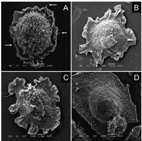
A leaf-like structure and cell flattening
Adherent NRK-52E cells displayed a thin leaf-like structure that closely resembled a lamellipodium, or an undulating ruffle, on the bottom surface of the cell culture vessel (Fig. 3A), and it was partially flattened at the periphery (Fig. 3B). The thickness of the flattened cellular part was greater than that of the leaf-like structure, and the area of the flattened part varied
(Fig. 3B, C, D). Our impression was that the cell shown in Fig. 3D had the longest attachment duration, compared with the cells appearing in Fig. 3A, B C. Because the leaf-like structure developed at the periphery of the flattened cellular part, it appeared to help expand the area of the flattened cellular part. When the cell density was small, the leaf-like structures developed circumferentially (Fig. 3A, B, C), indicating that the cells were not yet at the stage of directional migration. Interestingly, microvilli were seen on the flattened cellular parts but not on the leaf- like structures (Fig. 3B, C).
Formation of fine filopodia in later stages of cell adhesion
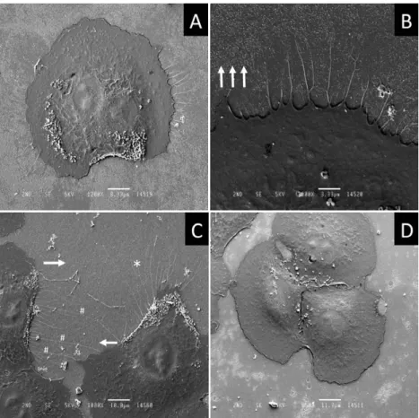
Seven hours after transfer to a new cell culture vessel, the NRK-52E cells had become almost completely flattened except for the bump of the nucleus and / or its surrounds (Fig. 4A). Some NRK-52E cells were at a stage of directional cell migration. One interesting observation was that long and branched filopodia developed, which were much larger in diameter (approx.
0.24 µm ; asterisk in Fig. 4C) compared to the filopodia that developed in the early stages of cell adhesion (approx. 0.05 µm in diameter ; arrows in Fig. 3A), or the filopodia that developed after scratching (manuscript in preparation). The maximum lengths of the filopodia appearing in
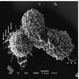
Fig. 2. Formation of membrane-like structures A representative scanning electron microscopy image of harvested and resuspended NRK-52E cells 15 min after transfer to a culture dish. Note that both cells are capable of developing a membrane- like structure : one with microvilli (arrows) and the other with small spherical structures (arrow-heads). Magnification : 3,000. Bar length : 3.33 µm.
Fig. 4 were about 50 µm. From a morphologic point of view, we had the impression that these filopodia were, at least partially, used to detect the position of the adjacent cell, and determine its direction of movement (Fig. 4C).
Discussion
In the present study, we used NRK-52E cells and employed SEM techniques to disclose the elaborate ultrastructure of cultured renal tubular epithelium at various time points in culture.
The major findings were : 1) a large number of small sphere-like structures and / or microvilli were apparent on the cell surface of the floating epithelium ; 2) cells with spherical structures, as well as cells with microvilli, were both capable of developing leaf-like structures after attaching to a cell culture vessel ; 3) the leaf-like structures resembling lamellipodia developed circumferentially at an early stage of culture ; and 4) long and branched filopodia developed which were much larger in diameter than the filopodia that developed at earlier stages.
Fig. 3. Representative scanning electron microscopy images of NRK-52E cells 30 min after attaching to the dish. The NRK-52E cell forms membrane- like structures closely resembling lamellipodia (A), and parts of the cell become flat (B). The flattened cellular areas expand (C) and (D). Arrows in (A) are filopodia. The diameters of filopodia and microvilli are similar.
Magnification : (A)5,500 ; (B)3,500 ; (C)2,700 ; (D)2,800. Bar length :
(A)1.81 µm ; (B)2.85 µm ; (C)3.70 µm ; (D)3.57 µm.
To our knowledge, small spherical structures on the cell surface of renal tubular epithelium have not been described previously, although SEM techniques have been used in cellular biological studies20, 21). The coexistence of microvilli with spherical structures (Fig. 1B), as well as the common ability to develop lamellipodia (Fig. 2) and a possible disappearance of the spherical structures on the cell surface (Fig. 3B, C, D), indicate the possibility that there is convertibility between the microvilli and spherical structures. Using light microscopic observation, the duration until detachment from the bottom surface of the culture vessel after exposure to trypsin varied considerably among NRK-52E cells. It is possible that this duration might be one of the major contributing factors determinating cell surface structures. Definitely, further studies are necessary to show a definite reversibility of these structures on the cell surface and elucidate the molecular mechanisms involved in these structures.
Lamellipodium formation at the periphery is an essential process in directional cell migration7)
Fig. 4. Representative scanning electron microscopy images of NRK-52E cells, seven hours after attaching to the culture dish. The NRK-52E cell becomes flat, and forms filopodia (A). At higher magnification, the filopodia appear to be branched (B). Apparently, two cells are connected by long filopodia (#)
that have a diameter of approximately 0.24 µm (C). The white arrows in Figs.
4B and 4C indicate the direction of cell migration. Magnification : (A)1,200 ;
(B)3,000 ; (C)1,000 ; (D)850. Bar length : (A) 8.33 µm ; (B) 3.33 µm ;
(C) 10.0 µm ; (D) 11.7 µm.
where dense actin polymerization is required to create the forces involved in migration8). In the present study, we observed thin leaf-like structures when the cell was attaching and spreading
(Fig. 3A, B, C). Lamellipodia are defined as the thinner regions located at the cell periphery where the actin fiber is much denser than in the remainder of the cell9, 22-24). However, in the lamellipodia definition, cellular polarity is not necessarily taken into account. Thus, there is a promising possibility that these leaf-like structures and the lamellipodia of colcemid-treated fibroblasts22) are essentially the same. Although we believe it is fair to refer to the leaf-like structures as lamellipodia, further confirmation of actin assembly is necessary before they can be defined as lamellipodia. There are striking similarities between lamellipodia and the membrane ruffles at the leading edge of moving cells, but how we scientifically differentiate between the two remains largely unknown25, 26). Again, molecular analyses of actin assembly are necessary before we can rightly refer to the membrane-like structures as membrane ruffles. Furthermore, it was apparent from a morphologic point of view that the leaf-like structures helped the cell to become flat (Fig. 3B, C).
According to our light microscopic observations, a single NRK-52E cell did not proliferate in culture. The NRK-52E cells interconnected with one another to form a cellular mass, and then they proliferated until reaching confluency (unpublished data). In the present culture system, it took as long as 72 hours to reach confluency, and then it was seven hours after transfer to a new cell culture vessel before we observed the cells that are seen in Fig. 4C.
Thus, we concluded that the cells were directionally migrating. Filopodia are defined as long, thin protrusions at the periphery of cells and growth cones9). We adopted this definition and referred to the thin, long and often branched projections as filopodia, although we did not examine the expression of F-actin bundles on a molecular basis.
The present study has its limitations. Firstly, it remains to be seen whether the ultrastructural changes observed in the NRK-52E cells are also seen in other renal tubular epithelium.
Secondly, our study was conducted in vitro, not in vivo, and thirdly, molecular analyses for actin assembly were not performed.
In conclusion, in spite of these limitations, our SEM analyses have shown a number of ultrastructural aspects of cultured renal tubular epithelium, which have not been previously documented. Understanding the elaborate ultrastructure of renal tubular epithelium may offer opportunities to design therapeutic interventions that might have a strong impact on the treatment of AKI.
Acknowledgements
We sincerely thank Mr. Hiroaki Oniki from the Laboratory of Electron Microscopy, Showa University Fujigaoka Hospital for his excellent technique.
Conflict of interest
The authors have declared no conflict of interest.
References
1) Lombardi D, Becherucci F, Romagnani P. How much can the tubule regenerate and who does it? An open ques- tion. Nephrol Dial Transplant. 2016;31:1243-1250.
2) Fujigaki Y. Different modes of renal proximal tubule regeneration in health and disease. World J Nephrol. 2012;1:92-99.
3) Cirio MC, de Groh ED, de Caestecker MP, et al. Kidney regeneration: common themes from the embryo to the adult. Pediatr Nephrol. 2014;29:553-564.
4) Yoshida M, Honma S. Regeneration of injured renal tubules. J Pharmacol Sci. 2014;124:117-122.
5) Maeshima A, Takahashi S, Nakasatomi M, et al. Diverse cell populations involved in regeneration of renal tubular epithelium following acute kidney injury. Stem Cells Int. 2015;2015:964849.
6) Petrie RJ, Doyle AD, Yamada KM. Random versus directionally persistent cell migration. Nat Rev Mol Cell Biol.
2009;10:538-549.
7) Mitchison TJ, Cramer LP. Actin-based cell motility and cell locomotion. Cell. 1996;84:371-379.
8) Lauffenburger DA, Horwitz AF. Cell migration: a physically integrated molecular process. Cell. 1996;84:359-369.
9) Parsons JT, Horwitz AR, Schwartz MA. Cell adhesion: integrating cytoskeletal dynamics and cellular tension. Nat Rev Mol Cell Biol. 2010;11:633-643.
10) Takenawa T, Suetsugu S. The WASP-WAVE protein network: connecting the membrane to the cytoskeleton. Nat Rev Mol Cell Biol. 2007;8:37-48.
11) Padrick SB, Rosen MK. Physical mechanisms of signal integration by WASP family proteins. Annu Rev Biochem.
2010;79:707-735.
12) Edwards M, Zwolak A, Schafer DA, et al. Capping protein regulators fine-tune actin assembly dynamics. Nat Rev Mol Cell Biol. 2014;15:677-689.
13) Krause M, Gautreau A. Steering cell migration: lamellipodium dynamics and the regulation of directional persis- tence. Nat Rev Mol Cell Biol. 2014;15:577-590.
14) Cazaux J. Recent developments and new strategies in scanning electron microscopy. J Microsc. 2005;217(Pt 1):16-35.
15) Fleck RA. Low-temperature electron microscopy: techniques and protocols. Methods Mol Biol. 2015;1257:243-274.
16) de Larco JE, Todaro GJ. Epithelioid and fibroblastic rat kidney cell clones: epidermal growth factor (EGF) recep- tors and the effect of mouse sarcoma virus transformation. J Cell Physiol. 1978;94:335-342.
17) Scherer WP, Scherer MD. Scanning electron microscope imaging of onychomycosis. J Am Podiatr Med Assoc.
2004;94:356-362.
18) Yue X, Li Q, Wang H, et al. An ultrastructural study of Trichophyton rubrum induced onychomycosis. BMC Infect Dis. 2015;15:532.
19) Zhang H, Cui YC, Tian Y, et al. A novel model for evaluating thrombolytic therapy in dogs with ST-elevation myocardial infarction. BMC Cardiovasc Disord. 2016;16:21.
20) Terryn S, Jouret F, Vandenabeele F, et al. A primary culture of mouse proximal tubular cells, established on collagen-coated membranes. Am J Physiol Renal Physiol. 2007;293:F476-F485.
21) Zhang K, Guo X, Zhao W, et al. Application of Wnt pathway inhibitor delivering scaffold for inhibiting fibrosis in urethra strictures: in vitro and in vivo study. Int J Mol Sci. 2015;16:27659-27676.
22) Vasiliev JM. Polarization of pseudopodial activities: cytoskeletal mechanisms. J Cell Sci. 1991;98(Pt 1):1-4.
23) Paluch EK, Raz E. The role and regulation of blebs in cell migration. Curr Opin Cell Biol. 2013;25:582-590.
24) Suetsugu S, Kurisu S, Takenawa T. Dynamic shaping of cellular membranes by phospholipids and membrane- deforming proteins. Physiol Rev. 2014;94:1219-1248.
25) Faix J, Rottner K. The making of filopodia. Curr Opin Cell Biol. 2006;18:18-25.
26) Steffen A, Koestler SA, Rottner K. Requirements for and consequences of Rac-dependent protrusion. Eur J Cell Biol. 2014;93:184-193.
[Received March 3, 2017 : Accepted April 5, 2017]