Specific action of prostacyclin on adipogenesis at different stages of cultured adipocytes
>ᇵ㣴⬡⫫⣽⬊ࡢ␗࡞ࡿࢫࢸ࣮ࢪ࡛ࡢ⬡⫫⣽⬊ᙧᡂ
ᑐࡍࡿࣉࣟࢫࢱࢧࢡࣜࣥࡢ≉␗ⓗస⏝@
Ferdous Khan
Student No.: D11A3105K
Research Field of Biochemistry and Molecular Cell Biology, Division of Applied Resources Chemistry,
The Course of Bioresources Science,
The United Graduate School of Agricultural Sciences, Tottori University
2016
Index
Index 2
Abbreviations 5
Chapter 1 Introduction 7
1.1 Different life stages of adipocytes 7
1.2 Transcriptional regulation of adipogenesis 8 1.3 Role of polyunsaturated fatty acids (PUFAs) and
cyclooxygenase cascade in adipogenesis
9
1.4 Role of prostacyclin in adipogenesis 10
1.5 Intertwined relationship of PUFAs, prostaglandins and cAMP in the life cycle of adipocytes
11
1.6 Mitotic clonal expansion and cAMP-PKA-dependent process 13 1.7 Prostaglandins can promote and block adipogenesis through
cell surface and uclear receptors
14
1.8 PPARJ the master regulator of adipogenesis 16
1.9 Aim of the present studies 16
References 17
Chapter 2 Pretreatment of cultured preadipocytes with arachidonic acid during the differentiation phase without a cAMP-elevating agent enhances fat storage after the maturation phase
26
1. Introduction 26
2. Materials and methods 28
2.1 Materials 28
2.2 Cell culture of 3T3-L1 cells and differentiation to adipocytes
29
2.3 Adipogenesis treatments 29
2.4 Prostanoid quantification by ELISA 29
2.5 Gene expression analysis 30
2.6 Triacylglycerol content and Oil Red O staining 32
2.7 Intracellular cAMP measurement 32
2.8 Other procedures 32
3. Results 33
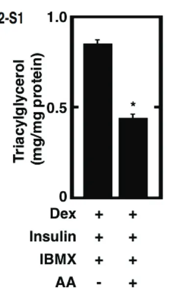
3.1 Pretreatment of cultured preadipocytes with AA during the differentiation phase without IBMX stimulate the storage of fats after the maturation phase
33
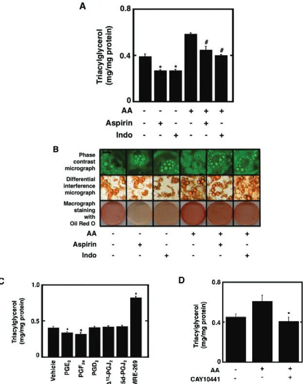
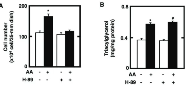
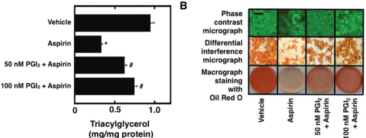
3.2 Effect of the pretreatment of cultured preadipocytes with AA together with either of COX inhibitors, a protein kinase A (PKA) inhibitor, and a IP receptor antagonist, or with prostanoids on the storage of fats
36
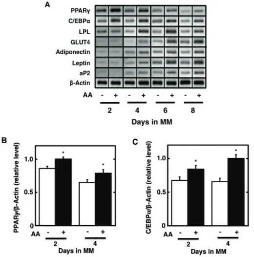
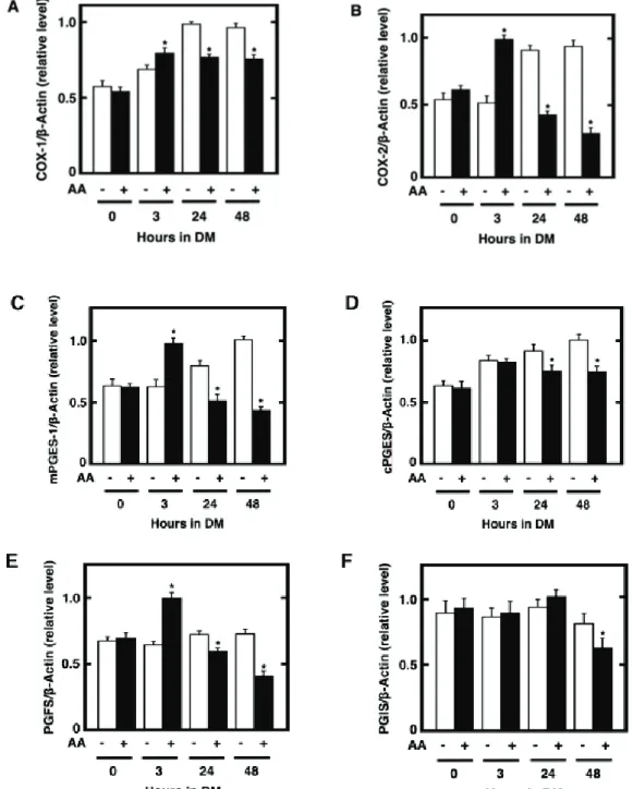
3.3 Gene expression of isoformic enzymes in the COX pathway in cultured preadipocytes treated with AA
39
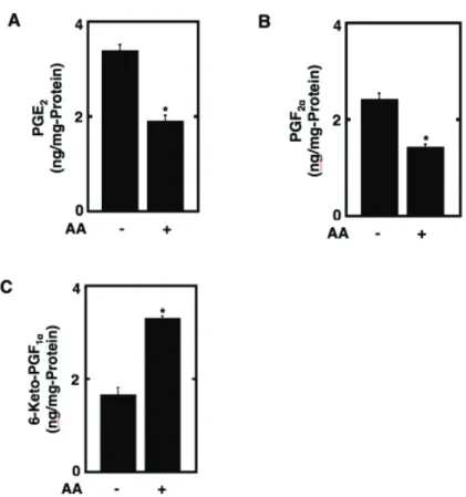
3.4 Biosynthesis of prostanoids by cultured preadipocytes treated with AA
41
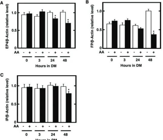
3.5 Gene expression of prostanoid receptors in cultured preadipocytes treated with AA
42
4. Discussion 44
References 47
Supplementary Figures 51
Chapter 3 Stimulation of fat storage by prostacyclin and selective agonists of prostanoid IP receptor during the maturation phase of cultured adipocytes
54
1. Introduction 54
2. Materials and methods 56
2.1 Materials 56
2.2 Cell culture of 3T3-L1 cells and adipogenesis during the maturation phase
56
2.3 Determination of cellular levels of triacylglycerol and protein
57
2.4 Microscopic and macroscopic observation of cultured adipocytes
57
2.5 Others 58
3. Results 58
3.1 Effect of prostacyclin and selective agonists or antagonist for IP receptor on the storage of fats attenuated by aspirin during the maturation phase of adipocytes
58
3.2 Combined effect of a selective agonist for IP receptor and an activator for PPARJ on adipogenesis along with aspirin during the maturation phase of adipocytes
61
3.3 Action of cAMP analogues, forskolin, and a protein kinase A (PKA) inhibitor on the storage of fats during the maturation phase of adipocytes
64
4. Discussion 67
References 72
Chapter 4 Conclusion 76
Summary 76
Summary in Japanese 77
Acknowledgement 80
List of Publications 81
Abbreviations
AA arachidonic acid
20:4 (n-6) arachidonic acid
aP-2 adipocyte protein 2
8-CPT-2’-O-Me-cAMP 8-(4-chlorophenyl)thio-2’-O-methyl-cAMP
8-CPT-cAMP 8-(4-chlorophenyl)thio-cAMP
C/EBP CCAAT enhancer-binding protein
COX cyclooxygenase
CRTH2 chemoattractant receptor-homologous molecule expressed on Th2 cells
Dex dexamethasone
DM differentiation medium
22:6 (n-3) 4, 7, 10, 13, 16, 19-docosahexaenoic acid
DMEM-HEPES Dulbecco’s modified Eagle medium with 25 mM HEPES 20:5 (n-3) 5, 8, 11, 14, 17-eicosapentaenoic acid
ELISA enzyme-linked immunosorbent assay
FBS fetal bovine serum
GLUT glucose transporter
GM growth medium
IBMX 3-isobutyl-1-methylxanthine
Indo indomethacin
18:2 (n-6) linoleic acid
18:3 (n-3) D-linolenic acid
LPL lipoprotein lipase
MM maturation medium
PBS (-) phosphate-buffered saline without Ca2+ and Mg2+
PCR polymerase chain reaction
PG prostaglandin
L-PGDS lipocalin-type PGD synthase
cPGES cytosolic PGE synthase
mPGES membrane-bound PGE synthase
PGFS PGF synthase
PGIS PGI synthase
15d-PGJ2 15-deoxy-'12,14-PGJ2
PKA protein kinase A
PPAR peroxisome proliferator-activated receptor
RT reverse transcriptase
Chapter 1
Introduction
1.1 Different life stages of adipocytes
Excess accumulation of fat is termed as obesity, which is characterized by the increase in the number and/or size of adipocytes (Flier 1995). The life cycle of adipocytes includes three distinct phases: the cell growth of preadipocytes, triggering of differentiation by appropriate hormonal and biochemical stimulation and the terminal differentiation. The differentiation of adipocyte is associated with the conversion of cells from fibroblastic to spherical shape (Rosen and MacDougald 2006). These morphological modifications are accomplished by changes in the level of extracellular matrix component and the level of cytoskeletal components (Gregoire et al. 1998). Recently, progression has been made in understanding the process of adipocytes differentiation and the cellular and molecular basis of adipose tissue growth. However, complete understanding of the cellular and molecular mechanism of adipose tissue growth in physiological and pathophysiological states is important to develop newer therapeutic strategies for the prevention and treatment of obesity. The most frequently employed cell lines to understand adipogenesis are
3T3-F442A and 3T3-L1. These were clonally isolated from Swiss 3T3 cells derived from disaggregated 17 to 19-days mouse embryos (Green and Kehinde 1975). 3T3-L1
preadipocyes spontaneously differentiate over a period of several weeks into fat-cell clusters when maintained in culture with fetal calf serum (Fig. 1-1).
During the growth phase, cells of preadipocyte lines as well as primary preadipocytes are morphologically similar to fibroblasts. At confluence, induction of differentiation by
appropriate combination of hormonal and biochemical stimulators leads to dramatic change in cell shape (Fan et al. 1983). For example, for triggering the differentiation of murine 3T3-L1 preadipocytes in vitro, an adequate combination of glucocorticoid, insulin and cyclic AMP (cAMP) elevating agent in presence of fetal bovine serum in Dulbecco’s modified Eagle’s medium (DMEM) is necessary (Student et al. 1980; Rubin et al. 1978).
The absence of any one of these abovementioned inducers in DMEM results in severe impediment of differentiation and eventually decreased lipid accumulation in 3T3-L1 cells (Madsen et al. 2005; Petersen et al. 2003). The preadipocyte converts to a spherical shape, accumulates lipid droplets, and progressively acquires the morphological and biochemical
characteristics of the mature adipocytes (Gregoire et al. 1998). During the terminal phase of differentiation, activation of the transcriptional cascade leads to increased activity, proteins, and mRNA levels for enzymes involved in triacylglycerol synthesis and degradation
(Gregoire 2001).
Fig. 1-1. Schematic presentation of life cycle of mouse embryogenic 3T3-L1 preadipocytes and the regulation by mouse embryonic 3T3-L1 cells and the regulation by C/EBPs and PPARJ
1.2 Transcriptional regulation of adipogenesis
During adipogenesis fibroblast-like preadipocytes differentiate into lipid-laden, insulin sensitive adipocyte via a complex processes including coordinated changes in hormone sensitivity and gene expression (Lefterova and Lazar 2009; Fujimori et al. 2012).
The treatment of confluent preadipocytes by the optimum combination of insulin,
glucocorticoid and cAMP elevating agent during the clonal expansion phase can optimally trigger several signal transduction events, which eventually culminates into the enhanced expression of adipocyte-specific marker genes, and hence formation of mature adipose cells. Genes differentially regulated during adipogenesis has been categorized into early, intermediate and late mRNA/protein markers (Rangwala and Lazzar, 2000; Morrison and Farmer, 2000; Rosen and Spiegelman, 2000; Boone et al. 2000; Ntambi and Young 2000).
This process involves the orchestrated activation of several signal transduction events in which numerous signaling molecules play very important roles through cell membrane receptors and nuclear receptors (Ailhaud 1999). Based on two-dimensional electrophoresis of cell extract before, during, and after adipose conversion, researchers have estimated that
at least 300 proteins are differentially expressed from the commencement of hormonal and biochemical induction up to the commitment of mature adipocyte phenotype (Sadowski et al. 1992).
Among numerous signaling molecules CCAAT/enhancer binding protein E and G (C/EBPE and C/EBPG) have been reported to be the first transcription factors induced after exposure of preadipocytes to differentiation cocktail containing dexamethasone, insulin and 3-isobutyl-1-methylxanthine (IBMX) in presence of fetal bovine serum (FBS) (Aubert et al.
2000; Cornelius et al. 1994; Ntambi and Young 2000; Otto and Lane 2005). Among these inducers IBMX increases cAMP, which in turn control the expression of numerous genes, including C/EBPE, through protein kinase A (PKA) mediated phosphorylation of cAMP responsive element binding protein (CREB) (Reusch et al. 2000; Zhang et al. 2004). The induction of C/EBPE and C/EBPG has been reported to mediate the expression of two master regulators (Lefternova and Lazar 2009) of adipogenesis: C/EBPD (Lane et al. 1999) and peroxisome proliferator-activated receptor gamma (PPARJ) (Clarke et al. 1997; Wu et al. 1995; Ham et al. 2001). Although the expression of C/EBPD is not strictly
adipocyte-specific, PPARJ is the most adipocyte-specific among the three PPAR isoforms (Gregorie et al. 1998). After activation PPARJ and C/EBPD can cross-regulate the
expression and activity of each other (Shao and Lazar 1997). Moreover combined expression of these two factors is reported to possess synergistic effect on the differentiation of 3T3-L1 adipocytes by up-regulating the expression of several
adipocyte-specific genes encoding proteins and enzymes involved in the accumulation of lipid (Tontonz et al. 1994a; Gregorie et al. 1998). For example, PPARJ and C/EBPD alone or in combination have been reported to co-ordinate the activation of genes encoding adipocyte-specific fatty acid binding protein (aP2) (Tontonoz et al 1994b; Herrera et al.
1989), GLUT4, adiponectin, (Long and Pekala 1996) and leptin (Hollenberg et al. 1997).
1.3 Role of polyunsaturated fatty acid (PUFA) and cyclooxygenase cascade in adipogenesis
Arachidonic acid, an omega-6 (n-6) polyunsaturated fatty acid, is released from the membrane phospholipids in response to a variety of stimuli. After release with the help of distinct enzymatic pathways such as cyclooxygenase and lipoxygenase pathways
arachidonic acid is converted into bioactive lipid mediators, which are also known as
eicosanoids (Needleman et al. 1986; Yamamoto 1992). The rate-limiting step in the biosynthesis of prostanoids is the conversion of arachidonic acid into prostaglandin H2, which is catalyzed by the prostaglandin endoperoxide H synthases-1 and 2 (PGHS-1 and PGHS-2) also known as cyclooxygenases-1 and 2 (COX-1 and COX-2) (Smith et al. 2000).
The constitutive COX-1 is mainly utilized in the immediate PG biosynthesis, which occurs within several minutes after stimulation with Ca2+ mobilizers, where as the inducible COX-2 is an absolute requirement for delayed PG biosynthesis, which last for several hours after triggering by proinflamatory stimuli (Rahman et al. 2013). PGH2 is subsequently used as substrate by several terminal prostaglandin synthases for the biosynthesis of corresponding prostaglandins. Prostaglandins produced by arachidonate cyclooxygenase pathway possess a diverse range of functions depending on the PG types and cell target. Among those effects, prostaglandins and their metabolites play either pro-adipogenic or anti-adipogenic role in controlling the differentiation of preadipocytes to mature adipocytes. For example, PGE2 and PGF2α are reported as anti-adipogenic (Serrero et al. 1992a and b) on the other hand, PGI2, PGD2, 15-deoxy Δ12,14-PGJ2 have been reported as pro-adipogenic (Forman et al. 1995a and b; Forman et al. 1997; Kliewer et al. 1995; Massiera et al. 2003) prostaglandins. Polyunsaturated fatty acids (PUFA) of n-3 series, for example eicosapentaenoic acid (EPA), docosahexaenoic acid decreased adipose tissue mass and suppress the development of obesity in rodents whereas n-6 PUFAs (arachidonic acid or linoleic acid) exerted either anti- or pro-adipogenic effects depending on the experimental conditions when used for triggering adipogenesis in growth arrested post-confluent preadipocytes, (Madsen et al. 2005).
1.4 Role of prostacyclin in adipogenesis
Prostacyclin also known as prostaglandin (PG) I2 is an unstable metabolite, which is converted rapidly to 6-keto-PGF1α by spontaneous hydrolysis (Kelton and Blajchman 1980).
Earlier studies have suggested that prostacyclin analogues can act as a potent inducer of pre-adipocyte differentiation. For example, carbaprostacyclin, a stable analogue of prostacyclin stimulates terminal differentiation of Ob1771 mouse pre-adipose cells and 3T3-F442A cells in serum-free hormone-supplemented medium (Negrel et al. 1989;
Catalioto et al. 1991). A recent study using a cell-based reporter gene assay, in HEK-293 cells stably expressing the IP receptor, has reported that PPARJ, a master regulator of adipogenesis, is activated through the IP receptor via a cAMP-independent mechanism by
stable prostacyclin analogues (Falcetti et al. 2007). Another study has described that fat mass gain is suppressed in IP-knockout mice compared with wild-type mice when mother mice were fed high-fat diet supplemented with linoleic acid, suggesting the contribution of the signaling through the IP receptor to adipose tissue development (Massiera et al. 2003).
In addition, our previous study supports the potential idea that the increased synthesis of endogenous prostacyclin by adipocytes during the maturation phase can stimulate adipogenesis in an autocrine manner by interacting with its specific IP receptor, the expression of which is regulated coordinately with PGIS (Rahman et al. 2014). But interestingly, Hopkins and Gorman reported that the repeated addition of exogenous PGI2
to cultured 3T3-L1 cells inhibited insulin- and indomethacin-mediated adipocyte differentiation (Hopkins and Gorman 1981).
Prostacyclin has been reported to be the primary product of COX-2 in many systems (McAdam et al. 1999) and preferentially synthesized by preadipocytes (Hyman et al. 1982).
In our earlier study, we reported that the biosynthesis of prostacyclin was gradually
suppressed after the induction of differentiation by dexamethasone, insulin and IBMX; but this suppression was rescued during the maturation phase, when the terminally
differentiating 3T3-L1 cells were exposed to DMEM containing 10% FBS and 5 Pg/ml insulin (Rahman et al. 2014). This suppressed biosynthesis of PGI2, during the induction of differentiation, might be attributed to the presence of dexamethasone, an anti-inflammatory synthetic compound acting through the glucocorticoid receptor (Goppelt-Struebe et al.
1989). Recently Lasa et al reported that dexamethasone can destabilize COX-2 mRNA by inhibiting mitogen-activated protein kinase (MAPK) p38 (Lasa et al. 2001).
1.5 Intertwined relationship of PUFAs, prostaglndins and cAMP in the life cycle of adipocytes
Besides prostaglandins another potent effector of adipogenesis is the second messenger molecule, cyclic AMP (cAMP) an important intracellular signaling molecule, generally control several signal transduction events via the activation of protein kinase A (PKA) (Kato et al. 2007). Arachidonic acid through the synthesis of prostacyclin (Négrel 1999) and PGE2 (Tsuboi et al. 2004) is able to increase the cAMP level (Hans-Erik 1980).
Conversely, Griesmacher and colleagues reported that the endothelial cells of human umbilical vein is able to release prostacyclin that is dependent on intracellular Ca2+ ion
mobilization but independent of intracellular cAMP level (Griesmacher et al. 1992).
Moreover, Williams and co-workers reported that, forskolin is able to stimulate prostacyclin synthesis in rabbit heart that was independent of cAMP but dependent on intracellular and extracellular calcium (Williams and Malik 1990). However, forskolin, a potent receptor-independent adenylyl cyclase activator, rapidly elevated cAMP synthesis and under this condition cultured mouse peritoneal macrophages were able to suppress the synthesis of prostacyclin measured as 6-keto-PGF1D (Chang et al. 1984). A widely used component of adipogenic cocktail, 3-isobutyl-1-methylxanthine (IBMX) through the inhibition of phosphodiesterases (PDE) elevates the level of cAMP (Fawcett et al. 2000).
Though baseline cAMP is necessary for triggering the differentiation of 3T3-L1 cells, elevation of cAMP above a critical level is reported to activate anti-adipogenic cellular signaling processes, which results in reduced fat accumulation (Li et al. 2008; Petersen et al. 2003).
The pro- or anti-adipogenic effect of n-6 polyunsaturated fatty acids (PUFAs) is attributed to the balance between carbohydrate and protein in diet of C57BL/6JBomTac mice. A high protein/carbohydrate ratio in diet is translated into a high glucagon/insulin ratio in blood, which enhances cAMP-dependent signaling. In this condition of augmented cAMP-dependent signaling, n-6 PUFAs are able to stimulate the production of
anti-adipogenic prostaglandins. This anti-adipogenic action can be restored by lowering the cAMP level either by the exclusion of n-6 PUFA or IBMX or by suppressing
anti-adipogenic prostanoid synthesis by inclusion of cyclooxygenase inhibitor (Madsen et al. 2008). These results also support the notion that augmented synthesis of PGE2 and PGF2D can thwart the conversion preadipocytes to mature adipocytes (Reginato et al. 1998).
Alternatively, Casimir and co-workers observed reduced production of endogenous PGF2D
in differentiating cells compared to un-stimulated preadipocytes and hence they concluded that PGF2D production by preadipocytes is one of the several prerequisites for the
maintenance of the undifferentiated state (Casimir et al. 1996). Moreover, during the early phase of adipogenesis, enhancement of COX-2 activity, through PGF2D-activated FP receptor/extracellular-signal-regulated kinase (ERK)/cyclic AMP response element binding protein (CREB) cascade, preadipocytes can enhance the production of PGF2D and PGE2, and thuscan form positive feed back loop of inhibitory action (Ueno and Fujimori 2011).
1.6 Mitotic clonal expansion and cAMP-PKA-dependent process
Growth-arrested confluent preadipocytes, if exposed to appropriate mitogenic and adipogenic signals, re-enter the cell cycle (Qiu et al. 2001) and undergo at least one round of DNA replication followed by the doubling of cell numbers (Gregorie et al. 1998), which is termed as mitotic clonal expansion (Lefterova and Lazar, 2009). Cyclic AMP (cAMP) is an important intracellular signaling molecule, generally control several signal transduction events via the activation of cAMP-dependent protein kinase A (PKA) (Kato et al. 2007).
The activation of PKA is necessary for mitotic clonal expansion (MCE) (Martini et al.
2009), and for cAMP responsive element binding protein (CREB)-dependent stimulation of CCAAT/enhancer binding protein E and G (C/EBPE and C/EBPG) gene expression
(Belmonte et al. 2001). Both MCE and C/EBPE and C/EBPG gene expression are very important events during first 2 days after induction of differentiation (Gregorie et al. 1998).
In the hierarchy of molecular and cellular events the expression of C/EBPE and C/EBPG is followed by the expression of PPARJ and C/EBPD: the master regulators (Lefternova and Lazar 2009) of adipocyte conversion (Gregorie et al. 1998; Cornelius et al. 1994; Ntambi and Young 2000; Clarke et al. 1997; Wu et al. 1995). C/EBPD through its antimitotic activity (Umek et al. 1991) and PPARJ through its ability to induce cell cycle withdrawal are strongly implicated in the growth arrest (Altiok et al. 1997). MCE is followed by the expression of adipogenic genes leading to adipocyte differentiation (Bernlohr et al. 1985;
Conrnelius et al. 1994; MacDougald and Lane 1995). Numerous studies support the
necessity of MCE in the differentiation of preadipocytes to mature adipocytes. For example, tumor necrosis factor alpha (TNFD) inhibits 3T3-L1 cell differentiation by blocking the MCE through the perturbation of p130 and p107 protein levels: two members of
retinoblastoma protein family, which are involved in the regulation of cell cycle events (Lyle et al. 1998). Consistent with this, Patel and Lane reported that inhibition of calpain activity by N-acetyl-Leu-Leu-norleucinal (ALLN), during the early stages, blocks both MCE and differentiation of 3T3-L1 cells (Patel and Lane 2000). However, controversial evidences made the role and necessity of MCE, and its mediator PKA, obscure in the differentiation of 3T3-L1 preadipocytes. For example, cAMP-PKA-dependent signaling is necessary for MCE but cAMP-PKA-independent signaling is necessary for the
differentiation of 3T3-L1 preadipocytes (Martini et al. 2009).
Although PKA was initially thought to be the only protein activated by cAMP;
PKA-independent cAMP-mediated cellular signaling processes have been reported (Chin and Abayasekara 2004; Holz et al. 2006; Kanda and Wantanabe 2007; Yin et al. 2006), which are also involved in controlling adipogenesis. For example, cAMP can activate Epac (exchange protein directly activated by cAMP); synergistic action between Epac and PKA is necessary to promote adipogenesis of 3T3-L1 cells (Petersen et al. 2008). Additionally, activated PKA can activate protein kinase B (PKB also known as Akt), through a
PI3-kinase-independent pathway (Filippa et al. 1999). Cyclic AMP mediated activation of PKA and PKB is necessary for the transcriptional activation of PPARγ (Kim et al. 2010).
Arachidonic acid through the synthesis of prostacyclin (Négrel 1999) and
3-isobutyl-1-methylxanthine (IBMX) through the inhibition of phosphodiesterases (PDE) (Fawcett et al. 2000) are able to elevate the level of cAMP; therefore co-administration of these two agents in the adipogenic cocktail may result in excessive elevation of cAMP level and activation PKA-dependent processes. All these evidences consolidate the notion that mitotic clonal expansion, a milestone in the highway of preadipocyte differentiation, is not an unavoidable step in the process of adipogenesis.
1.7 Prostaglandins can promote and block adipogenesis through cell surface and nuclear receptors
Certain cell surface receptors (Tsuboi et al. 2004) and nuclear receptors (Yu et al. 1995) are involved in controlling the series of signal transduction events leading to the
commitment of preadipose cells to mature adipocytes (Ailhaud 1999). Extensive studies over last few decades have reveled that prostaglandins, which are naturally produced in different life stages of adipocytes, play a variety of roles in adipogenesis through the cell-surface receptors and nuclear receptors (Clarke et al. 1997). For example, prostacyclin produced near the cell membrane can act as an autocrine or a paracrine effector molecule by activating the G-protein coupled seven transmembrane IP-receptor of the same cell or neighboring cells respectively, to activate adenylyl cyclase and increase cAMP (Narumiya et al. 1999; Wise 2003). On the other hand, prostacyclin produced near the nucleus if transported into the nucleus may act as a ligand for nuclear receptors: peroxisome proliferator-activated receptors (PPARs), the nuclear receptor superfamily of
ligand-activated transcription factors, both in presence and in absence of functional IP receptor and can influence gene expression and thus may be considered as an intracrine mediator of cell signaling process (Lim and Dey 2002). For example, prostacyclin
analogues are able to induce the activation of PPARD and PPARG in monkey kidney CV-1 and human hepatoma HepC2 cells not expressing the IP receptor (Hertz et al. 1996). In a recent study, Falcetti and co-workers reported that human embryonic kidney (HEK-293) cells stably expressing the IP receptor are able to activate PPARJ through the cell surface IP receptor via a cAMP-independent mechanism by stable prostacyclin analogues (Falcetti et al. 2007). In our earlier study we reported that cell surface IP-receptor, which is expressed in preadipocytes, is temporarily suppressed after hormonal induction of differentiation and up-regulated during the terminal differentiation and eventually down-regulated in the matured adipocytes. This peaks and valleys in the expression of IP receptor was also reflected as synchronized augmentation and diminution of the expression of prostacyclin synthase (PGIS) and hence prostacyclin itself. This enhancement of IP receptor mediated activity was also confirmed by the ability of IP agonists to partially rescue cyclooxygenase inhibitor suppressed adipogenesis during the maturation phase of 3T3-L1 cells (Rahman et al. 2014).
Prostaglandins are also able to interact with PPARJ, the master regulator of adipogenesis and thus can promote or block adipogenesis (Reginato et al. 1998). For example, derivatives of prostaglandin D2 (PGD2) including 15-deoxy-'12,14-prostaglandin J2 (15d-PGJ2) and '12-prostaglandin J2 ('12-PGJ2) are ligands of PPARJ (Forman et al.
1995b; Mazid et al. 2006). In our earlier studies, we have also reported that the
biosynthesis of 15d-PGJ2 (Mazid et al. 2006) and '12-PGJ2 (Syeda et al. 2012a), natural ligands of PPARJ, are increased during the maturation phase of adipogenesis when the gene expression of PPARJ also reach to its maximum level. Conversely, anti-adipogenic prostaglandin PGE2, which is synthesized mainly by undifferentiated preadipocytes, through EP4 receptor, is able to increase intracellular cAMP level (Fujimori, 2012), which inhibits PPARJ activity and eventually down-regulates the early stages of 3T3-L1
preadipocytes differentiation (Tsuboi et al. 2004). Another antiadipogenic prostaglandin, PGF2D, blocks adipogenesis through the activation of mitogen-activated protein kinase, resulting in inhibitory phosphorylation of PPARJ. Both mitogen-activated protein kinase activation and PPARJ phosphorylation are required for the anti-adipogenic effects of PGF2D. Thus, PG signals, generated through cell surface receptor, regulate the activities of a
nuclear hormone receptor that can also be directly activated or inhibited by other PGs. The balance among PGI2, PGE2, PGF2D, 15d-PGJ2 and '12-PGJ2, and their signaling may be
central to the development of obesity and diabetes (Reginato et al. 1998).
1.8 PPARJJ the master regulator of adipogenesis
The three peroxisome proliferator-activated receptor (PPAR) isotypes, PPARD, PPARE (also known as PPARG), and PPARγ form a subfamily of nuclear receptors mainly involved in lipid and glucose homeostasis, regulation of food intake and body weight, control of inflammation and wound healing (Chawla et al. 1994; Farmer 2005). Among these three, PPARγ is preferentially expressed expressed in adipose tissue (Tontonz et al. 1994b).
Ectopic expression of PPARJ in non-adipogenic mouse fibroblasts, which is known as gain of function study, revealed that PPARγ alone can initiate the entire adipogenic program with the accomplishment of mature adipocyte phenotype (Tontonz et al. 1994a).
Conversely, loss of function studies reconfirms the necessity of PPARJ in fat cell formation (Farmer 2006; Kim et al. 2010). Transfection of dominant-negative mutant of PPARγ into mature 3T3-L1 adipocytes causes loss of lipid accumulation and decreased expression of adipocyte markers, suggesting the requirement of PPARγ for the maintenance of
differentiated state (Tamori et al. 2002). PPARJ null fibroblasts and embryonic stem cells as well as dominant negative PPARJ mutant were failed to differentiate in vitro (Kubota et al. 1999; Rosen et al. 1999; Barroso et al. 1999). Numerous reports gathered in last few years consolidated the notion that no transcriptional regulator has been discovered that promotes adipocyte differentiation in the absence of PPARJ; hence all pro-adipogenic factors somehow exert their effect through stimulating PPARJ gene expression or activity (Christodoulides and Vidal 2010). Conversely, many of the anti-adipogenic factors exert their activity by suppressing the gene expression of PPARJ or by deactivating PPARJ by post-translational modification (Reginato et al. 1998; Fujimori 2012; Anghel 2007). Thus numerous studies uphold the notion that PPARγ activity is absolutely necessary for
adipogenesis, and no factor has been discovered until now which can promote adipogenesis in the absence of PPARγ activity (Christodoulides and Vidal 2010).
1.9 Aim of the present studies
By considering the intertwined role of cAMP, PUFAs and their products prostaglandins and the receptors of prostaglandins in adipogenesis, in this study, we attempted to
determine the effect of the pretreatment of cultured 3T3-L1 preadipocytes with AA during the differentiation phase without IBMX. Our aim of this study was to investigate the impact
of this pre-treatment upon adipogenesis of cultured adipocytes after the maturation phase, and to seek the cellular mechanisms underlying the effect of AA. Moreover, archidonic acid can be converted to prostacyclin and the prostacyclin can elevate cAMP level as well as can interact with not only cell surface IP receptor but also nuclear receptor PPARJ. The gene expression of these two receptors reaches to the maximum level during the maturation phase of 3T3-L1 cells. Hence in the subsequent part of our study, we attempt to determine the specific action of the parent prostacylin and the related agonists or antagonists for the specific IP receptor on adipogenesis in combination with the agents that influence the activation of PPARJ and the elevation of cAMP. We discuss the mode of how prostacylin affects adipogenesis through the IP receptor during the maturation phase of adipocytes.
Reference
Ailhaud G (1999) Cell surface receptors, nuclear receptors and ligands that regulate adipose tissue development. Clin Chim Acta 286: 181-90
Altiok S, Xu M, Spiegelman BM (1997) PPAR gamma induces cell cycle withdrawal:
inhibition of E2F/DP DNA-binding activity via down-regulation of PP2A. Genes Dev 11:1987-98
Anghel SI, Wahli W. Fat poetry: a kingdom for PPAR gamma (2007) Cell Res 17: 486-511 Barroso I, Gurnell M, Crowley VE, Agostini M, Schwabe JW, Soos MA, Maslen GL,
Williams TD, Lewis H, Schafer AJ, Chatterjee VK, O'Rahilly S (1999) Dominant negative mutations in human PPARgamma associated with severe insulin resistance, diabetes mellitus and hypertension. Nature 402:880-883
Belmonte N, Phillips BW, Massiera F, Villageois P, Wdziekonski B, Saint-Marc P, Nichols J, Aubert J, Saeki K, Yuo A, Narumiya S, Ailhaud G, Dani C (2001) Activation of extracellular signal-regulated kinases and CREB/ATF-1 mediate the expressionof CCAAT/enhancer binding proteins beta and -delta in preadipocytes. Mol Endocrinol 11:2037-49
Bernlohr DA, Bolanowski MA, Kelly TJ Jr, Lane MD (1985) Evidence for an increase in transcription of specific mRNAs during differentiation of 3T3-L1 preadipocytes. J Biol Chem 260:5563-7
Boone C, Mourot J, Grégoire F, Remacle C (2000) The adipose conversion process:
regulation by extracellular and intracellular factors. Reprod Nutr Dev 40:325-58 Casimir DA, Miller CW, Ntambi JM (1996) Preadipocyte differentiation blocked by
prostaglandin stimulation of prostanoid FP2 receptor in murine 3T3-L1 cells.
Differentiation 60:203-10
Catalioto RM, Gaillard D, Maclouf J, Ailhaud G, Negrel R (1991) Autocrine control of adipose cell differentiation by prostacyclin and PGF2 alpha. Biochim Biophys Acta 1091:364-9
Chang J, Cherney ML, Moyer JA, Lewis AJ (1984) Effect of forskolin on prostaglandin synthesis by mouse resident peritoneal macrophages. Eur J Pharmacol 103:303-12 Chawla A, Schwartz EJ, Dimaculangan DD, Lazar MA (1994) Peroxisome
proliferator-activated receptor (PPAR) J: adipose-predominant expression and induction early in adipocyte differentiation. Endocrinology 135:798-800
Chin EC, Abayasekara DRE, (2004) Progesterone secretion by luteinizing human granulosa cells: a possible cAMP-dependent but PKA-independent mechanism involved in its regulation. Journal of Endocrinology 183:51–60
Christodoulides C, Vidal-Puig A (2010) PPARs and adipocyte function. Mol Cell Endocrinol 318:61-68
Clarke SL, Robinson CE, Gimble JM (1997) CAAT/enhancer binding proteins directly modulate transcription from the peroxisome proliferator-activated receptor gamma 2 promoter. Biochem Biophys Res Commun 240:99-103
Cornelius P, MacDougald OA, Lane MD (1994) Regulation of adipocyte development.
Annu Rev Nutr 14:99-129
Falcetti E, Flavell DM, Staels B, Tinker A, Haworth SG, Clapp LH (2007) IP
receptor-dependent activation of PPARJ by stable prostacyclin analogues. Biochem Biophys Res Commun 360:821-827
Fan JY, Carpentier JL, van Obberghen E, Grunfeld C, Gorden P, Orci L (1983) Morphological changes of the 3T3-L1 fibroblast plasma membrane upon differentiation to the adipocyte form. J Cell Sci 61:219-30
Farmer SR (2005) Regulation of PPARgamma activity during adipogenesis. Int J Obes 29 Suppl 1:S13-16
Farmer SR (2006) Transcriptional control of adipocyte formation. Cell Metab 4:263-273 Fawcett L, Baxendale R, Stacey P, McGrouther C, Harrow I, Soderling S, Hetman J, Beavo
JA, Phillips SC (2000) Molecular cloning and characterization of a distinct human phosphodiesterase gene family: PDE11A. Proc Natl Acad Sci USA 97:3702-3707 Feige JN, Gelman L, Michalik L, Desvergne B, Wahli W (2006) From molecular action to
physiological outputs: peroxisome proliferator-activated receptors are nuclear receptors at the crossroads of key cellular functions. Prog Lipid Res 45:120-59 Filippa N, Sable CL, Filloux C, Hemmings B, Van Obberghen E (1999) Mechanism of
protein kinase B activation by cyclic AMP-dependent protein kinase. Mol Cell Biol 19:4989-5000
Flier JS (1995) The adipocyte: storage depot or node on the energy information superhighway? Cell 80:15-8
Forman BM, Chen J, Evans RM (1997) Hypolipidemic drugs, polyunsaturated fatty acids, and eicosanoids are ligands for peroxisome proliferator-activated receptors D and G.
Proc Natl Acad Sci USA 94:4312-4317
Forman BM, Tontonoz P, Chen J, Brun RP, Spiegelman BM, Evans RM (1995a) 15-Deoxy- Ǽ12,14-PGJ2 is a ligand for the adipocyte determination factor PPARJ. Cell 83:803-812 Forman BM, Tontonoz P, Chen J, Brun RP, Spiegelman BM, Evans RM (1995b)
15-Deoxy-delta 12, 14-prostaglandin J2 is a ligand for the adipocyte determination factor PPAR gamma. Cell 83:803-812
Fujimori K (2012) Prostaglandins as PPARγ Modulators in Adipogenesis. PPAR Res.
2012:527607
Fujimori K, Yano M, Ueno T (2012) Synergistic suppression of early phase of adipogenesis by microsomal PGE synthase-1 (PTGES1)-produced PGE2 and aldo-keto reductase 1B3-produced PGF2α. PLoS One 7:e44698
Goppelt-Struebe M, Wolter D, Resch K (1989) Glucocorticoids inhibit prostaglandin synthesis not only at the level of phospholipase A2 but also at the level of cyclo-oxygenase/PGE isomerase. Br J Pharmacol 98:1287-95
Green H, Kehinde O (1975) An established preadipose cell line and its differentiation in culture. II. Factors affecting the adipose conversion. Cell 5:19-27
Gregoire FM (2001) Adipocyte differentiation: from fibroblast to endocrine cell. Exp Biol 226:997-1002
Gregoire FM, Smas CM, Sul HS (1998) Understanding adipocyte differentiation. Physiol Rev 78:783-809
Griesmacher A, Weigel G, David M, Horvath G, Mueller MM (1992) Functional
implications of cAMP and Ca2+ on prostaglandin I2 and thromboxane A2 synthesis by human endothelial cells. Arterioscler Thromb 12:512-518
H. Wise (2003) Multiple signalling options for prostacyclin, Acta Pharmacol Sin
24:625–630
Hamm JK, Park BH, Farmer SR (2001) A role for C/EBPbeta in regulating peroxisome proliferator-activated receptor gamma activity during adipogenesis in 3T3-L1 preadipocytes. J Biol Chem 276(21):18464-71
Hans-Erik Claesson (1980) Prostaglandin I2 synthesis and elevation of cAMP levels in 3T3 fibroblasts. Biochim Biophys Acta 618:399-406
Herrera R, Ro HS, Robinson GS, Xanthopoulos KG, Spiegelman BM (1989) A direct role for C/EBP and the AP-I binding site in gene expression linked to adipocyte
differentiation. Mol Cell Biol 9:5331-5339
Hertz R, Berman I, Keppler D, Bar-Tana J (1996) Activation of gene transcription by prostacyclin analogues is mediated by the peroxisome-proliferators-activated receptor (PPAR). Eur J Biochem 235:242-247
Hollenberg AN, Susulic VS, Madura JP, Zhang B, Moller DE, Tontonoz P, Sarraf P,
Spiegelman BM, Lowell BB (1997) Functional antagonism between CCAAT/Enhancer binding protein-alpha and peroxisome proliferator-activated receptor-gamma on the leptin promoter. J Biol Chem 272:5283-90
Holz GG, Kang G, Harbeck M, Roe MW, Chepurny OG (2006) Cell physiology of cAMP sensor Epac. Journal of Physiology 577:5–15
Hopkins NK, Gorman RR (1981) Regulation of 3T3-L1 fibroblast differentiation by prostacyclin (prostaglandin I2). Biochim Biophys Acta 663:457-66
Hyman BT, Stoll LL, Spector AA (1982) Prostaglandin production by 3T3-L1 cells in culture. Biochim Biophys Acta 713: 375-385
Kanda Y, Watanabe Y, (2007) Adrenaline increases glucose transport via a Rap1- p38MAPK pathway in rat vascular smooth muscle cells. British Journal of Pharmacology 151:476–482
Kato Y, Ozaki N, Yamada T, Miura Y, Oiso Y (2007) H-89 potentiates adipogenesis in 3T3-L1 cells by activating insulin signaling independently of protein kinase A. Life Sci 80:476-83
Kelton JG, Blajchman MA (1980) Prostaglandin I2 (prostacyclin). Can Med Assoc J 122:175–179
Kim SP, Ha JM, Yun SJ, Kim EK, Chung SW, Hong KW, Kim CD, Bae SS (2010) Transcriptional activation of peroxisome proliferator-activated receptor-gamma requires activation of both protein kinase A and Akt during adipocyte differentiation.
Biochem Biophys Res Commun 399:55-9
Kliewer SA, Lenhard JM, Wilson TM, Patel I, Morris DC, Lehmann JM (1995) A prostaglandin J2 metabolite binds peroxisome proliferator-activated receptor J and promotes adipocyte differentiation. Cell 83:813-819
Kubota N, Terauchi Y, Miki H, Tamemoto H, Yamauchi T, Komeda K, Satoh S, Nakano R, Ishii C, Sugiyama T, Eto K, Tsubamoto Y, Okuno A, Murakami K, Sekihara H,
Hasegawa G, Naito M, Toyoshima Y, Tanaka S, Shiota K, Kitamura T, Fujita T, Ezaki O, Aizawa S, Kadowaki T (1999) PPAR gamma mediates high-fat diet-induced adipocyte hypertrophy and insulin resistance. Mol Cell 4:597-609
Lane MD, Tang QQ, Jiang MS (1999) Role of the CCAAT enhancer binding proteins (C/EBPs) in adipocyte differentiation. Biochem Biophys Res Commun 266:677-83 Lasa M, Brook M, Saklatvala J, Clark AR (2001) Dexamethasone destabilizes
cyclooxygenase 2 mRNA by inhibiting mitogen-activated protein kinase p38. Mol Cell Biol 21:771-80
Lefterova MI, Lazar MA (2009) New developments in adipogenesis. Trends Endocrinol Metab 20:107-114
Li F, Wang D, Zhou Y, Zhou B, Yang Y, Chen H, Song J (2008) Protein kinase A suppresses the differentiation of 3T3-L1 preadipocytes. Cell Res 18:311-23
Lim H, Dey SK (2002) A novel pathway of prostacyclin signaling-hanging out with nuclear receptors. Endocrinology 143:3207-10
Long SD, Pekala PH (1996) Regulation of GLUT4 gene expression by arachidonic acid.
Evidence for multiple pathways, one of which requires oxidation to prostaglandin E2. J Biol Chem 271:1138-1144
Lyle RE, Richon VM, McGehee RE (1998) TNFalpha disrupts mitotic clonal expansion and regulation of retinoblastoma proteins p130 and p107 during 3T3-L1 adipocyte differentiation. Biochem Biophys Res Commun 247:373-8
MacDougald OA, Lane MD (1995) Transcriptional regulation of gene expression during adipocyte differentiation. Annu Rev Biochem 64:345-73
Madsen L, Pedersen LM, Liaset B, Ma T, Petersen RK, van den Berg S, Pan J,
Müller-Decker K, Dülsner ED, Kleemann R, Kooistra T, Døskeland SO, Kristiansen K (2008) cAMP-dependent signaling regulates the adipogenic effect of n-6
polyunsaturated fatty acids. J Biol Chem 283:7196-205
Madsen L, Petersen RK, Kristiansen K (2005) Regulation of adipocyte differentiation and
function by polyunsaturated fatty acids. Biochim Biophys Acta 1740:266-86
Martini CN, Plaza MV, Vila MC (2009) PKA-dependent and independent cAMP signaling in 3T3-L1 fibroblasts differentiation. Mol Cell Endocrinol 298:42-47
Massiera F, Saint-Marc P, Seydoux J, Murata T, Kobayashi T, Narumiya S, Guesnet P, Amri EZ, Négrel R, Ailhaud G (2003) Arachidonic acid and prostacyclin signaling promote adipose tissue development: a human health concern? J Lipid Res 44: 271-279
Mazid MA, Chowdhury AA, Nagao K, Nishimura K, Jisaka M, Nagaya T, Yokota K (2006) Endogenous 15-deoxy-'12,14-PGJ2 synthesized by adipocytes during maturation phase contributes to upregulation of fat storage. FEBS Lett 580:6885-6890
McAdam BF, Catella-Lawson F, Mardini IA, Kapoor S, Lawson JA, FitzGerald GA (1999) Systemic biosynthesis of prostacyclin by cyclooxygenase (COX)-2: the human
pharmacology of a selective inhibitor of COX-2. Proc Natl Acad Sci USA 96:272-7 Morrison RF, Farmer SR (2000) Hormonal signaling and transcriptional control of
adipocyte differentiation. J Nutr 130:3116S-3121S
Narumiya S, Sugimoto Y, Ushikubi F (1999) Prostanoid receptors: structures, properties, and functions. Physiol Rev 79:1193-1226
Needleman P, Turk J, Jakschik BA, Morrison AR, Lefkowith JB (1986) Arachidonic acid metabolism. Annu Rev Biochem 55:69-102
Négrel R (1999) Prostacyclin as a critical prostanoid in adipogenesis. Prostaglandins Leukot Essent Fatty Acids 60:383-6
Négrel R, Gaillard D, Ailhaud G (1989) Prostacyclin as a potent effector of adipose-cell differentiation. Biochem J 257:399-405
Ntambi JM, Young-Cheul K (2000) Adipocyte differentiation and gene expression. J Nutr 130:3122S-3126S
Otto TC, Lane MD (2005) Adipose development: from stem cell to adipocyte. Crit Rev Biochem Mol Biol 40:229-42
Patel YM, Lane MD (2000) Mitotic clonal expansion during preadipocyte differentiation:
calpain-mediated turnover of p27. J Biol Chem 275:17653-17660
Petersen RK, Jørgensen C, Rustan AC, Frøyland L, Muller-Decker K, Furstenberger G, Berge RK, Kristiansen K, Madsen L (2003) Arachidonic acid-dependent inhibition of adipocyte differentiation requires PKA activity and is associated with sustained expression of cyclooxygenases. J Lipid Res 44:2320-2330
Petersen RK, Madsen L, Pedersen LM, Hallenborg P, Hagland H, Viste K, Døskeland SO,
Kristiansen K (2008) Cyclic AMP (cAMP)-mediated stimulation of adipocyte differentiation requires the synergistic action of Epac- and cAMP-dependent protein kinase-dependent processes. Mol Cell Biol 28:3804-16
Qiu Z, Wei Y, Chen N, Jiang M, Wu J, Liao K (2001) DNA synthesis and mitotic clonal expansion is not a required step for 3T3-L1 preadipocyte differentiation into adipocytes. J Biol Chem 276:11988-11995
Rahman MS, Khan F, Syeda PK, Nishimura K, Jisaka M, Nagaya T, Shono F, Yokota K (2014) Endogenous synthesis of prostacyclin was positively regulated during the maturation phase of cultured adipocytes. Cytotechnology 66:635-646
Rahman MS, Syeda PK, Khan F, Nishimura K, Jisaka M, Nagaya T, Shono F, Yokota K (2013) Cultured preadipocytes undergoing stable transfection with cyclooxygenase-1 in the antisense direction accelerate adipogenesis during the maturation phase of adipocytes. Appl Biochem Biotechnol 171:128-44
Rangwala SM, Lazar MA (2000) Transcriptional control of adipogenesis. Annu Rev Nutr 20:535-59
Reginato MJ, Krakow SL, Bailey ST, Lazar MA (1998) Prostaglandins promote and block adipogenesis through opposing effects on peroxisome proliferator-activated receptor gamma. J Biol Chem 273:1855-1858
Reusch JE, Colton LA, Klemm DJ (2000) CREB activation induces adipogenesis in 3T3-L1 cells. Mol Cell Biol 20:1008-20
Rosen ED, Hsu CH, Wang X, Sakai S, Freeman MW, Gonzalez FJ, Spiegelman BM (2002) C/EBPalpha induces adipogenesis through PPARgamma: a unified pathway. Genes Dev 16:22-6
Rosen ED, MacDougald OA (2006) Adipocyte differentiation from the inside out. Nat Rev Mol Cell Biol 7:885–896
Rosen ED, Spiegelman BM (2000) Molecular regulation of adipogenesis. Annu Rev Cell Dev Biol 16:145-71
Rosen, ED et al, (1999) PPAR gamma is required for the differentiation of adipose tissue in vivo and in vitro. Mol. Cell 4: 611–617
Rubin CS, Hirsch A, Fung C, Rosen OM (1978) Development of hormone receptors and hormonal responsiveness in vitro. Insulin receptors and insulin sensitivity in the preadipocyte and adipocyte forms of 3T3-L1 cells. J Biol Chem 253:7570-8
Sadowski HB, Wheeler TT, Young DA (1992) Gene expression during 3T3-L1 adipocyte
differentiation. Characterization of initial responses to the inducing agents and changes during commitment to differentiation. J Biol Chem 267:4722-4731
Serrero G, Lepak NM, Goodrich SP (1992a) Paracrine regulation of adipose differentiation by arachidonate metabolites: prostaglandin F2 alpha inhibits early and late markers of differentiation in the adipogenic cell line 1246. Endocrinology 131:2545-51
Serrero G, Lepak NM, Goodrich SP (1992b) Prostaglandin F2 alpha inhibits the differentiation of adipocyte precursors in primary culture. Biochem Biophys Res Commun 183:438-42
Shao D, Lazar MA (1997) Peroxisome proliferator activated receptor gamma,
CCAAT/enhancer-binding protein alpha, and cell cycle status regulate the commitment to adipocyte differentiation. J Biol Chem 272:21473-8
Smith WL, DeWitt DL, Garavito RM (2000) Cyclooxygenases: structural, cellular, and molecular biology. Annu Rev Biochem 69:145-82
Student AK, Hsu RY, Lane MD (1980) Induction of fatty acid synthetase synthesis in differentiating 3T3-L1 preadipocytes. J Biol Chem 255:4745-50
Syeda PK, Hossain MS, Chowdhury AA, Rahman MS, Khan F, Nishimura K, Jisaka M, Nagaya T, Shono F, Yokota K (2012) A monoclonal antibody specific for
Δ12-prostaglandin J2 and its utilization in the immunological assay in cell culture system of adipocytes. Hybridoma 31:364-71
Tamori Y, Masugi J, Nishino N et al (2002) Role of peroxisome proliferator-activated receptor-g in maintenance of the characteristics of mature 3T3-L1 adipocytes. Diabetes 51:2045–2055
Tontonoz P, Hu E, Graves RA, Budavari AI, Spiegelman BM (1994a) mPPAR gamma 2:
tissue-specific regulator of an adipocyte enhancer. Genes Dev 8:1224-34
Tontonoz P, Hu E, Spiegelman BM (1994b) Stimulation of adipogenesis in fibroblasts by PPAR gamma 2, a lipid-activated transcription factor. Cell 79:1147-56
Tsuboi H, Sugimoto Y, Kainoh T, Ichikawa A (2004) Prostanoid EP4 receptor is involved in suppression of 3T3-L1 adipocyte differentiation. Biochem Biophys Res Commun 322:1066-1072
Ueno T, Fujimori K (2011) Novel suppression mechanism operating in early phase of adipogenesis by positive feedback loop for enhancement of cyclooxygenase-2 expression through prostaglandin F2α receptor mediated activation of
MEK/ERK-CREB cascade. FEBS J 278:2901-12
Umek RM, Friedman AD, McKnight SL (1991) CCAAT-enhancer binding protein: a component of a differentiation switch. Science 251:288-92
Wise H (2003) Multiple signalling options for prostacyclin. Acta Pharmacol Sin 24:625-630
Wu Z, Bucher NL, Farmer SR (1996) Induction of peroxisome proliferator-activated receptor gamma during the conversion of 3T3 fibroblasts into adipocytes is mediated by C/EBPbeta, C/EBPdelta, and glucocorticoids. Mol Cell Biol 16:4128-36
Wu Z, Xie Y, Bucher NL, Farmer SR (1995) Conditional ectopic expression of C/EBP beta in NIH-3T3 cells induces PPAR gamma and stimulates adipogenesis. Genes Dev 9:2350-63
Yamamoto S (1992) Mammalian lipoxygenases: molecular structures and functions.
Biochim Biophys Acta 1128:117-31
Yin, F, Wang YY, Du JH, Li C, Lu ZZ, Han C, Zhang YY (2006) Noncanonical cAMP pathway and p38 MAPK mediate beta2-adrenergic receptor-induced IL-6 production in neonatal mouse cardiac fibroblasts. Journal of Molecular and Cellular Cardiology 40:384–393
Yu K, Bayona W, Kallen CB, Harding HP, Ravera CP, McMahon G, Brown M, Lazar MA (1995) Differential activation of peroxisome proliferator-activated receptors by eicosanoids. J Biol Chem 270:23975-83
Zhang JW, Klemm DJ, Vinson C, Lane MD (2004) Role of CREB in transcriptional regulation of CCAAT/enhancer-binding protein beta gene during adipogenesis. J Biol Chem 279:4471-4478
Chapter 2
Pretreatment of cultured preadipocytes with arachidonic acid during the
differentiation phase without a cAMP-elevating agent enhances fat storage after the maturation phase
1. Introduction
Adipose tissue functions as the storage site of triacyglycerols, which can be used for the mobilization of free fatty acids depending on nutritional status and hormonal response.
The body fat mass is controlled by adipogenesis involving the differentiation of
preadipocytes into mature adipocytes. Excess uptake of fuel molecules and lower energy expenditure are well known to generate obesity with an increase in the number or size of white adipocytes. In addition, adipocytes serve as an endocrine organ to secrete various bioactive molecules called adipokines, such as leptin, adiponectin, and resistin, to affect other tissues in vivo (Camfield et al. 1955; Kadowaki and Yamaguchi 2005; Sethi and Hotamisligul 1999). Hypertrophic adipocytes from obese tissue show alterations in the profiles of adipokines to secrete pro-inflammatory factors including tumor necrosis factor- Ș, interleukin-6, and monocyte chemoattractant protein-1, which are associated with adipocyte inflammation and insulin resistance (Permana et al. 2006). Under these conditions, free fatty acids are released to impair the oxidation of glucose though the inhibition of glucose uptake in adipose tissue and other organs, contributing to the onset of insulin resistance (Boden et al. 1994). However, certain types of fatty acids and their metabolites have been shown to act as endogenous ligands for peroxisome
proliferator-activated receptor (PPAR) J, which serves as a master regulator of adipogenesis and a positive regulator of insulin sensitivity (Yu et al. 1995). Different classes of free fatty acids, such as saturated, monounsaturated, n-6 and n-3 polyunsaturated fatty acids, are considered to affect the adipogenic process in different manners. Nevertheless, the cellular mechanisms underlying these opposite effects have not been understood fully.
Arachidonic acid (AA), a member of n-6 polyunsaturated fatty acids, can be
converted to several prostanoids with pro-adipogenic or anti-adipogenic effects through the arachidonate cyclooxygenase (COX) pathway with two types of COX isoforms, the
rate-limiting enzymes of this pathway (Smith et al. 2000). A previous animal study
described that heterozygous mice deficient for the COX-2 gene exhibit more increased fat
mass as compared with wild-type mice, suggesting the functional coupling of COX-2 with the generation of anti-adipogenic prostanoids (Fain et al. 2001). Preadipogenic mouse 3T3-L1 cells have been utilized as a useful model cell culture system for studying different life stages of adipogenesis under the defined cultured conditions including growth,
differentiation, and maturation phases (Green and Kehinde 1974; Green and Kehinde 1975).
Recent studies have established that prostaglandin (PG) E2 and PGF2D are synthesized preferentially in cultured 3T3-L1 preadipocytes and serve as anti-adipogenic prostanoids (Xu et al. 2006). PGE2 has been shown to inhibit the differentiation of cultured 3T3-L1 cells through its specific EP4 receptor, one of the receptor subtypes for PGE2 (Tsuboi et al.
2004). Alternatively, PGF2D can also inhibit the differentiation of 3T3-L1 preadipocytes into the adipocytes by interacting with its FP receptor (Reginato et al. 1998). On the other hand, previous studies have reported the selective expression of lipocalin-type PGD synthase (L-PGDS) necessary for the biosynthesis of PGD2 after the maturation phase of cultured 3T3-L1 cells (Jowsey et al. 2003; Xie et al. 2006). PGD2 readily undergo the non-enzymatic dehydration to give biologically active PGJ2 derivatives including
15-deoxy-'12,14-PGJ2 (15d-PGJ2) and '12-PGJ2 (Fitzpatrick and Wynalda 1983; Shibata et al. 2002). Of these, 15d-PGJ2 is the most potent natural activator for the nuclear hormone receptor PPARJ (Forman et al. 1995; Kliewer et al 1995). We have also shown that cultured adipocytes after the maturation phase have the ability to increasingly produce endogenous PGs of J2 series and contribute to the up-regulation of adipogenesis (Mazid et al. 2006).
Therefore, PGD2 and the related PGJ2 derivatives can be regarded as pro-adipogenic prostanoids. More recently, we have reported that endogenous synthesis of prostacyclin, PGI2, is also positively regulated after the maturation phase of cultured 3T3-L1 adipocytes (Rahman et al. 2014).
Cultured 3T3-L1 preadipocytes have been usually exposed to the differentiation medium supplemented with 3-isobutyl-1-methylxanthine (IBMX), insulin, and
dexamethasone to induce the program to drive the resting cells into adipocytes (Xu et al.
2006; Tsuboi et al. 2004; Mazid et al. 2006; Rahman et al. 2014; Casimir et al. 1996;
Kamon et al. 2001; Petersen et al. 2003). The addition of exogenous AA to the
differentiation medium has been shown to suppress the differentiation of cultured 3T3-L1 preadipocytes (Casimir et al. 1996; Kamon et al. 2001; Petersen et al. 2003). On the other hand, earlier reports described that exogenous AA in the culture medium without IBMX was effective to induce the differentiation of Ob1771 preadipose cells (Gaillard et al 1989;
Catalioto et al. 1991) and 3T3-F442A cells (Gaillard et al 1989). Hence, we hypothesized that the opposite effects of exogenous AA on adipogenesis in different cell lines could be explained by presence or absence of IBMX, a cAMP elevating agent. In this study, we attempted to determine the effect of the pretreatment of cultured 3T3-L1 preadipocytes with AA during the differentiation phase without IBMX on adipogenesis of cultured adipocytes after the maturation phase, and to seek the cellular mechanisms underlying the effect of AA.
2. Materials and methods
2.1 Materials
Dulbecco’s modified Eagle’s medium with HEPES (DMEM-HEPES), penicillin G potassium salt, streptomycin sulfate, dexamethasone, recombinant human insulin, fatty acid-free bovine serum albumin, and ExtraAvidin-peroxidase conjugate were obtained from Sigma (St. Louis, MO, USA). Fetal bovine serum (FBS) was the product of MP
Biochemicals (Solon, OH, USA). L-Ascorbic acid phosphate magnesium salt n-hydrate, 3-isobutyl-1-methylxanthine (IBMX), and Triglyceride E-Test Kit were provided by Wako (Osaka, Japan). Biotin-conjugated rabbit anti-mouse IgG antibody was supplied by Jackson ImmunoResearch Laboratories (West Grove, PA, USA). AA, aspirin, indomethacin, H-89, authentic fatty acids, PGs, MRE-269, CAY10441, and cAMP EIA kit were purchased from Cayman Chemical (Ann Arbor, MI, USA). M-MLV reverse transcriptase (RT)
(Ribonuclease H minus, point mutant) and polymerase chain reaction (PCR) MasterMix were obtained from Promega (Madison, WI, USA). Oligonucleotides used for the PCR amplification were provided by Sigma Genosys Japan (Ishikari, Japan). 96-Well
microplates for enzyme-linked immunosorbent assay (ELISA) were supplied by BD Falcon (Durham, NC, USA), and other Petri dishes for cull culture were from Asahi Glass (Tokyo, Japan). Antibodies specific for PGE2, PGF2D, and 6-keto-PGF1D were prepared in our laboratory and used for the development of ELISA for each of them as described earlier (Xu et al. 2006; Rahman et al. 2014; Shono et al. 1988; Yokota et al. 1996). All other chemicals used here are of reagent or tissue culture grade.
2.2 Cell culture of 3T3-L1 cells and differentiation to adipocytes
The mouse 3T3-L1 preadipogenic cell line (JCRB9014) was obtained from JCRB Cell Bank (Osaka, Japan). The cells were plated at 5x10⁴ cells/ml in the growth medium (GM) containing DMEM-HEPES, 10% FBS, 100 units/ml penicillin G, 100 Pg/ml streptomycin sulfate, and 200 ȣM ascorbic acid, and grown until confluence at 37 rC under 7% CO2. Under the standard culture conditions, the confluent monolayer cells were exposed to the differentiation medium (DM) supplemented with 1 PM dexamethasone, 0.5 mM IBMX, and 10 ȣg/ml insulin for 2 days to enter the differentiation phase as described earlier (Xu et al. 2006; Rahman et al. 2014; Lu et al. 2004). To promote the storage of fats in adipocytes during the maturation phase, the treated cells were cultured furthermore in the maturation medium (MM) with 5 ȣg/ml insulin for a total of 10 days by replacing with the fresh MM every 2 days.
2.3 Adipogenesis treatments
To assess the influence of the pretreatment of cultured preadipocytes with AA or other compounds during the differentiation phase without IBMX on the storage of fats after the maturation phase, the confluent monolayer cells were mainly exposed to DM
supplemented with 1 PM dexamethasone and 10 Pg/ml insulin without IBMX for 48 h during the differentiation phase in the presence or absence of 50 PM AA or either of other compounds including various polyunsaturated fatty acids at 50 PM, prostanoids and MRE-269 at 1 PM, 100 nM PGI2, 500 PM aspirin, 1 PM indomethacin, 0.1 PM or 1 PM CAY10441, and 20 PM H-89. The compounds to be tested were dissolved in ethanol as a vehicle and added to DM to adjust the volume of ethanol to 0.2%. After this pretreatment, the cells were cultured furthermore in the standard MM as above up to 10 days of the maturation phase to determine the accumulation of triacylglycerols. Moreover, gene expression levels of adipocyte marker proteins were evaluated by harvesting the cells at different days after the maturation phase. Cell number of cultured cells attached to the surface of Petri dishes was counted by suspending the cells after the incubation with 0.05%
trypsin and 0.53 mM EDTA in phosphate-buffered saline for 5 min.
2.4 Prostanoid quantification by ELISA
PGE2, PGF2D, and 6-keto-PGF1D levels were measured in DM of preadipocytes exposed to vehicle or 50 PM AA for 48 h during the differentiation phase. Briefly, for the
quantification of PGE2 by its specific ELISA, the conjugate of PGE2 and fatty acid-free bovine serum albumin was used as an immobilized antigen in 96-well microplates for ELISA. The immobilized antigen in each 96-well was incubated competitively with a diluted mouse monoclonal antibody specific for PGE2 in a standard or a sample to be tested.
The resulting immunocomplex was detected spectrophotometrically by monitoring the peroxidase activity using 0-phenylenediamine as a substrate after binding to
biotin-conjugated rabbit anti-mouse IgG antibody and the subsequent
ExtrAvidin-peroxidase conjugate as described previously (Yokota et al. 1996). Alternatively, to determine the amounts of PGF2D and 6-keto-PGF1D reflecting the biosynthesis of PGI2, a polyclonal mouse antiserum specific for each of them was prepared and used for the development of the respective solid-phase ELISA using the corresponding immobilized antigen as reported earlier (Rahman et al. 2014). For the measurement of the levels of each prostanoid biosynthesized during the differentiation phase, the fresh culture medium of DM without IBMX was used for the generation of a standard curve for each prostanoid species.
2.5 Gene expression analysis
Total RNA was extracted from cultured 3T3-L1 cells during the differentiation phase or the cultured adipocytes after the maturation phase by the method of acid guanidium thiocyanate/phenol/chloroform mixture (Chomczynski and Sacchi 1987). For the specific detection of mRNA levels of target genes, total RNA (1 Pg) from each dish was subjected to the amplification of desired DNA fragments by RT-PCR using M-MLV
reverse-transcriptase (Ribonuclease H minus, point mutant) and 1 x PCR MasterMix as described previously (Xu et al. 2006; Lu et al. 2004). In brief, for the synthesis of single stranded cDNA by the RT reaction, oligo-(dT)15 and a random 9 mer (Promega) were used as primers. The cDNA fragments for the target genes from mouse were amplified by PCR in a semi-quantitative manner using a combination of 5’- and 3’-primers specific for each of PPARJ, C/EBPD, LPL, GLUT4, adiponectin, leptin, aP2, and E-actin as reported earlier (Xu et al. 2006; Mazid et al. 2006; Chu et al. 2009). The amplified DNA fragments were separated by 1.5% agarose gel electrophoresis and detected by staining with ethidium bromide. For the confirmation of the target genes, the nucleotide sequences of those genes were determined by an ABI Prism 3100 Genetic Analyzer (Applied Biosystems, Foster City, CA, USA) after the sequencing reaction with BigDye Terminator v.1.1 Cycle Sequence Kit (Applied Biosystems) according to our previous methods (Xu et al. 2006; Chu et al. 2009).
For the determination of the transcript levels of COX isoforms, other biosynthetic enzymes, receptors, and adipogenic markers, quantitative real-time PCR was conducted with the kit of SYBR Premix Ex Taq (Tli RNaseH Plus) (Takara Bio Inc., Ohtsu, Japan) and analyzed by a Thermal Cycler Dice Real Time System Single (Takara Bio Inc.) according to the manufacturer’s recommended procedures. Oligonucleotides used here were mouse sequences corresponding to-
5’-ATCGGCCTGGCCTTCTAAAC-3’ as 5’-primer and 5’- CTGCCGAAGGTCCACCATTT-3’ as 3’-primer for PPARJ, 5’- GCCAAGAAGTCGGTGGACA-3’ as 5’-primer and
5’-GTCTCCACGTTGCGTTGTTT-3’ as 3’-primer for C/EBPD, 5’-GGATTCCATCCCACAAGGCA-3’ as 5’-primer and
5’-CCAACACGGCCAAGACATTG-3’ as 3’-primer for GLUT4, 5’-TTTCACACACGCAGTCGGTA-3’ as 5’-primer and
5’-CACATTTTGGGAAGGCAGGC-3’ as 3’-primer for leptin, 5’-ACCATCTCTATCACTGGCATC-3’ as 5’-primer and
5’-TATTCATTGAAGGGCTGTAGG-3’ as 3’-primer for COX-1, 5’-GTTTGTTGAGTCATTCACCAG-3’ as 5’-primer and
5’-CAGAATTGAAAGCCCTCTACA-3’ as 3’-primer for COX-2, 5’-TTTCTGCTCTGCAGCACACT-3’ as 5’-primer and
5’-GGGTCCCAGGAATGAGTACA-3’ as 3’-primer for mPGES-1, 5’-TTGGAAAAACTGGGAGGATG-3’ as 5’-primer and
5’-AAAATCCAGGCGATGACAAC-3’ as 3’-primer for cPGES, 5’-CAAGCCTGAAGATCCGTCTC-3’ as 5’-primer and
5’-CACCCTCCAGTTCCTGTTGT-3’ as 3’-primer for PGF synthase (PGFS), 5’-TGCCAGCTTCCTTACCAGGAT-3’ as 5’-primer and
5’-TTCTCCTCACTGGGGTTGAAA-3’ as 3’-primer for PGI synthase (PGIS), 5’-GATGTTCATCTTCGGGGTGGT-3’ as 5’-primer and
5’-GTGCTATAGTCACACAGTGCC-3’ as 3’-primer for EP4, 5’-CATCTTCATGACAGTGGGGAT-3’ as 5’-primer and 5’-GACTGATCAAAGCGGATCCAG-3’ as 3’-primer for FP, 5’-CCTGCTGGAATATCACCTACG-3’ as 5’-primer and 5’-GCATAGGCCACAAACACTGCA-3’ as 3’-primer for IP, 5’-GAGCAAGAGAGGTATCCTGAC-3’ as 5’-primer and
5’-GTGTTGAAGGTCTCAAACATG-3’ as 3’-primer for E-actin
The reaction was carried out at 950C for 30 s and followed by 40 cycles of amplification at 950C for 5s and 600C for 30 s. Following further reaction at 950C for 15 s and 600C for 30 s, the transcript levels of each target gene were determined by the normalization to those of E-actin as a control.
2.6 Triacylglycerol content and Oil Red O staining
The storage of fats was determined as the accumulation of triacylgycerols in adipocytes after the maturation phase using the Triglyceride E-Test Kit Chu et al. 2009;
Chu et al. 2010). Data of triacylglycerol content were expressed as mg triacylglycerol per mg protein. The storage of oil droplets in adipocytes was stained with Oil Red O for the observation of cultured cells by differential-interference microscopy and macroscopic views of cultured dishes as described earlier (Kuri-Harcuch and Geen 1978).
2.7 Intracellular cAMP measurement
For the measurement of intracellular cAMP in cultured 3T3-L1 cells, the cells were plated at 5x10⁴ cells/ml and grown to confluence. The confluent cells were treated with vehicle, 50 PM AA, 100 nM PGI2, 1 PM MRE-269, or a mixture of 50 PM AA and 0.1 PM CAY10441 for 30 min in DM without IBMX. After removing DM, cAMP was extracted from the cells with 0.1 M HCl and measured using a cAMP EIA kit according to
manufacturer̓s instructions.
2.8 Other procedures
Cellular proteins were quantified by the method of Lowry et al. (Lowry et al. 1951) using bovine serum albumin as a standard after the proteins to be assayed were precipitated with cold trichloroacetic acid to remove the interfering substances (Markwell et al. 1981).
Statistical significance was evaluated by the Student’s t test, and the difference was considered to be significant when P<0.05.
3. Results
3.1 Pretreatment of cultured preadipocytes with AA during the differentiation phase without IBMX stimulate the storage of fats after the maturation phase
Fig. 2-1. Effect of the pretreatment of cultured preadipocytes with AA or either of n-6 and n-3
polyunsaturated fatty acids on the storage of fats after the maturation phase. 3T3-L1 cells were plated at 5 x 10⁴ cells/ml in a 35-mm Petri dish containing 2 ml of GM and grown until 100% confluence. The
resulting confluent cells were pretreated for 48 h during the differentiation phase with 2 ml of DM without IBMX in the presence or absence of 50 PM AA. The cells were furthermore cultured for a total of 10 days of the maturation phase by replacing every 2 days with 2 ml of fresh MM. The mature adipocytes at the terminal differentiation were harvested and then subjected to the determination of the amounts of cellular
triacylglycerols per mg protein (A). Data represent the mean r S.E.M. of three independent experiments.
*p<0.05 compared with the cells treated with vehicle in DM without IBMX. #p<0.05 compared with the cells treated with vehicle in DM without IBMX and dexamethasone. Moreover, the microscopic views of cultured adipocytes were observed by phase-contrast microscopy (upper panels) (B). Alternatively, the cultured adipocytes were stained with Oil Red O and then subjected to the observation of microscopic views by differential-interference microscopy (middle panels) or macroscopic views (lower panels) (B). Pictures are