Neurotoxic Effects of Lactational Exposure to Perfluorooctane Sulfonate on Learning and Memory in Adult Male Mouse
授乳期におけるぺルフルオロオクタンスルホン酸曝露による神経毒性の 生体期の学習と記憶への影響
指導教員 鯉淵 典之 教授 令和2年7月
群馬大学大学院医学系研究科 平成28年度入学
基礎・基盤医学領域 応用生理学
Abdallah Mshaty
1
Abstract
The present study aims to examine the effect of early lactational perfluorooctane sulfonate (PFOS) exposures on learning and memory in male mice and reveal the underlying mechanisms involved.
PFOS solution was orally administered to dams from the postpartum days 1 to 14, so that pups would be exposed through breast milk. At 8–10 weeks of age, we performed object location test (OLT), object recognition test (ORT), and pairwise visual discrimination (VD) task. We also performed in vivo microdialysis, and mRNA and protein analysis of genes responsible for hippocampal development and function. In both OLT and ORT, the performance of mice in the PFOS-exposed group was significantly lower than those in the control group. In the VD task, the PFOS-exposed group learned significantly slower than the control group. Concentrations of glutamate and gamma-aminobutyric acid in the dorsal hippocampus were significantly higher in the PFOS-exposed group than in the control group. No notable differences were shown in our mRNA and protein studies. The present study showed that lactational PFOS exposure has a profound, long-lasting neurotoxic effect in the hippocampus and consequently leads to learning and memory deficits.
Keywords
developmental neurotoxicity, behavior, hippocampus, postnatal exposure, object recognition, object location, visual discrimination
2
Abbreviations
α-amino-3-hydroxy-5-methyl-4-isoxazole propionic acid (AMPA), Brain Growth Spurt (BGS), Discrimination Index (DI), gamma-aminobutyric acid (GABA), glutamate (Glu), glutamine (Gln), glycine (Gly), N-methyl-D-aspartate (NMDA), Object-location test (OLT), object recognition memory test (ORT), Tris-buffered saline containing 0.1% Tween 20 (TBS-T), visual discrimination (VD), Perfluorooctane sulfonate (PFOS).
1 Introduction
Perfluorooctane sulfonate (PFOS) is one of the most well-known perfluorinated compounds and has been produced in large amounts since the 1950s (Prevedouros et al., 2006).
Because of its surfactant properties and chemical stability, it was particularly useful in many industrial applications such as electronicmanufacturing as well as consumer products that include firefighting foams, cookware, and food containers (Wang et al., 2014). Moreover, itsamphiphilic nature enabled it to be used in water and oil repellents for clothes, carpets, and leather products (Thompson et al., 2015). This compound has been detected in all types of environmental media including soil, water, air, and biota (Kannan et al., 2001a). Consequently, PFOS has become recognized as a major environmental pollutant in recent decades. Because of the ecotoxic effect of PFOS, 3M (i.e., the primary global manufacturer of PFOS) voluntarily announced phasing out the production of PFOS and PFOS-related products in 2000 (3M, 2000). Moreover, PFOS production in the United States and Europe gradually tapered off between 2000 and 2002 (OECD, 2002).
PFOS has been suspected to induce various toxic effects such as hepatotoxicity (Cui et al., 2009; Gallo et al., 2012); systemic, reproductive, and developmental toxicity (Han and Fang, 2010;
3
Jensen and Leffers, 2008; Chen et al., 2013); endocrine disruption (Austin et al., 2003; Shi et al., 2009);
and carcinogenicity (Chang et al., 2014) in humans and animals. Because of its bioaccumulation, PFOS was also detected in the serum of occupational and non-occupational people (Miralles-marco and Harrad, 2015). Furthermore, the lipophilic characteristic of this amphiphilic molecule leads to its accumulation in lipid-rich matrices like the adipose tissue, liver, and brain of mammals including humans (Gallo et al., 2012; Pérez et al., 2013; Tang et al., 2014). These reports on PFOS-related health effects highlight a significant concern despite the considerable reduction in its production.
Previous studies have reported that gestational and lactational exposure to PFOS may influence brain development (Lau et al., 2003a, 2003b; Lee et al., 2013; Wang et al., 2015; Yu et al., 2009; Q. Zhang et al., 2019) and induce behavioral abnormalities (Butenhoff et al., 2009;
Fuentes et al., 2007a; Johansson et al., 2008). The hippocampus is considered to be one of the main target regions of PFOS neurotoxicity. A previous study also reported that perinatal PFOS exposure disrupts mRNA and protein expression of factors that are important for calcium signaling (Liu et al., 2010a). Although the hippocampus is a brain region that plays a critical role in learning and memory (Bird and Burgess, 2008), few studies have investigated the effects of PFOS exposure on these cognitive functions.
In epidemiological and rodent studies, prenatal PFOS exposure has been considered to be more toxic compared with postnatal exposure (Butenhoff et al., 2009; Høyer et al., 2015). However, considering the developmental period difference among species, this definition may not always accurately explain the possible critical period for PFOS toxicity during brain development. In rodents, the main brain development processes, including cell proliferation, migration, synaptogenesis, astrocyte maturation and myelination, take place mainly in the first two to three postnatal weeks (Semple et al., 2013). Thus, it is considered that the brain is extremely vulnerable
4
to neurotoxicants during the first two weeks after birth. Furthermore, regional differences in brain developmental period should be considered. In the mouse hippocampus, for example, dendritogenesis and synaptogenesis persist from the prenatal to postnatal period (Grove and Tole, 1999). Postnatal neurogenesis is also well documented (Gilley et al., 2011; Semënov, 2019), with 85% of the granule cells in the dentate gyrus generated mainly in the first postnatal week before this neurogenesis tapers off during the second and third week, leaving the dentate gyrus immature until the time of weaning (day 21) (Bayer, 1980). It was also well documented through an MRI study of the C57BL/6 mice brain development anatomy that the volume growth of the hippocampal region increases dramatically during the first two weeks of life (Chuang et al., 2011). Moreover, developmental PFOS exposure can affect the thyroid hormone system (Lau et al., 2003a), which is crucial for early postnatal neurodevelopment (Koibuchi et al., 2003; Morreale de Escobar et al., 2004). Thus, it is important to examine the effects of postnatal PFOS exposure on brain function.
However, at present, a limited amount of data is available concerning the effect of PFOS exposure during early postnatal development on cognitive functions. An analysis of behavior and underlying mechanisms is necessary to clarify the developmental neurotoxicity of PFOS.
Besides being a crucial period for the hippocampal development, hence the formation of memory, the first two weeks of life are the best period to expose pups to PFOS solution and study the future behavioral changes, since mouse pups start to eat food pellets at around two weeks of age (Murofushi et al., 2018), which could make the individual difference of neurotoxicity larger after two weeks of age should the pups be exposed to the toxicant after this period. To the best of our knowledge, since the start of studying the PFOS toxic effects, exposure to 1 mg/kg/day during lactational period has been the lowest dose of PFOS exposure to be accompanied with toxic effects (Cui et al., 2009; Kim et al., 2011). Thus, we wanted to examine the lowest possible neurotoxic
5
dose where signs of neurotoxicity start. Therefore, we designed our experiment using 0.1, 0.25 and 1 mg/kg of body weight (b.w.) as the set of doses we want to use in studying the effect of lactational PFOS exposure on learning and memory function.
We are aware that, despite using low doses compared with other animal studies (Ishida et al., 2017; Lau et al., 2003), these doses are still significantly higher than human daily PFOS intake through drinking water, as an example of one source of exposure (Zhang et al., 2019). Nevertheless, PFOS half-life is significantly higher in humans (about 5 years) compared with rodents (1-2 months) (Chang et al., 2012); besides, humans are exposed to PFOS daily and continuously through many resources (Kannan et al., 2001b). These factors make the rate of PFOS accumulation to a certain exposure dose is much higher in humans compared to mice (Loccisano et al., 2013;
Loccisano et al., 2012). The mentioned points make our study design reliable as a reference of the possible effect of PFOS exposure on learning and memory in human population.
In the present study, we examined the effect of low-dose PFOS exposure during the early lactational period on memory and learning in male mice in adulthood. Moreover, we examined the possible underlying molecular mechanism responsible for these behavioral changes.
2 Materials and Methods
2.1 Chemical acquisition and preparation
PFOS (purity > 98%) was purchased from Fluka Chemical (Buchs, Switzerland). It was dissolved in deionized water containing 0.5% Tween 20 (Sigma-Aldrich Chemical Co., St. Louis, USA).
2.2 Animal care and treatment
The animal experimentation protocol used for this study was approved (15-026) by the
6
Animal Care and Experimentation Committee, Gunma University Showa Campus. Time-mated pregnant C57BL/6J mice at gestation day 14 (n = 15 per group) were obtained from Japan SLC (Hamamatsu, Japan). They were singly housed and allowed to give birth. The day of delivery was designated as postnatal day (P) 0 for the pup and postpartum day 0 for the dam. They were housed in a temperature- and humidity-controlled room (22°C–24°C, 30%–60% humidity) and were maintained on a 12 h light–dark cycle (lights on at 7:00–19:00). Food and water were provided to the animals ad libitum except during the visual discrimination (VD) task.
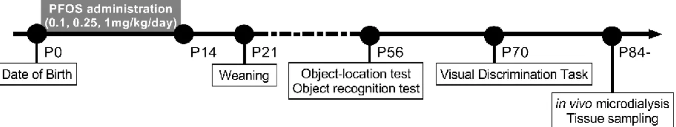
From postpartum days 1 to 14, dams were randomly assigned into four groups, one of which served as control, and given PFOS by gavage in amounts of 0.1 (PF-0.1), 0.25 (PF-0.25), or 1 mg/kg b.w. (PF-1) (Figure 1). Control dams received an equivalent volume of vehicle via gavage. Pups were housed with their biological mothers until weaning at P 21. At 8–10 weeks of age, 1–2 male offsprings per litter (n = 15 per group) were used for behavioral experiments. At 12 weeks of age, 1–2 male offsprings per litter of the control and PF-1 groups (n = 10 per group) were used for in vivo microdialysis to measure extracellular concentrations of amino acids in the dorsal hippocampus. At 12 weeks of age, 1–3 male offsprings per litter of the control and PF-1 groups (n = 5–6 per group) were used for mRNA and protein analysis of the hippocampus.
Figure 1. Schematic drawing of the experimental design. The experimental group dams were
7
exposed to PFOS through gavage from postpartum days 1 to 14. All pups were permitted free access to normal food and water from P 21.
2.3 Object location test and object recognition memory test
Mice were selected randomly from each group for testing. Mice were tested in an open field arena (length, 30 cm; width, 30 cm; height, 39 cm) made of acryl, as previously described (Amano et al., 2018). An overhead camera and a video recorder were used to monitor and record behavior (Figure 2A). Prior to testing, mice were allowed to explore the arena without objects for 30 min per day for 5 days to become habituated to the environment.
After the completion of the habituation, mice were tested for object location and object recognition memories. These tests consisted of three phases. In the first phase, two identical objects were placed near the two diagonal corners at the end of one side of the arena (7.5 cm from each adjacent wall), and the mouse was allowed to explore for 5 min. Then, 24 h after the first phase, in the second phase or the object location recognition memory test (OLT), one of the objects used in the first phase was placed in a novel location (i.e., any one of the corners adjacent to the familiar one), and the mouse was allowed to explore for 5 min. Finally, 24 h after the second phase, in the third phase or the object recognition memory test (ORT), one of the objects was replaced by a novel object, and, again, the mouse was allowed to explore for 5 min. Both objects were placed at the same locations in the second phase. The assignments of objects and object locations were counterbalanced. Objects used in this experiment were made of glass, metal, or plastic, and their size ranged from 10.3 to 21.6 cm high and from 5.2 to 7.5 cm wide. The arena and the objects were cleaned using 70% ethanol between phases to eliminate odor cues.
8
Time spent exploring each object during each phase was measured via stopwatch by the experimenters, who were blind to the group assignment, who observed the animal’s behavior on a monitor screen. Exploration was defined as the touching of an object with the nose and/or the nose directed toward the object within 1 cm. Touching the object with other parts of the body or climbing over the object was not included in this definition. A discrimination index (DI) representing the preference for a novel location or object compared with a familiar one was calculated as (timenovel − timefamiliar)/(timenovel + timefamiliar) (Cavoy and Delacour, 1993). A positive DI indicates a preference for the novel location or object.
2.4 Pairwise VD task
After 1 week of the ORT, we started the VD task in a touchscreen-equipped trapezoidal- shaped operant chamber, housed within a sound-attenuating box containing a ventilation fan, two small speakers, and a house light (O’hara & Co., Ltd., Tokyo, Japan), as previously described (Yajima et al., 2018). One side of the chamber (width 24 cm, height 19.5 cm) was equipped with a touchscreen liquid crystal display monitor, which was connected to a computer. To restrict the touch area, the touchscreen was covered by a black board that had two windows (width, 6 cm;
height 6 cm) that were 3 cm apart and located at a height of 3 cm from the floor of the chamber.
The opposite side of the chamber (width, 5.5 cm; height 19.5 cm) was equipped with a pellet tray (width, 3 cm; height, 19.5 cm; depth, 3 cm), a pellet dispenser, and an infrared nose poke detector.
The apparatus was controlled by Visual Task Studio (O’hara). The sidewalls of the chamber were equipped with water bottles.
Following 5 days of food restriction (85%–90% of free feeding body weight), mice were trained to learn a task procedure for 3 days. In the training session, a trial commenced when the mouse would nose poke the empty food tray. This was followed by the sounding of a tone for 1 s,
9
illuminating the house light and presenting identical stimuli at both windows. The stimuli remained at the windows until a response was made or 5 min elapsed. The mouse was rewarded a pellet via the pellet dispenser when it touched either stimuli. If the mouse did not touch the stimuli within 5 min, the house light was extinguished, and no reward was given. This indicated the end of a trial.
Each trial was separated by a 15 s interval, and a new trial began following a nose poke by the mouse. This training session was conducted for 30 min each day.
After the completion of the training sessions, mice were trained to discriminate between two different novel stimuli that were vertical and horizontal stripes. One of these stimuli was assigned as the correct stimulus (S+), whereas the other was the incorrect one (S−). Assignment of S+ stimulus is counterbalanced. A response to the S+ resulted in a reward pellet, whereas a response to the S− resulted in the extinguishing of the house light and no reward. The stimuli were simultaneously presented at each window, and the left–right presentation of the S+ was randomized across a daily session, which consisted of 50 trials. This trial procedure was identical to that of the training trial, except that the mouse was only rewarded a pellet after touching S+ but not S−. The stimuli remained at the windows until a response was made or 5 min elapsed. If the mouse did not touch the stimuli within 5 min, the house light was extinguished, and the trial was excluded from the experiment. The VD task was conducted for 9 days.
2.5 In vivo microdialysis
Mice were selected randomly from control and PF-1 groups for testing. A guide cannula (CXG-2; Eicom, Kyoto, Japan) was surgically implanted as described previously (Takatsuru et al., 2013). While under ketamine/xylazine anesthesia, the cannula was targeted at the dorsal hippocampus (2.8 mm lateral to the sagittal suture, 2.0 mm posterior to the bregma) through the cranial window (diameter 0.5 mm) of the mouse. The mice recovered from the surgery for at least
10
2 to 3 days before the experiments commenced. A microdialysis probe (CX-I; Eicom) was inserted into the guide cannula 2 h before the dialysis. Microdialysis was conducted under a free moving condition in the test chamber. During the experiment, the probe was perfused at a rate of 2 mL/min with HEPES buffer (NaCl, 140.0 mM; KCl, 2.5 mM; CaCl2, 2.0 mM; MgCl2, 1.0 mM; HEPES, 5.0 mM; glucose, 10.0 mM; pH 7.4). To analyze neurotransmitters, we collected dialysates for 15 min (2 mL/min) during each session. The concentrations of glutamate (Glu), glutamine (Gln), glycine (Gly), and gamma-aminobutyric acid (GABA) were immediately measured via high- performance liquid chromatography (Eicom).
2.6 Quantitative RT-PCR
Total RNA of the hippocampus was extracted using a RNeasy kit (Qiagen, Hilden, Germany). We synthesized cDNA from 250 ng of total RNA using ReverTra Ace® qPCR RT Master Mix with gDNA Remover (Toyobo Co., Ltd. Life Science Department, Osaka, Japan).
Real-time RT-PCR was conducted as described in the instruction manual of the Thunderbird®
Sybr qPCR Mix (Toyobo) and the Step One Real-Time PCR System (Thermo Fisher Scientific, Waltham, MA, USA). The real-time RT-PCR protocol for all genes was conducted as follows:
50°C for 2 min and 95°C for 10 min, followed by 40 cycles of amplification at 95°C for 15 s and 60°C for 1 min. Levels of measured mRNA were normalized by Gapdh. Primers are described in Table 1.
Table 1: Primer sequence for quantitative RT-PCR
Gene symbol Gene name Primer sequence
Grin1 NMDAR1 F: TTATGGCTTCTGCGTTGACC
R: TGTGTGCCAAACTTGCCATC
11
2.7 Western blot analysis
The protein levels for α-amino-3-hydroxy-5-methyl-4-isoxazole propionic acid (AMPA) receptor subunits were determined via western blot analysis. The dissected hippocampus was homogenized in a lysis buffer containing 10 mM of Tris-HCl (pH 7.8), 150 mM of NaCl, 1 mM of EDTA, 1% Nonidet P40, and cOmplete™ protease inhibitor cocktail (Roche, Mannheim, Germany). After centrifugation, the proteins were separated by sodium dodecylsulphate- polyacrylamide gel electrophoresis, and the separated proteins were later transferred to a
Grin2a NMDAR2A F: ATCCGACATCCACGTTCTTC
R: AGATGGTGGTGACCAAGGAG Grin2b
NMDAR2B F: GTGAGAGCTCCTTTGCCAAC
R: GTCAGGGTAGAGCGACTTGC
Gria1 AMPAR1 F: AGGTGCGCTTTGAAGGTTTG
R: TGCGGATTCCATCATGCTTC
Gria2 AMPAR2 F: TTTCAAGAGGCGTCTATGCG
R: ACAAATGGATGCGTGCCATC
Gria3 AMPAR3 F: ACTTTCTGGGCGCATTGTTG
R: ACAGTCAGGAAAGCAGCAAG Gabra1 GABAR ALPHA1 F: AAGGTGGCTTATGCAACAGC
R: TTCTGGAACCACGCTTTTGC Gabra2 GABAR ALPHA2 F: ATGCCAAACAAGCTGCTTCG
R: TTCAGTGGGCATGAATGAGC Gabrb2 GABAR BETHA2 F: TGCCTGCATGATGGACCTAAG
R: TGTGACTGCATTGTCATCGC Gabrg2 GABAR GAMMA2 F: TTGATGCTGAGTGCCAGTTG R: ACAGAACTGCGCTTCCATTG
Gapdh GAPDH F: TGCGACTTCAACAGCAACTC
R: ATGTAGGCCATGAGGTCCAC
12
nitrocellulose membrane. The membranes were blocked with 5% nonfat dry milk in Tris-buffered saline containing 0.1% Tween 20 (TBS-T), followed by overnight incubation with the appropriately diluted primary antibodies. After washing with TBS-T, the membranes were incubated with a horseradish peroxidase-conjugated antirabbit IgG secondary antibody (1:10000 dilution ratio) for 1 h at room temperature and detected using an ECL detection system. After obtaining the data of AMPA receptor subunits, membranes were washed with TBS-T, stripped and then washed again with TBS-T. Then, the same procedure was followed for β-actin, which was used as a loading control. For stripping, we used Restore™ Western Blot Stripping Buffer (Thermo Fisher Scientific). Membranes were stripped for one hour at 37°C, then washed with TBS-T before applying the primary antibody of β-actin. The antibody information is provided in Table 2.
Table 2: Antibodies used in the Western Blot
Antibody specificity Host Source Catalogue
Number
Dilution GluA1 Rabbit Cell Signaling Technology 8850S 1:1,000 GluA2 Rabbit Cell Signaling Technology 5306S 1:1,000 GluA3 Rabbit Cell Signaling Technology 4676S 1:1,000 β-Actin Rabbit Cell Signaling Technology 4967 1:5,000
2.8 Statistical analysis
All data in the text and figures are expressed as the standard deviation (SD). Statistical comparisons were performed using Student’s t-test, or one- or two-way analysis of variance
13
(ANOVA) followed by Dunnett’s test or Bonferroni test for post hoc analysis. P values less than 0.05 were considered to be statistically significant. Data were analyzed using GraphPad Prism version 8.2.1 for Windows (GraphPad Software, San Diego, CA, USA).
3 Results
3.1 General development was not affected by PFOS
There was no significant difference in the body weight of dams and pups in control and PF-1 groups at P 21 (dam: control 28.61 g ± 0.44, n = 15; PF-1 29.04 gram ± 0.58, n = 15;
p = 0.561 by t-test), (pup: control 6.68 gram ± 0.14, n = 15; PF-1 6.64 gram ± 0.27, n = 15;
p = 0.898 by t-test). There was no significant difference in the number of surviving pups by P 21 in control and PF-1 groups (data not shown). Furthermore, the day of eye opening did not vary between control and PF-1 groups (data not shown), reflecting no signs of general developmental delay in PFOS-exposed mice.
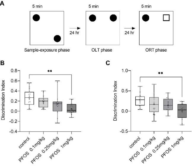
3.2 Object location and object recognition memory are impaired by PFOS
We evaluated the function of object location and object recognition memory using a modified spontaneous object recognition test. In each phase, there was no significant difference on the total exploration time among groups (supplementary figure 1). Figure 2B shows the DI in the second phase for the object location recognition memory. The DIs of PFOS-exposed groups were lower than that of the control group, and a one-way ANOVA showed a significant main effect of group (F(3, 56) = 4.95, p = 0.004). Post hoc analysis by Dunnett’s test showed a significant difference between PF-1 and the control group (p = 0.002). Figure 2C shows the DI in the third phase for the object recognition memory. The DIs in PFOS-exposed groups were lower than that of the control group, and a one-way ANOVA showed a significant main effect of group (F(3,
14
56) = 5.34, p = 0.003). Post hoc analysis by Dunnett’s test showed a significant difference between the PF-1 and control groups (p = 0.001).
Supplementary figure 1. Box-and-Whisker plot showing total exploration time (s) in (A) first, (B) second, and (C) third phase. There is no significant difference among all groups in each phase by one-way ANOVA.
15
Figure 2. (A) Schematic drawings of the object location recognition memory test (OLT) and object recognition memory test (ORT). Square-shaped symbols represent the test objects. The test began with a 5 min sample exposure phase followed by a 5 min test phase for both OLT and ORT, after a 24 h interval in the home cage. (B) Box-and-whisker plot showing the results of OLT in the control (n = 15; 25th percentile, 0.13; mean, 0.27; 75th percentile, 0.38), PF-0.1 (n = 15; 25th percentile, 0.08; mean, 0.19; 75th percentile, 0.26), PF-0.25 (n = 15; 25th percentile, 0.00; mean, 0.14; 75th percentile, 0.19) and PF-1 (n = 15; 25th percentile, -0.02;
mean, 0.05; 75th percentile, 0.14). + indicates mean. (C) Box-and-whisker plot showingthe
16
results of ORT in the control (n = 15; 25th percentile, 0.14; mean, 0.28; 75th percentile, 0.36), PF-0.1 (n = 15; 25th percentile, -0.02; mean, 0.1693; 75th percentile, 0.34), PF-0.25 (n = 15;
25th percentile, 0.03; mean, 0.13; 75th percentile, 0.3) and PF-1 (n = 15; 25th percentile, - 0.16; mean, 0.002; 75th percentile, 0.17). + indicates mean. Discrimination index shown in Box-and-whisker plotwas calculated as the exploration time of new location or a new object divided by the exploration time of all objects. The PF-1 (i.e., PFOS 1 mg/kg) group showed impairment in both object and location memory. **: p < 0.01 compared with the control group, determined by the Dunnett’s test.
3.3 Visual discrimination task for learning ability is impaired by PFOS
Next, we assessed learning ability using a VD task in a touchscreen-equipped operant system. Although the proportion of correct responses gradually increased in all groups, PFOS- exposed mice learned at a slower rate compared with the control group (Figure 3). A two-way ANOVA showed main effects of group (F(3, 56) = 7.21, p < 0.001) and day (F(8, 448) = 96.61, p < 0.001). Post hoc analysis by Bonferroni test showed that the value of PF-1 group was significantly lower than that of the control group (p < 0.001).
17
Figure 3. Learning curves of mice during the visual discrimination task using a touch screen. All groups showed discrimination around chance level (50%) on day 1. Discrimination levels of the PF-1 group were lower than other groups and significantly lower than that of the control group during experimental period. The two other PFOS groups showed a lower discrimination tendency compared with the control. Data are presented as mean andstandard deviation (SD) of n=15 in each group. *: p < 0.05 for comparison of mice in the PF-1 group with the control group by Bonferroni test.
3.4 PFOS exposure affects neurotransmitter levels in the dorsal hippocampus
Mice in PF-1 group showed significant learning and memory impairments, suggesting
18
hippocampal dysfunction. Thus, we measured neurotransmitter levels for Glu, Gln, Gly, and GABA in the dorsal hippocampus of the control and PF-1 groups using in vivo microdialysis.
Figure 4 shows the levels of amino acid neurotransmitters. Glu and GABA in PF-1 group were significantly higher than those in the control group (Glu: t(18) = -2.52, p = 0.021; GABA:
t(18) = −2.42, p = 0.026). No significant difference was observed in Gln and Gly levels.
Figure 4. Neurotransmitter concentration in the dorsal hippocampus of PF-1 (n=10) and control (n=10) groups. Representative neurotransmitter levels were analyzed from collected samples under free moving conditions for 15 min during each session. Data are presented as mean and standard deviation (SD). *: p < 0.05, control vs. PF-1 group mice by t-test.
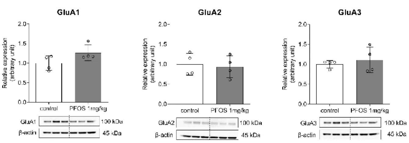
3.5 Expression levels of neurotransmitter receptors in the hippocampus
The mRNA levels of the GABAergic and glutamatergic receptors-related genes in the hippocampus were examined (Figure 5). The mRNA levels of the AMPA receptor subunit GluA3
19
(Gria3) were significantly downregulated in the PF-1 group compared with the control (Gria3:
t(10) = 2.76, p = 0.02). Other mRNA levels were not altered by PFOS treatment. Then, the expression levels of AMPA receptor subunits were examined via western blot analysis (Figure 6, supplementary figure 2). However, there was no significant difference in the protein levels of AMPA receptor subunits between the PF-1 and control groups.
Figure 5. The mRNA levels of (A) the N-methyl-D-aspartate (NMDA) receptor subunits, (B) α- amino-3-hydroxy-5-methyl-4-isoxazole propionic acid (AMPA) receptor subunits, and (C) gamma-aminobutyric acid (GABA) receptor subunits in the hippocampus of adult male
20
offspring. Gria3 expression levels significantly decreased in PF-1 group. The expression levels were normalized to those of Gapdh mRNA. Data are presented as mean and standard deviation (SD) of n=6 in each group. *: p < 0.05 for comparison of PFOS-treated mice with control, by t-test.
Figure 6. Western blot analysis of expression levels of α-amino-3-hydroxy-5-methyl-4-isoxazole propionic acid (AMPA) receptor subunits GluA1, GluA2, and GluA3 in the hippocampus of PF-1 and control groups. There is no significant difference in the expression levels of AMPA receptor subunits among both groups. The expression levels were normalized to β-actin expression levels. Data are presented as mean and standard deviation (SD) of n=4 in each group. *: p < 0.05 for comparison of mice in the PF-1 and control groups, by t-test.
21
Supplementary figure 2. Representative western blot membranes showing the bands for (A) α- amino-3-hydroxy-5-methyl-4-isoxazole propionic acid (AMPA) receptor subunits GluA1, (B) GluA2, and (C) GluA3 (upper panels), and β-actin (lower panels) in the hippocampus
of PF-1 and control groups. Western blot for β-actin was performed after stripping each membrane. Each blot contains data of 3 control and 3 PF-1 group mice.
4 Discussion
In the present study, we investigated the effect of lactational PFOS exposure on learning and memory in adult male mice. Early postnatal exposure to PFOS induced object location and object recognition memory impairments and learning deficits. Moreover, the concentrations of extracellular glutamate and GABA in the dorsal hippocampus were elevated in PFOS-exposed
22
group. These results indicate that lactational PFOS exposure induces learning and memory damage, at least in part through the disruption of hippocampal functions.
During the OLTs and ORTs, the DIs of PFOS-exposed mice significantly decreased dose- dependently. Since the spontaneous object recognition test is based on the natural tendency of rodents to explore novel stimuli (Ennaceur and Aggleton, 1994), the animals did not need to be trained to learn the task. Moreover, all groups of animals showed a similar level of object exploration in each phase, indicating that they were equally familiarized with the sample locations and objects and they paid equal attention to the objects. Thus, the impairments in the PFOS- exposed mice are not attributable to decreased familiarization or to the dysfunction of attention to the objects. Therefore, the present results indicate that lactational PFOS exposure induced an impairment in spatial and non-spatial recognition memory. To the best of our knowledge, our study is the first to examine the effect of low-dose lactational PFOS exposure on learning memory function in rodents using specific memory function tests. Nevertheless, epidemiological studies have reported rather controversial effects of prenatal and childhood PFOS exposure on memory and other cognitive functions in humans (Gallo et al., 2013; Hoffman et al., 2010; Oulhote et al., 2017). In epidemiological studies, because many genetic and environmental factors as well as PFOS may affect the development of higher brain function, it may be difficult to clarify the effect of exposure to a particular chemical. The present study, which uses mouse models, suggests the possibility that lactational PFOS exposure may cause adverse effects in brain development.
In the VD task, although PFOS-exposed mice gradually selected more correct responses as sessions progressed, improvement was delayed compared with that of the control group. Because the touchscreen-equipped operant system required less physical and emotional demands (Bussey et al., 2012; Yajima et al., 2018), the influence of motor and/or emotional abnormalities were
23
minimum. Moreover, since animals of all groups were able to continue the task throughout the sessions, several factors other than learning ability (e.g., visual function, motivation, and attention) may not have affected the task performance. However, PFOS-exposed groups still improved task performance, indicating that their learning ability may be able to persist to some extent. This result is consistent with a previous cross-fostering study that showed partial delay of learning during water maze tests by rats with postnatal PFOS exposure (Wang et al., 2015). Taken together, lactational PFOS exposure may only partially influence learning ability.
The in vivo microdialysis experiment showed elevated extracellular glutamate and GABA levels in the dorsal hippocampus of the PFOS exposed group, compared with that of the control group. This shows that PFOS exposure in this study may disrupt GABAergic and glutamatergic functions despite the insignificant change in the mRNA levels of the GABA and glutamate receptor genes found in our qRT-PCR and the protein analysis of AMPA receptor subunits via western blot analysis. Our results are similar to the in vitro findings of other group (Li et al., 2017) regarding extracellular glutamate levels. They found that there was an increase in the extracellular glutamate in the primary hippocampal neurons exposed to PFOS. Furthermore, they found that PFOS exposure decreased the mRNA levels of glutamine synthase, and this was reported to slowdown the uptake of glutamate by astrocytes from the synaptic cleft and, thus, leads to the accumulation of extracellular glutamate (Zou et al., 2010).
The extracellular glutamate excess in the synaptic cleft has an excitotoxic effect on synaptic function by increasing the inward calcium currents to the neuron through the continuous stimulation of N-methyl-D-aspartate (NMDA) glutamatergic receptors (Liu et al., 2010). This eventually, along with other mechanisms of PFOS that lead to an increase in intracellular calcium, inhibits the synaptogenesis that is crucial for memory formation. Moreover, the increase in GABA
24
levels after PFOS exposure in our study is in lines with one previous report that examined GABA concentration in the hypothalamus, although we used a much lower dose (Salgado et al., 2015).
It has been known that the glutamate and GABA homeostasis in the hippocampus are important for cognitive functions (Amaral and Witter, 1989; Paulsen and Moser, 1998). Moreover, previous studies have reported that the hippocampus is involved in OLTs (reviewed in Warburton and Brown, 2015) and VD tasks (Yajima et al., 2018). Thus, the present result may reflect the hippocampal dysfunction, and this may lead to our behavioral results, as described above. The hippocampus is suspected to be a major target for PFOS. Lactational PFOS exposure decreases mRNA levels of the NMDA receptor type 2B, which plays an important role in learning and memory functions, and calmodulin, which has key role in calcium signaling in the hippocampus (Liu et al., 2010). Moreover, developmental PFOS exposure inhibits long-term potentiation induction and disrupted AMPA receptors trafficking (Zhang et al., 2019). Furthermore, PFOS exposure in the primary cultured hippocampal neurons induced an increase in extracellular glutamate levels (Li et al., 2017). These findings are consistent with those in the present study.
Taken together, lactational PFOS exposure may affect hippocampal development and disrupt hippocampal function. Moreover, since it has been reported that the perirhinal cortex has a critical role in ORTs (Aggleton and Brown, 2005), and PFOS exposure affects the cerebral cortex (Lee and Viberg, 2013), lactational exposure to PFOS may also affect brain regions other than the hippocampus. This hypothesis is supported further by the fact that limited changes in the AMPA glutamate and GABA receptor levels were observed in the hippocampus samples in the present study, despite the profound changes in the OLT and VD tests for the PF-1 and control groups. This suggests that hippocampal function may be indirectly altered because of the influence of other brain regions such as the entorhinal cortex. The axons of the pyramidal cells in the entorhinal
25
cortex form the glutamatergic perforant pathway, which is the main source of input to the hippocampal formation, and transmit signals from the parahippocampal gyrus to all subregions of the hippocampal formation (Van Strien et al., 2009). This entorhinal–hippocampal network plays a crucial role in regulating cognitive function (Kitamura, 2017). Thus, disruption of the entorhinal cortex via PFOS exposure may influence hippocampal function. The changes in Glu levels in the hippocampus may reflect a change in neuronal functions of the entorhinal cortex. Further investigations are needed to uncover the extent of regional developmental neurotoxicity caused by PFOS lactational exposure.
Although previous studies have suggested that the neurotoxicity of prenatal PFOS exposure has more adverse effects compared with that of postnatal exposure from epidemiological and rodent studies (Butenhoff et al., 2009; Fei et al., 2008; Lau et al., 2003b), this hypothesis may not correctly describe the toxicity of PFOS, since species and regional differences related to developmental periods need to be considered. In the present study, PFOS exposure during the postnatal lactational period affects the development of cognitive functions. However, the mechanisms of PFOS-induced cognitive impairment cannot be fully clarified. Although PFOS exposure may have altered hippocampal neurotransmitter secretion or turnover, it is not clear whether such an alteration is caused by direct PFOS action in the hippocampal neurons.
Furthermore, molecular mechanisms of PFOS action to alter brain function have not yet been clarified. Because previous studies have reported that PFOS may affect the thyroid hormone system (Ballesteros et al., 2017; Kim et al., 2011; Lau et al., 2003; Thibodeaux et al., 2003; Yu et al., 2009), which plays a crucial role in brain development during prenatal and postnatal developmental periods (Gilbert et al., 2012; Koibuchi and Chin, 2000; Koibuchi, 2013), PFOS may exert its action by disrupting thyroid hormone action in the brain. Indeed, many other systems
26
may be disrupted by PFOS. Further investigations are needed to elucidate the mechanisms of PFOS action.
Summary
In conclusion, the present study revealed that lactational PFOS exposure induced spatial and non- spatial memory impairment and learning disability in adult mice offspring. Moreover, because PFOS-exposed animals have elevated concentrations of glutamate and GABA in the dorsal hippocampus, PFOS exposure could affect hippocampal development, resulting in disruption of the hippocampal function.
Conflict of interest
The authors declare that there is no conflict of interest.
Acknowledgment
This study was supported in part by JSPS KAKENHI Grant Numbers JP24710068 (to AH) and JP25281024 (to NK).
References
Aggleton, J.P., Brown, M.W., 2005. Contrasting Hippocampal and Perirhinalcortex Function using Immediate Early Gene Imaging. Q. J. Exp. Psychol. B 58, 218–233.
https://doi.org/10.1080/02724990444000131
Amano, I., Takatsuru, Y., Khairinisa, M.A., Kokubo, M., Haijima, A., Koibuchi, N., 2018. Effects of Mild Perinatal Hypothyroidism on Cognitive Function of Adult Male Offspring.
27
Endocrinology 159, 1910–1921. https://doi.org/10.1210/en.2017-03125
Amaral, D.G., Witter, M.P., 1989. The three-dimensional organization of the hippocampal formation: A review of anatomical data. Neuroscience 31, 571–591.
https://doi.org/10.1016/0306-4522(89)90424-7
Austin, M.E., Kasturi, B.S., Barber, M., Kannan, K., MohanKumar, P.S., MohanKumar, S.M.J., 2003. Neuroendocrine effects of perflurooactane sulfonate in rats. Environ. Health Perspect.
111, 1485–1489. https://doi.org/10.1289/ehp.6128
Bayer, S.A., 1980. Development of the hippocampal region in the rat I. Neurogenesis examined with3H-thymidine autoradiography. J. Comp. Neurol. 190, 87–114.
https://doi.org/10.1002/cne.901900107
Bird, C.M., Burgess, N., 2008. The hippocampus and memory: Insights from spatial processing.
Nat. Rev. Neurosci. 9, 182–194. https://doi.org/10.1038/nrn2335
Butenhoff, J.L., Ehresman, D.J., Chang, S., Parker, G.A., Stump, D.G., 2009. Gestational and lactational exposure to potassium perfluorooctanesulfonate (K + PFOS) in rats : Developmental neurotoxicity. Reprod. Toxicol. 27, 319–330.
https://doi.org/10.1016/j.reprotox.2008.12.010
Chang, E.T., Adami, H.O., Boffetta, P., Cole, P., Starr, T.B., Mandel, J.S., 2014. A critical review of perfluorooctanoate and perfluorooctanesulfonate exposure and cancer risk in humans. Crit.
Rev. Toxicol. 44, 1–81. https://doi.org/10.3109/10408444.2014.905767
Chang, S.-C., Noker, P.E., Gorman, G.S., Gibson, S.J., Hart, J.A., Ehresman, D.J., Butenhoff, J.L., 2012. Comparative pharmacokinetics of perfluorooctanesulfonate (PFOS) in rats, mice, and monkeys. Reprod. Toxicol. 33, 428–440. https://doi.org/10.1016/j.reprotox.2011.07.002 Chen, M., Ha, E., Liao, H., Jeng, S., Su, Y., Wen, T., 2013. Perfluorinated Compound Levels in
28
Cord Blood and Neurodevelopment at 2 Years of Age. Epidemiology 24, 800–808.
https://doi.org/10.1097/EDE.0b013e3182a6dd46
Chuang, N., Mori, S., Yamamoto, A., Jiang, H., Ye, X., Xu, X., Richards, L.J., Nathans, J., Miller, M.I., Toga, A.W., Sidman, R.L., Zhang, J., 2011. An MRI-based atlas and database of the
developing mouse brain. Neuroimage 54, 80–89.
https://doi.org/10.1016/j.neuroimage.2010.07.043
Cui, L., Zhou, Q., Liao, C., Fu, J., Jiang, G., 2009. Studies on the Toxicological Effects of PFOA and PFOS on Rats Using Histological Observation and Chemical Analysis. Arch. Environ.
Contam. Toxicol. 56, 338–349. https://doi.org/10.1007/s00244-008-9194-6
Fei, C., McLaughlin, J.K., Lipworth, L., Olsen, J., 2008. Prenatal exposure to perfluorooctanoate (PFOA) and perfluorooctanesulfonate (PFOS) and maternally reported developmental milestones in infancy. Environ. Health Perspect. 116, 1391–1395.
https://doi.org/10.1289/ehp.11277
Gallo, V., Leonardi, G., Brayne, C., Armstrong, B., Fletcher, T., 2013. Serum perfluoroalkyl acids concentrations and memory impairment in a large cross-sectional study. BMJ Open 3, 1–9.
https://doi.org/10.1136/bmjopen-2012-002414
Gallo, V., Leonardi, G., Genser, B., Lopez-Espinosa, M.-J., Frisbee, S.J., Karlsson, L., Ducatman, A.M., Fletcher, T., 2012. Serum Perfluorooctanoate (PFOA) and Perfluorooctane Sulfonate (PFOS) Concentrations and Liver Function Biomarkers in a Population with Elevated PFOA Exposure. Environ. Health Perspect. 120, 655–660. https://doi.org/10.1289/ehp.1104436 Gilley, J.A., Yang, C.-P., Kernie, S.G., 2011. Developmental profiling of postnatal dentate gyrus
progenitors provides evidence for dynamic cell-autonomous regulation. Hippocampus 21, 33–47. https://doi.org/10.1002/hipo.20719
29
Grove, E.A., Tole, S., 1999. Patterning events and specification signals in the developing hippocampus. Cereb. Cortex 9, 551–561. https://doi.org/10.1093/cercor/9.6.551
Han, J., Fang, Z., 2010. Estrogenic effects, reproductive impairment and developmental toxicity in ovoviparous swordtail fish (Xiphophorus helleri) exposed to perfluorooctane sulfonate (PFOS). Aquat. Toxicol. 99, 281–290. https://doi.org/10.1016/j.aquatox.2010.05.010
Hoffman, K., Webster, T.F., Weisskopf, M.G., Weinberg, J., Vieira, V.M., 2010. Exposure to polyfuoroalkyl chemicals and attention deficit/hyperactivity disorder in U.S. children 12-15 years of age. Environ. Health Perspect. 118, 1762–1767. https://doi.org/10.1289/ehp.1001898 Høyer, B.B., Ramlau-hansen, C.H., Obel, C., Pedersen, H.S., Hernik, A., Ogniev, V., Jönsson, B.A.G., Lindh, C.H., Rylander, L., Rignell-hydbom, A., Bonde, J.P., 2015. Pregnancy serum concentrations of perfluorinated alkyl substances and offspring behaviour and motor development at age 5–9 years – a prospective study. Environ. Heal. 14, 1–11.
Ishida, K., Tsuyama, Y., Sanoh, S., Ohta, S., Kotake, Y., 2017. Perfluorooctane sulfonate induces neuronal vulnerability by decreasing GluR2 expression. Arch. Toxicol. 91, 885–895.
https://doi.org/10.1007/s00204-016-1731-x
Jensen, A.A., Leffers, H., 2008. Emerging endocrine disrupters: Perfluoroalkylated substances. Int.
J. Androl. 31, 161–169. https://doi.org/10.1111/j.1365-2605.2008.00870.x
Kannan, K., Franson, J.C., Bowerman, W.W., Hansen, K.J., Jones, P.D., Giesy, J.P., 2001a.
Perfluorooctane sulfonate in fish-eating water birds including bald eagles and albatrosses.
Environ. Sci. Technol. 35, 3065–3070. https://doi.org/10.1021/es001935i
Kannan, K., Franson, J.C., Bowerman, W.W., Hansen, K.J., Jones, P.D., Giesy, J.P., 2001b.
Perfluorooctane Sulfonate in Fish-Eating Water Birds Including Bald Eagles and Albatrosses.
Environ. Sci. Technol. 35, 3065–3070. https://doi.org/10.1021/es001935i
30
Kim, H.-S., Jun Kwack, S., Sik Han, E., Seok Kang, T., Hee Kim, S., Young Han, S., 2011.
Induction of apoptosis and CYP4A1 expression in Sprague-Dawley rats exposed to low doses of perfluorooctane sulfonate. J. Toxicol. Sci. 36, 201–210. https://doi.org/10.2131/jts.36.201 Kitamura, T., 2017. Driving and regulating temporal association learning coordinated by entorhinal-hippocampal network. Neurosci. Res. 121, 1–6.
https://doi.org/10.1016/j.neures.2017.04.005
Koibuchi, N., Jingu, H., Iwasaki, T., Chin, W.W., 2003. Current perspectives on the role of thyroid hormone in growth and development of cerebellum. Cerebellum 2, 279–289.
https://doi.org/10.1080/14734220310011920
Lau, C., Thibodeaux, J.R., Hanson, R.G., Rogers, J.M., Grey, B.E., Stanton, M.E., Butenhoff, J.L., Stevenson, L.A., 2003. Exposure to Perfluorooctane Sulfonate during Pregnancy in Rat and Mouse. II: Postnatal Evaluation. Toxicol. Sci. 74, 382–392.
https://doi.org/10.1093/toxsci/kfg122
Lau, C., Thibodeaux, J.R., Hanson, R.G., Rogers, J.M., Grey, B.E., Stanton, M.E., Buttenhoff, J.L., Stevenson, L.A., 2003a. Exposure to perfluorooctane sulfonate during pregnancy in rat and mouse. II: Postnatal evaluation. Toxicol. Sci. 74, 382–392.
https://doi.org/10.1093/toxsci/kfg122
Lau, C., Thibodeaux, J.R., Hanson, R.G., Rogers, J.M., Grey, B.E., Stanton, M.E., Buttenhoff, J.L., Stevenson, L.A., 2003b. Exposure to perfluorooctane sulfonate during pregnancy in rat and mouse. II: Postnatal evaluation. Toxicol. Sci. 74, 382–392.
https://doi.org/10.1093/toxsci/kfg122
Lee, Y.J., Lee, H.G., Yang, J.H., 2013. Perfluorooctane sulfonate-induced apoptosis of cerebellar granule cells is mediated by ERK 1/2 pathway. Chemosphere 90, 1597–1602.
31
https://doi.org/10.1016/j.chemosphere.2012.08.033
Li, Z., Liu, Q., Liu, C., Li, C., Li, Y., Li, S., Liu, X., Shao, J., 2017. Evaluation of PFOS-mediated neurotoxicity in rat primary neurons and astrocytes cultured separately or in co-culture.
Toxicol. in Vitro. 38, 77–90. https://doi.org/10.1016/j.tiv.2016.11.002
Liu, X., Liu, W., Jin, Y., Yu, W., Wang, F., Liu, L., 2010a. Effect of gestational and lactational exposure to perfluorooctanesulfonate on calcium-dependent signaling molecules gene expression in rats’ hippocampus. Arch. Toxicol. 84, 71–79. https://doi.org/10.1007/s00204- 009-0467-2
Liu, X., Liu, W., Jin, Y., Yu, W., Wang, F., Liu, L., 2010b. Effect of gestational and lactational exposure to perfluorooctanesulfonate on calcium-dependent signaling molecules gene expression in rats’ hippocampus. Arch. Toxicol. 84, 71–79. https://doi.org/10.1007/s00204- 009-0467-2
Loccisano, A.E., Campbell, J.L., Butenhoff, J.L., Andersen, M.E., Clewell, H.J., 2012. Evaluation of placental and lactational pharmacokinetics of PFOA and PFOS in the pregnant, lactating, fetal and neonatal rat using a physiologically based pharmacokinetic model. Reprod. Toxicol.
33, 468–490. https://doi.org/10.1016/j.reprotox.2011.07.003
Loccisano, A.E., Longnecker, M.P., Campbell, J.L., Andersen, M.E., Clewell, H.J., 2013.
Development of pbpk models for pfoa and pfos for human pregnancy and lactation life stages, Journal of Toxicology and Environmental Health - Part A: Current Issues.
https://doi.org/10.1080/15287394.2012.722523
Miralles-marco, A., Harrad, S., 2015. Perfluorooctane sulfonate : A review of human exposure , biomonitoring and the environmental forensics utility of its chirality and isomer distribution.
Environ. Int. 77, 148–159. https://doi.org/10.1016/j.envint.2015.02.002
32
Morreale de Escobar, G., Obregon, M., Escobar del Rey, F., 2004. Role of thyroid hormone during early brain development. Eur. J. Endocrinol. 151, U25–U37.
https://doi.org/10.1530/eje.0.151U025
Murofushi, W., Mori, K., Murata, K., Yamaguchi, M., 2018. Functional development of olfactory tubercle domains during weaning period in mice. Sci. Rep. 8, 1–15.
https://doi.org/10.1038/s41598-018-31604-1
OECD [WWW Document], 2002. URL http://www.oecd.org/chemicalsafety/risk- management/perfluorooctanesulfonatepfosandrelatedchemicalproducts.htm
Oulhote, Y., Steuerwald, U., Debes, F., Weihe, P., Grandjean, P., 2017. Behavioral difficulties in 7- year old children in relation to developmental exposure to perfluorinated alkyl substances.
Environ. Int. 97, 237–245. https://doi.org/10.1016/j.physbeh.2017.03.040
Paulsen, O., Moser, E.I., 1998. A model of hippocampal memory encoding and retrieval:
GABAergic control of synaptic plasticity. Trends Neurosci. 21, 273–278.
https://doi.org/10.1016/S0166-2236(97)01205-8
Pérez, F., Nadal, M., Navarro-Ortega, A., Fàbrega, F., Domingo, J.L., Barceló, D., Farré, M., 2013.
Accumulation of perfluoroalkyl substances in human tissues. Environ. Int. 59, 354–362.
https://doi.org/10.1016/j.envint.2013.06.004
Prevedouros, K., Cousins, I.T., Buck, R.C., Korzeniowski, S.H., 2006. Sources, fate and transport of perfluorocarboxylates. Environ. Sci. Technol. 40, 32–44.
https://doi.org/10.1021/es0512475
Salgado, R., Pereiro, N., López-Doval, S., Lafuente, A., 2015. Initial study on the possible mechanisms involved in the effects of high doses of perfluorooctane sulfonate (PFOS) on prolactin secretion. Food Chem. Toxicol. 83, 10–16. https://doi.org/10.1016/j.fct.2015.05.013
33
Semënov, M. V., 2019. Adult Hippocampal Neurogenesis Is a Developmental Process Involved in Cognitive Development. Front. Neurosci. 13, 1–5. https://doi.org/10.3389/fnins.2019.00159 Semple, B.D., Blomgren, K., Gimlin, K., Ferriero, D.M., Noble-Haeusslein, L.J., 2013. Brain
development in rodents and humans: Identifying benchmarks of maturation and vulnerability to injury across species. Prog. Neurobiol. 1–16, 106–107.
https://doi.org/10.1016/j.pneurobio.2013.04.001
Shi, X., Liu, C., Wu, G., Zhou, B., 2009. Waterborne exposure to PFOS causes disruption of the hypothalamus–pituitary–thyroid axis in zebrafish larvae. Chemosphere 77, 1010–1018.
https://doi.org/10.1016/j.chemosphere.2009.07.074
Takatsuru, Y., Eto, K., Kaneko, R., Masuda, H., Shimokawa, N., Koibuchi, N., Nabekura, J., 2013.
Critical Role of the Astrocyte for Functional Remodeling in Contralateral Hemisphere of Somatosensory Cortex after Stroke. J. Neurosci. 33, 4683–4692.
https://doi.org/10.1523/JNEUROSCI.2657-12.2013
Thompson, J., Eaglesham, G., Reungoat, J., Poussade, Y., Bartkow, M., Lawrence, M., Mueller, J.F., 2015. Removal of PFOS , PFOA and other perfluoroalkyl acids at water reclamation plants in South East Queensland Australia. Chemosphere 82, 9–17.
https://doi.org/10.1016/j.chemosphere.2010.10.040
van Strien, N.M., Cappaert, N.L.M., Witter, M.P., 2009. The anatomy of memory: An interactive overview of the parahippocampal- hippocampal network. Nat. Rev. Neurosci. 10, 272–282.
https://doi.org/10.1038/nrn2614
Wang, Y., Liu, W., Zhang, Q., Zhao, H., Quan, X., 2015. Effects of developmental perfluorooctane sulfonate exposure on spatial learning and memory ability of rats and mechanism associated with synaptic plasticity. Food Chem. Toxicol. 76, 70–76.
34
https://doi.org/10.1016/j.fct.2014.12.008
Wang, Z., Cousins, I.T., Scheringer, M., Buck, R.C., Hungerbühler, K., 2014. Global emission inventories for C 4 – C 14 per fl uoroalkyl carboxylic acid ( PFCA ) homologues from 1951 to 2030 , Part I : production and emissions from quanti fi able sources. Environ. Int. 70, 62–
75. https://doi.org/10.1016/j.envint.2014.04.013
Yu, W.-G., Liu, W., Jin, Y.-H., Liu, X.-H., Wang, F.-Q., Liu, L., Nakayama, S.F., 2009. Prenatal and Postnatal Impact of Perfluorooctane Sulfonate (PFOS) on Rat Development: A Cross- Foster Study on Chemical Burden and Thyroid Hormone System. Environ. Sci. Technol. 43, 8416–8422. https://doi.org/10.1021/es901602d
Zhang, Q., Liu, W., Zhao, H., Zhang, Z., Qin, H., Luo, F., Niu, Q., 2019. Developmental perfluorooctane sulfonate exposure inhibits long-term potentiation by affecting AMPA receptor trafficking. Toxicology 412, 55–62. https://doi.org/10.1016/j.tox.2018.11.015 Zhang, S., Kang, Q., Peng, H., Ding, M., Zhao, F., Zhou, Y., Dong, Z., Zhang, H., Yang, M., Tao,
S., Hu, J., 2019. Relationship between perfluorooctanoate and perfluorooctane sulfonate blood concentrations in the general population and routine drinking water exposure. Environ.
Int. 126, 54–60. https://doi.org/10.1016/j.envint.2019.02.009
Zou, J., Wang, Y.-X., Dou, F.-F., Lü, H.-Z., Ma, Z.-W., Lu, P.-H., Xu, X.-M., 2010. Glutamine synthetase down-regulation reduces astrocyte protection against glutamate excitotoxicity to neurons. Neurochem. Int. 56, 577–584. https://doi.org/10.1016/j.neuint.2009.12.021
35
発表予定論文
Neurotoxic Effects of Lactational Exposure to Perfluorooctane Sulfonate on Learning and Memory in Adult Male Mouse
(授乳期におけるぺルフルオロオクタンスルホン酸曝露 による神経毒性の生体期の学習と記憶への影響)
雑誌名
Food and Chemical Toxicology (submitted)
著者名
Abdallah Mshaty, Asahi Haijima, Yusuke Takatsuru, Ayane Ninomiya, Hiroyuki Yajima, Michifumi Kokubo, Miski Aghnia Khairinisa, Wataru Miyazaki, Izuki Amano, Noriyuki Koibuchi