Conservative treatment of retained placenta in six patients:
a retrospective case series
Analysis of conservative medical treatment against retained placenta was possible in six cases
Risa Fujishima1, Masao Shimaoka2, Kiko Yamamoto3, Chiho Miyagawa4, Yoshie Yo1, Akiko Kanto1, Yasushi KOTANI1, Ayako Suzuki1, Noriomi Matsumura1
1 Department of Obstetrics & Gynecology, Kindai University faculty of Medicine
2 Department of Obstetrics and Gynecology, Kobe University Graduate School of medicine
3 Chibune General Hospital
4 Osaka Red Cross Hospital
Abstract
Retained placenta is associated with postpartum hemorrhage and intrauterine infection and can be comorbid with placenta accreta. Although critical obstetric hemorrhages necessitating massive blood transfusion, intensive care, or total hysterectomy occur in some cases of retained placenta, conser- vative treatment was possible in many recently re- ported cases. This retrospective study investigated six cases of retained placenta treated conservative- ly at our hospital. We report a comparative eval-
uation of the clinical course and serum levels of human chorionic gonadotropin in each case. The retained placenta was expelled spontaneously in three of the six cases, while rebleeding occurred in the other three cases, necessitating hemostatic treatment. Finally, the uterus was successfully pre- served in all six cases.
Key words : Conservative treatment, retained pla- centa, postpartum hemorrhage
Introduction
Retained placenta is associated with postpartum hemorrhage and intrauterine infection and can be comorbid with placenta accreta. Although critical obstetric hemorrhages necessitating massive blood transfusion, intensive care, or total hysterectomy occur in some cases of retained placenta, conserva- tive treatment was possible without additional pre- ventive, invasive treatment measures after achiev- ing primary hemostasis in many recently reported cases. According to past references, Timmerman et al 1 reported that conservative treatment enabled the preservation of fertility, albeit in a limited num- ber of cases. Ramoni et al 2 reported that the blood flow to remaining placental tissue was reduced and tissue mass became small over time. However, in a
clinical setting, there is a potential for major hem- orrhage to occur during conservative treatment.
It is understood that conservative treatment can be effective, but the type of cases where rebleed- ing occurs has not been well defined. Against this background, we conducted this retrospective com- parative study of cases of retained placenta treated at our hospital to assess when conservative treat- ment might be indicated.
Cases and Methods
Cases: Six cases were selected for this investigation from patients who were diagnosed with retained placenta after vaginal delivery and for whom serum human chorionic gonadotropin (S-hCG) levels (from detailed pregnancy hormone
Received October 16, 2018; Accepted February 27, 2019
test results) and chronological images could be re- viewed. These patients delivered at our hospital between 2000 and 2016. The patient ages ranged from 31 to 38 years, with three patients giving birth for the first time and three patients having previously given birth. The deliveries occurred be- tween the 37th and 41st week of gestation. Birth was achieved by natural delivery in three cases, in- duced delivery in two cases, and vacuum extraction in one case.
Methods: The major axis of the remaining placental tissue (hereafter, remnant diameter) was measured on ultrasound and/or magnetic resonance imaging (MRI) images taken at the time of diag- nosis for the six cases, and the subsequent clinical course of each case was evaluated. The items for evaluating the clinical course were occurrence of rebleeding/requirement of hemostatic treatment;
manner of expulsion/removal of retained tissue;
and the need for blood transfusion. Those who de- veloped fever and/or lower abdominal pain associ- ated with elevated inflammation markers on labo- ratory tests were diagnosed as having intrauterine infection. In addition, two further parameters were evaluated before and after an event (rebleeding or spontaneous expulsion): S-hCG levels and the change or lack of change in blood flow to placen- tal tissue as determined using color Doppler ultra- sound imaging.
Results
Rebleeding occurred and hemostatic treat- ment was required in three cases (Patients A, B, and C). Rebleeding was noted at 13, 14, and 22 days after delivery in Patients A, B, and C, respective- ly. The details concerning the cases with rebleed- ing were as follows. The remnant diameter was 107 mm in Patient A. The S-hCG level just before rebleeding was abnormally high, at 15,891 mIU/
mL. Patient A underwent manual removal of the re- maining tissue when the rebleeding occurred. The remnant size was 74mm in Patient B. The S-hCG level just before rebleeding was 331 mIU/mL. Pa- tient B underwent curettage, uterine artery embo- lization (UAE), and methotrexate (MTX) therapy, after which the S-hCG result was negative and the remaining placental tissue was removed via hys- teroscopy. The remnant diameter was small, at 25 mm in Patient C. Patient C developed rebleeding when transvaginal removal was attempted after the S-hCG level showed a decreasing trend to 43 mIU/
mL. This patient accordingly underwent UAE, af- ter which the remaining placental tissue shrunk to 5mm in diameter and spontaneously disappeared by day41. (Figure 1)
Spontaneous expulsion occurred in the other three cases (Patients D, E, and F) (Table 1).
Spontaneous expulsion of retained placenta was noted at 20, 27, and 46 days after delivery in Pa-
A) T2-weighted MRI Image on day 7. Part
of placenta was observed in the uterine B) T2-weighted MRI image on day 29, an
examination 7 days after UAE. The re- C) T2-weighted MRI image on day 69. Most of the retained placenta was absorbed re- Figure 1. MRI image of the retained placenta in Patient C.
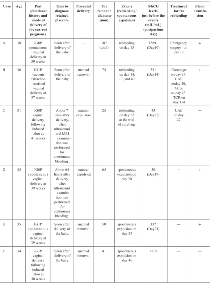
Table 1. Summary of the six cases with retained placenta. S-hCG; serum human chorionic gonadotrophin, G; gravidity, P; parity, UAE;
uterine artery embolization, MTX; methotrexate, TCR; transcervical resectoscope.
Case Age Past
gestational history and mode of delivery of the current pregnancy
Time to diagnose retained placenta
Placental
delivery The remnant diameter
(mm)
Events (rebleeding/
spontaneous expulsion)
S-hCG levels just before the
events (mIU/mL) (postpartum
day)
Treatment for the rebleeding
Blood transfu-
sion
A 38 2G0P,
spontaneous vaginal delivery at
39 weeks
Soon after delivery of the baby
― 107
(total) rebleeding
on day 13 15891
(Day10) Emergency surgery on
day 13
+
B 36 1G1P,
vacuum extraction -assisted vaginal delivery at
37 weeks
Soon after delivery of the baby
manual
removal 74 rebleeding
on day 14, 17, and 49
(Day14)331 Curettage on day 14, onday 20, UAE on day 23,MTX TCR on day 114
+
C 31 0G0P,
vaginal delivery following
induced labor at 41 weeks
About 7 days after delivery, when ultrasound
and MRI examina-
tion was performed continuousfor bleeding .
natural
expulsion 25 rebleeding on day 22 at the trial of curettage
(Day22)43 UAE on day
22
―
D 35 0G0P,
spontaneous vaginal delivery at
39 weeks
About 64 hours after delivery,
when ultrasound examina- tion was performed continuous for bleeding .
natural
expulsion 65 spontaneous expulsion on
day 20
(Day19)50 ― +
E 35 1G1P,
spontaneous vaginal delivery at
39 weeks
Soon after delivery of
the baby.
manual
removal 50 spontaneous
expulsion on day 27
(Day24)137 ― +
F 34 2G1P,
vaginal delivery following
induced labor at 40 weeks
Soon after delivery of the baby.
manual
removal 41 spontaneous
expulsion on day 46
< 0.5 ― ―
tients D, E, and F, respectively. The remnant diam- eters in the three cases of spontaneous expulsion (with no bleeding) tended to be small, at 65 mm (Patient D), 50 mm (Patient E), and 41 mm (Patient F). The S-hCG levels just before spontaneous ex- pulsion of the remaining tissue were 50 mIU/mL in Patient D, 137 mIU/mL in Patient E, and <0.5 mIU/
mL in Patient F.
Findings on color Doppler ultrasound im- aging just before an event were positive for blood flow to the remaining tissue in two of the three cases with rebleeding (Patients A on day17 and B on day12). However, the corresponding ultrasound findings in two of the three cases with spontaneous expulsion of the remaining tissue (Patients D on day 46 and E on day 50) also showed blood flow, and no clear relationship was established between blood flow to placental tissue as measured by ul- trasound and rebleeding. Larger remnant diameters were more frequent in cases where blood transfu- sion was needed before primary hemostasis (A, B, D, E), irrespective of the presence or absence of rebleeding. The uterus was preserved in all six cas- es. No intrauterine infection was noted under con- servative treatment in the present cases.
Discussion
A correlation between risk of rebleeding during conservative treatment of retained placenta and the size of the remaining tissue was suggested in this study. Timmerman et al 1 reported that pres- ervation of the uterus depended on the total volume of remaining placental tissue, which we consider to be similar to the findings of this study.
It is believed that S-hCG levels reflect via- bility and blood flow of retained placenta 3, thus the retained placenta may be successfully removed in patients with low S-hCG levels. However, S-hCG level to predict successful removal has not been determined. Placenta was removed at 33 IU/L 3 or 52~1551 IU/L 4 level of S-hCG, while removal of a placenta was associated with massive hemorrhage requiring blood transfusion at a S-hCG level of 366 IU/L4. Notably, we experienced massive hem- orrhage in Patient C during attempted transvaginal removal of the remaining tissue after a confirmed decrease of S-hCG to 43 IU/L. Patient C had the smallest remaining tissue and low level of S-hCG.
The massive hemorrhage in this case was caused
were predominantly abnormally high, at 15,891 mIU/mL (Patient A) and 331 mIU/mL (Patient B).
The S-hCG levels just before spontaneous expul- sion of the retained placenta were 50 mIU/mL (Pa- tient D), 137 mIU/mL (Patient E), and 0.5 mIU/mL (Patient F), suggesting that S-hCG levels are usu- ally low when spontaneous expulsion occurs. How- ever, we should also note that predicting the timing of spontaneous expulsion based on pre-expulsion S-hCG levels is difficult. In previous reports, low or negative S-hCG levels were followed by spon- taneous expulsion 5, while other patients with low (15 IU/L) or negative S-hCG levels required hys- terectomy due to massive hemorrhage 5,6. There- fore, it is difficult to know whether conservative care would be successful from S-hCG level.
Blood flow as measured on ultrasound was considered to be unrelated to the occurrence of re- bleeding. Rebleeding developed in a case where blood flow to the remaining tissue had disappeared, and spontaneous expulsion occurred in a case where a plentiful blood supply to the remaining tissue was suspected. Variability in the evaluation procedure was considered an explanatory factor for this phenomenon: this study was a retrospective investigation, and a different technician carried out the ultrasound examination in each case. MRI could be used when objectivity is critical; however, there are difficulties in conducting frequent MRI imaging and in using the images to predict the clin- ical course. Alternatively, pulsatility index (PI) of uterine artery could be useful to assess the blood flow of placenta, although we did not examine in our cases. PI increased after removal of placenta 4. In another report, spontaneous expulsion of placen- ta was observed in cases with PI >1, whereas PI ≦1 was associated with rebleeding and hysterectomy 5.
In this study, rebleeding occurred at 13 (Patient A), 14 (Patient B), and 22 (Patient C) days after delivery, suggesting the likelihood of rebleed- ing in cases of retained placenta is highest within one month of delivery, particularly around a few weeks after delivery. Spontaneous expulsion of the retained placenta occurred approximately one month after delivery, at 20, 27, and 46 days (Pa- tients D, E, and F, respectively), and it is consid- ered that conservative treatment of retained placen- ta involves an extended period of hospitalization.
It was reported that spontaneous expulsion was observed in 75% of retained placenta cases, which
teroscope surgery 8,9. This procedure would shorten the duration of hospitalization, although the risk of massive hemorrhage requiring hysterectomy should be considered. There was no significant pathological difference in spontaneous expulsion tissue and operation sample in this study. Both of them were only necrotic tissues.
Conclusion
This study suggested conservative treat- ment of retained placenta without total hysterec- tomy would be possible. However, we consider that such conservative treatment should be carried out in advanced medical facilities with multidis- ciplinary therapeutic capabilities given that emer- gency hemostatic treatment is required in cases of rebleeding. We also consider that invasive handling is related to risk of re-bleeding even at low level of S-hCG. The collection of further data from addi- tional retained placenta cases and the elucidation of factors that influence the clinical course of this condition are necessary in future research.
There is no financial support and no relationship that poses a conflict of interest.
This article is secondary publication of Con- servative treatment of retained placenta in six patients; Obstetrical and Gynecological Practice Vol.65 No.12 2016 p.1685-1688.
References
1. Timmermans S, van Hof AC, Duvekot JJ (2007) Conser- vative management of abnormally invasive placentation.
Obset Gynecol Surv 62: 529-539
2. Ramoni A, Strobl EM, Tiechl J, Ritter M, Marth C (2013) Conservative management of abnormally invasive pla- centa: four case reports. Acta Obstet Gynecol Scand 92:
468-471
3. Kulkarni A, Draycott T (2005) The use of serial betaH- CG to plan surgical evacuation of retained placenta in a case of placenta accreta. J Matern Fetal Neonatal Med 17(4):295-297
4. Matsumura N, Inoue T, Fukuoka M, Sagawa N, Fujii S (2000) Changes in the serum levels of human chorionic gonadotropin and the pulsatility index of uterine arteries during conservative management of retained adherent placenta. J Obstet Gynaecol Res 26(2):81-87
5. Dueñas-Garcia OF, Diaz-Sotomayor M, Rico-Olvera H (2011) Utility of the pulsatility index of the uterine arter- ies and human chorionic gonadotropin in a series of cases of placenta accreta. J Obstet Gynaecol Res 37(8):1112- 6. El-Bialy G, Kassab A, Armstrong M (2007) Magnetic 1116
resonance imagining (MRI) and serial beta-human cho-
rionic gonadotrophin (beta-hCG) follow up for placenta percreta. Arch Gynecol Obstet 276(4):371-373
7. Sentilhes L, et al (2010) Maternal outcome after con- servative treatment of placenta accreta. Obstet Gynecol 115(3):526-534
8. Ganer Herman H, et al (2018) Pregnancies Following Hysteroscopic Removal of Retained Products of Con- ception after Delivery Versus Abortion. Gynecol Obstet Invest 26:1-7
9. Legendre G, Zoulovits FJ, Kinn J, Senthiles L, Fernan- dez H (2014) Conservative management of placenta ac- creta: hysteroscopic resection of retained tissues. J Minim Invasive Gynecol 21(5):910-913