CASE REPORT
Osteolytic primary bone lymphoma in the multiple bones
Shusuke Yagi1,2,3, Robert Zheng1, Seiichi Nishiyama3, Yutaka Kawabata1, Takayuki Ise1, Kosuke Sugiura4, Haruhiko Yoshinari4, Toshihiko Nishisho4, Yoshimi Bando5, Kumiko Kagawa6, Daiju Fukuda1, Tomohiro Soga2,3,7, Yoshihito Saijo1, Kenya Kusunose1, Koji Yamaguchi1, Hirotsugu Yamada1, Takeshi Soeki1, Tetsuzo Wakatsuki1, Shinji Kawahito2,3,7, Masashi Akaike8, and Masataka Sata11Department of Cardiovascular Medicine, Tokushima University Graduate School of Biomedical Sciences, Tokushima, Japan, 2Department of Community Medicine and Human Resource Development, Tokushima University Graduate School of Biomedical Sciences, Tokushima, Japan, 3Shikoku Central Hospital, Ehime, Japan, 4Department of Orthopedic Surgery, Tokushima University Graduate School of Biomedical Sciences, Tokushima, Japan, 5Division of Pathology, Tokushima University Hospital, Tokushima, Japan, 6Department of Hematology, Endocrinology and Metabolism, Tokushima University Graduate School of Biomedical Sciences, Tokushima, Japan, 7Department of Anesthesiology, Tokushima University Graduate School of Biomedical Sciences, Tokushima, Japan, 8Department of Medical Education, Tokushima University Graduate School of Biomedical Sciences, Tokushima, Japan
Abstract : Primary non-Hodgkin bone lymphoma (PBL) can involve solitary or multiple destructive bone lesions such as those of the femur or pelvis humerus, and some cases have osteolytic lesions. PBL is a rare disease in adults. Thus, PBL is rarely considered a differential diagnosis of the osteolytic tumor. In addition, PBL can be un-derdiagnosed because patients do not experience symptoms or show objective abnormalities in the early stage. Here, we reported an elderly patient with PBL in multiple bones, including the cranial and femoral bones that were fractured due to falling. J. Med. Invest. 66 : 347-350, August, 2019
Keywords : diffuse large B-cell lymphoma, head tumor, differential diagnosis
INTRODUCTION
Primary non-Hodgkin bone lymphoma (PBL) is a rare dis-ease that accounts for < 2% of all lymphomas in adults (1). PBL is estimated to account for 3%–7% of primary bone tumors and 3%–5% of all extranodal non-Hodgkin lymphomas (2). PBL can involve solitary or multiple destructive bone lesions such as those of the femur or pelvis humerus, and some cases have os-teolytic lesions. However, PBL in the cranial vault is extremely rare (3). PBL is rarely considered a differential diagnosis of the osteolytic tumor. PBL, especially diffuse large B-cell lympho-ma (DLBCL), exhibits a better response rate for therapy and better prognosis if treated early with chemotherapy or radio-therapy than primary nodal or extranodal DLBCL at other sites (4). Thus, accurate early diagnosis is crucial for treatment. However, PBL can be underdiagnosed because patients do not experience symptoms or show objective abnormalities in the early stage.
Here, we reported an elderly patient with PBL in multiple bones, including the cranial and femoral bones that were frac-tured due to falling.
CASE
An 89-year-old woman presented to our hospital because of pain in her right hip after falling. She had severe aortic valve stenosis without symptoms. No abnormal neurological and ob-jective findings on the head were noted. Computed tomography (CT) revealed that her right femoral shaft was completely broken
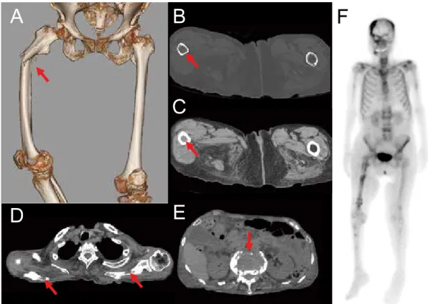
and that her thin bone cortex was filled with a high-intensity tumor at the site of fracture ; osteolytic tumors were also noted in the bilateral scapula and lumbar spine bones (Figure 1A, B, C, D, E). Whole-body bone scintigram showed accumulation of technetium-99m-methylene-diphosphonate (MDP) in the head, shoulders, lumbar spine bones, and right femur (Figure 1F) ; this indicated that the fracture in her fragile femur was a pathologi-cal fracture caused by tumor invasion and induced by the trauma of falling. She received skeletal traction of the right leg, followed by transcatheter aortic valve implantation before the operation of the fracture, which was successful. Her broken femoral shaft was fixed with a nail device. After the operation for the fracture, she experienced drowsiness without any focal neurological signs. Head CT showed an osteolytic tumor on the right temporal and left back of the head (Figure 2 A, B, C, D). The head tumor was mildly tender and overlaid by healthy skin. T1- (Figure 2E) and T2-weighted (Figure 2F) magnetic resonance images showed iso-intensity and low intensity of the tumor, respectively, and the dura structure was maintained. A CT image of the whole body showed neither lymphadenopathy nor any evidence of sys-temic involvement and pathological fracture. Blood examination results revealed elevated ALP, LDH, and soluble interleukin-2 receptor levels at 423 IU/L, 400 IU/L, and 5490 U/mL (upper limit < 519 pmol/L), respectively. Decreased serum total protein and albumin levels of 5.9 g/dL and 2.9 g/dL, respectively, were noted, with no M-protein (IgG 903 mg/dL, IgA 200 mg/dL, IgM 38 mg/dL, kappa 18 mg/dL, or lamda 78 mg/dL). No monoclonal protein was detected by protein electrophoresis of the serum and urine. Normocalcemia (albumin-adjusted calcium 9.6 mg/dL) with increased parathyroid hormone-related protein-C levels at 6.5 pmol/L (upper limit < 1.1 pmol/L) were also noted. Increased bone markers of type I collagen carboxyterminal telopeptide (ICTP) of 20 ng/mL (upper limit < 4.5 ng/mL), tartrate-resistant acid phosphatase isoform 5b (TRACP 5b) of 534 mU/dL (upper limit 420 mU/dL), and urinary deoxypyridinoline of 79 nmol/ mM・creatinine (upper limit < 7.6 nmol/mM・creatinine) were detected, although bone-APL was within normal limits of 20.3
The Journal of Medical Investigation Vol. 66 2019
347
Received for publication January 29, 2019 ; accepted May 26, 2019. Address correspondence and reprint requests to Shusuke Yagi, MD, PhD, Department of Cardiovascular Medicine, Tokushima University Graduate School of Biomedical Sciences, Tokushima, 3-18-15 Kuramo-to-cho, Tokushima 770-8503, Japan and Fax : +81-88-633-7894.
348
S. Yagi, et al. Osteolytic bone lymphomaFigure 1. Computed tomography showing fracture of the right femoral shaft (A : arrow), the thin bone cortex (B : arrow) filled with a high-intensity tumor (C : arrow) at the fracture site, and osteolytic tumors in the bilateral scapula (D: arrow) and lumbar spine bones (E : arrow). Whole-body bone scintigram showing accumulation of technetium-99m-methylene-diphosphonate in the head, shoulders, lumbar spine bones, and right femur (F).
Figure 2. Computed tomography showing osteolytic tumors in the right temporal bone, left occipital bone (A, B, C), and sphenoid bone (D : arrow). T1-weighted (E) and T2-weighted magnetic resonance imaging (F) showing iso-intensity and low intensity of the tumor, respectively.
349
The Journal of Medical Investigation Vol. 66 August 2019
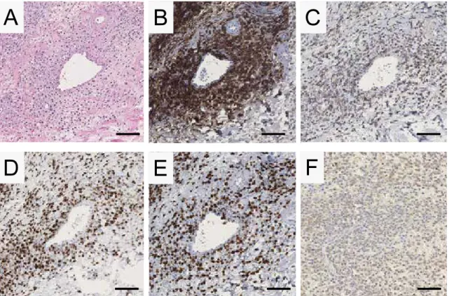
μg/L (upper limit < 22.6 μg/L). Serological test results werenegative for Epstein-Barr (EB) virus early antigen (EBEA) an-tibody (IgG) and anti-viral capsid antigen (VCA) IgM and were positive for EB virus nuclear antigen IgG and anti-VCA IgG, indicating that the patient was infected with EB virus, although PCR of EB virus-DNA with the peripheral blood sample was not performed. Biopsy of the right temporal soft tissue revealed diffuse growth pattern with large cells (Figure 3A) and positiv-ity for CD20 (Figure 3B), a B-cell marker ; B-cell lymphoma 2 (BCL-2), an anti-apoptosis factor regulating B-cell development and differentiation (Figure 3C). Furthermore, multiple myeloma oncogene 1 (MUM-1) (Figure 3D) and negativity for CD3, CD5, CD10, CD30, and PD-L1 were noted. The tumor cells included Ki-67-positive cells with a Ki labeling index of 80% (Figure 3E), indicating highly proliferative potential. In situ hybridization of small RNAs of EB virus showed positive cells for EB virus infection (Figure 3F). Bone marrow aspiration and bone mar-row biopsy were not performed. The lesion was localized in the bones ; thus, we diagnosed her as having PBL of EBV-positive DLBCL of the elderly. Clinical stage of the patient was IV EB with a B-symptom of weight loss. The international prognostic index was 5, indicating a high risk. The best supportive care rather than chemotherapy or radiation therapy was selected, and her general condition worsened after the successful operation of the femoral fracture. The level of ALP increased to 661 IU/L from 571 IU/L during the course, indicating progression of bone disease. She died 9 weeks after admission.
DISCUSSION
The differential diagnosis of the osteolytic lesion includes secondary bone involvement of the systemic lymphoma, bone metastasis from neoplasm, and multiple myeloma. The patient was infected with EB virus, and thus, the differential diagnosis of EB-related malignancy also included Hodgkin lymphoma and Burkitt lymphoma. Imaging studies, including CT and scintig-raphy, revealed no primary lesion of the malignant tumor, and blood examination and results showed no evidence of multiple myeloma. In addition, tumor cells were negative for CD138, indi-cating no differentiation of plasma cells. Immunohistochemistry for light chain kappa and lambda staining showed no dominancy for either immunoglobulin. Thus, we excluded CD20-positive myeloma. Histopathological analysis showed no presence of Reed-Sternberg cells and no appearance of a “starry-sky” pat-tern. Immunophenotype analysis distinguished DLBCL from Hodgkin lymphoma and Burkitt lymphoma. Thus, we diagnosed her as PBL of EBV-positive DLBCL of the elderly.
T2-weighted MR imaging characteristics of primary lym-phoma of bone vary and do not seem to be a simple reflection of histologic findings of lymphoma (5). In this case, the intensity of tumorous lesions was low in T2-weighted MRI. MRI of DLBCL with a high nuclear/cytoplasm ratio and hypercellularity could cause decreased extracellular fluid concentration and small intratumoral edema which results in low signal on t2-weighted images (6).
The precise mechanism of osteolysis remains unclear ; how-ever, the increased expression of osteoclast-activating factors,
Figure 3. Hematoxylin and eosin staining (A) of the biopsy obtained from the head tumor showing diffuse growth patterns with large cells. Immunohistochemical analysis showing positive results for CD20 (B), BCL-2 (C), MUM-1 (D), and high Ki-67 labeling index (E). In situ hybridization of the small RNAs of EB virus showing positive cells for EB virus infection (F) (Barr : 100 μm).
350
S. Yagi, et al. Osteolytic bone lymphoma including MIP-1α, MIP-1β, and RANKL, was observed in theRNA derived from the patient’s lymphoma cells. In addition, increased protein levels of RANKL and osteoprotegerin, a key regulator of osteoclastogenesis, from the patient’s lymphoma cells were sharply decreased after the chemotherapy (7, 8). Thus, the secretion of osteoclast-activating factors by tumor cells (and/ or bone marrow stromal cells) might be involved in the mecha-nism of osteolysis in some malignant lymphoma cases (9).
Anthracycline-based chemotherapy and radiation therapy are recommended for treating PBL (4). The destructive bone involve-ment in the cranial vault could be regenerated after therapy (10). However, we did not recommend such aggressive therapies, but suggested best supportive care to our patient because of her older age.
The prognosis of PBL is relatively better than that of other lymphoma types (4). Patients with advanced-stage DLBCL and skeletal involvement show a 65% complete remission rate (11), with 5-year progression-free and overall survival rates of 54% and 59%, respectively (12), and bone involvement does not seem to be an adverse prognostic factor (4). However, her general con-dition progressively worsened after the operation of the fracture. High proliferative potentials with high Ki-67 labeling index might be involved in her progressive poor general condition, which was different from gradual proliferative tumors, such as multiple myeloma.
Although the patient visited several clinics for medical prob-lems, including common cold and aortic valve stenosis, the head tumor was not detected. Furthermore, PBL can be underdi-agnosed because skull tumors may be covered by hair. Little symptoms are exhibited because the dura structure was main-tained and brain invasion by the tumor is rare (13). Thus, careful inspection and palpation of the head, especially during the first visit, are essential even when patients have no head complaints in order to make an early diagnosis. In addition, osteolytic PBL should be included as a differential diagnosis in cases of femoral shaft fractures, even if these fractures occur after falling.
CONFLICT OF INTEREST
The authors have no conflicts of interest to disclose.
REFERENCES
1. Hayase E, Kurosawa M, Suzuki H, Kasahara K, Yamakawa T, Yonezumi M, Suzuki S, Teshima T : Primary Bone Lymphoma : A Clinical Analysis of 17 Patients in a Single Institution. Acta Haematol 134 : 80-85, 2015
2. Pilorge S, Harel S, Ribrag V, Larousserie F, Willems L, Franchi P, Legoff M, Biau D, Anract P, Roux C, Blanc-Autran E, Delarue R, Gisselbrecht C, Ketterer N, Recher C, Bonnet C, Peyrade F, Haioun C, Tilly H, Salles G, Brice P, Bouscary
D, Deau B, Tamburini J : Primary bone diffuse large B-cell lymphoma : a retrospective evaluation on 76 cases from French institutional and LYSA studies. Leuk Lymphoma 57 : 2820-2826, 2016
3. Israel O, Mekel M, Bar-Shalom R, Epelbaum R, Hermony N, Haim N, Dann EJ, Frenkel A, Ben-Arush M, Gaitini D : Bone lymphoma : 67Ga scintigraphy and CT for predic-tion of outcome after treatment. J Nucl Med 43 : 1295-1303, 2002
4. Messina C, Ferreri AJ, Govi S, Bruno-Ventre M, Gracia Medina EA, Porter D, Radford J, Heo DS, Park HY, Pro B, Jayamohan J, Visco C, Scarfo L, Zucca E, Gospodarowicz M, Christie D, International Extranodal Lymphoma Study G : Clinical features, management and prognosis of multifo-cal primary bone lymphoma : a retrospective study of the in-ternational extranodal lymphoma study group (the IELSG 14 study). Br J Haematol 164 : 834-840, 2014
5. White LM, Schweitzer ME, Khalili K, Howarth DJ, Wunder JS, Bell RS : MR imaging of primary lymphoma of bone : variability of T2-weighted signal intensity. AJR Am J Roentgenol 170 : 1243-1247, 1998
6. Zimny A, Neska-Matuszewska M, Bladowska J, Sasiadek MJ : Intracranial Lesions with Low Signal Intensity on T2-weighted MR Images - Review of Pathologies. Pol J Ra-diol 80 : 40-50, 2015
7. Kong YY, Yoshida H, Sarosi I, Tan HL, Timms E, Capparelli C, Morony S, Oliveira-dos-Santos AJ, Van G, Itie A, Khoo W, Wakeham A, Dunstan CR, Lacey DL, Mak TW, Boyle WJ, Penninger JM : OPGL is a key regulator of osteoclas-togenesis, lymphocyte development and lymph-node organo-genesis. Nature 397 : 315-323, 1999
8. Chen P, Li B, Zhuang W, Huang H, Zhang H, Fu J : Multiple bone lesions and hypercalcemia presented in diffuse large B cell lymphoma : mimicking multiple myeloma? Int J Hema-tol 91 : 716-722, 2010
9. Matsuhashi Y, Tasaka T, Uehara E, Fujimoto M, Fujita M, Tamura T, Honda T, Kuwajima M, Shimoura Y, Mano S, Nagai M, Ishida T : Diffuse large B-cell lymphoma pre-senting with hypercalcemia and multiple osteolysis. Leuk Lymphoma 45 : 397-400, 2004
10. Akamatsu Y, Kanamori M, Uenohara H, Tominaga T : Bone regeneration after chemotherapy for vault lymphoma. BMJ Case Rep 2016 : 2016
11. Achemlal L, Mikdame M, Nouijai A, Bezza A, El Maghraoui A : Dramatical improvement of chemoresistant bone lym-phoma with rituximab. Clin Rheumatol 25 : 394-395, 2006 12. Lee HY, Kim SJ, Kim K, Ko YH, Kim WS : Bone
involve-ment in patients with stage IV diffuse large B-cell lym-phoma : does it have a prognostic value? Leuk Lymlym-phoma 53 : 173-175, 2012
13. Ciarpaglini R, Otten P : Primary cranial vault lymphoma with brain infiltration : case report and review of the litera-ture. Br J Neurosurg 26 : 756-758, 2012