Osteological and functional development of the flyingfish,
Cypselurus heterurus doederleini (Teleostei: Exocoetidae)
Juanito C. DAsiLAo, Jr.i'2 & Kosaku YAMAoKA2
i Laboratoip, ofAquatic Ecology, F`nulty ofAgriculture, Kochi Universdy, 200 Otsu, Monobe, Nanleoku, Kochi 783-O093, laPan2 Usa Man'ne Biological lnstitute, Kochi Universdy, 194 Inoshiri, Usa-cho Tosa, Kochi 780-1164, laPan (e-mail: [email protected] V
Abstract: The osteological development and functional processes in relation to feeding, swimming and flying of the laboratory-reared fiyiTigfish, C)IPselums hetemras doederleini were examined. Ossincation started upon hatch-ing at 5.54 mm SL (standard length) and almost fully ossdied at 15.80 mm SL. Early ossfication of cranial ele-ments suggests the impact of strength on the neurocraniurn, thereby toler.ating pressures from the developing brain, muscle mass, vertebral colum and the trunk region. In the early stage, larvae were actively feeding which was attributable to the early ossfication of the feedmg apparatus. Active swimming and darting above the water surface were attributable to the rudder-iike structure of the hypurals, providing more attachment for muscles re-sulting to sufiicient force, and to the development of the pectoral and the ventral fin rudiments. Generally, the early on-set of osteological development in the early stage and the short peried of ossfication process in C. heternnes cloederleini, suggest an early feeding, higher growth rate and the abruty to escape from predators by darting and swimming, resulting to higher survival rate.
Key words: Clmpselums, osteotogical develoPment, function.
I]swtRODUCTION
FIyingfish C)IPselurus heterurus doederleini (Steindachner) is an abundant, highly oceanic, and
one of the most important commercial species in Japan. It belongs to the 1argest fiyingfishes
that thrives in the water of Japan and its meat is one of the favorite delicacies as sliced raw fish
called "sashirni". Developmental informations of this species are found in many literatures (Nayudu, 1923; Bruun, 1935; Breder, 1938; Hubbs and Kampa, 1946; lmai, 1959). However, its osteological development has not been published yet. Hence, the present study was con-ducted to describe the developmental pattern from the initial appearance to ful1 ossdication and their relationhips to swimming, feeding and fiying functions which are vitally important for the survival.
MATERIALS AND METHODS
Samples of larvae were taken from the artficially inseminated eggs of Cl)abselums heterurus doedoleini taken from the parents collected off Iburi, Tosashimiztt, Kochi Prefecture. Reared in 2 tons rearing tank, 10 larvae were sampled every other day and iixed mitially in 109o neutral formalin solution. Clearing and staining procedures follow the method of Dingerkus and Uhler (1977) for both canilage and bone. Osteological developoment was observed and sketched using a Nikon S]Vrz-10 stereoscopic dissecting microscope with a camera lucida. Osteological terminology follows Weitzman (1962), Rosen (1964), Fujita (1990, 1992) and Fujita and Oozeki (1994).
RESULTS
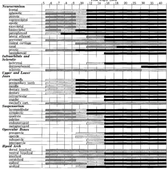
Neurocranium .
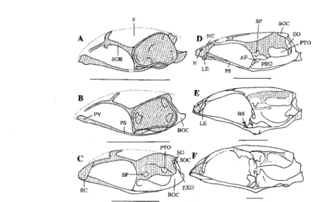
Shortly after hatching at 5.54 mm standard length (SL) specimen (O day), the skulI was fu11y cartilaginous. The ethmoid, lateral ethmoid, rostral cartilage, nasal and basisphenoid were ab-sent (Figs. 2 and 3). Prevomerine plate and parachondral cartilage were bridged by fibrous
ossfication of parasphenoid (Figs. 2A and 3A). ln this stage, the frontal was fibrously ossified
and became fully ossdied in 11.43 mrn SL specimen (16 days) (Figs. 1 and 2A-D). The pre-formed cartilaginous sphenotic, pterotic, supraoccipital, epiotic and exoccipital, started ossifYing
fibrously in 9.43 mm SL specimen (13 days) and became fully ossified in 11.43 mm SL speci-men (16 days) (Figs. 1, 2C-D). The basioccipital became fibrously ossified in 7.36 mm SL
Total length (mm)
Fig• 1. 0steological developmental patterns in CIJipseJurus heterurus doederleini. '::"s•tsI'E::':I'. cartilaginous; il{{{{•i{igl:i{i, fibrously ossified; -, fully ossfied.
specimen (8 days) and became fully ossified in 9.43 SL specimen (13 days) (Figs. 1, 2B-C and 3B-C). The parasphenoid became fu11y ossified in 7.36 mm SL specirnen (8 days) (Figs. 1, 2B and 3B). In 11.43 mm SL specimen (16 days), cartilaginous lateral ethmoid started to be formed laterally to the mass of rostral cartilage and became fully ossdied in 15.80 mm SL specimen (22 days) (Figs. 1 and 2D-E). In 15.80 mm SL specimen (22 days), cartilaginous prevomer became ossified (Figs. 1 and 3E-F). The rostral cartilage started foming in 9.43 mm SL spicemen (13 days) (Figs. 1 and 2C-F). The nasal started foming fibrously in 11.43 mm SL specimen (16 days) and became fUlly ossified in 15.80 rrrm SL specimen (22 days) (Figs. 1 and 2D-E). The cartilaginous prootic became fu11y ossMed in 9.43 mm SL specirnen (13 days) (Figs. 1, 2C-D and 3C-D). The basisphenoid appeared to be ossified in 15.80 mm SL specimen (22 days) and developed with subsequent growth (Figs. 1 and 2E-F).
F
.. 1
' ' i ' '4>>seeSX
sp socr
-• "A .71f ' D NC t{:-1-N LE ps AF V PRQ EO rroB-• Pv
ps:
va
al
E di BStf.r' .
''
t rvt. .. t" :=:=) 11 BOC rroC.
RC sp .EO F soc tr ...-N-
,' C"K. EXO Iv/ r ijK
L
BOCFig. 2. Development of neurocranium (lateral view) in CyPselurus heterarus doederleini. Stippled area, cartilage. Broken line, fibrously ossdied. Open area, ossdied portion. A) 5.54 mm; B) 7.36 mm;
C) 9.43 mm; D) 11.43 mm; E) 15.80 mm; F) 30.04 mm. A.F-auditory foramen; pital; BS-basisphenoid; EO-epiotic; EXO-exoccipital; F-frontal; LE-lateral ethrnoid;
nasal; NC-nasal canal; PRO-prootic; PS-parasphenoid; PTO-pterotic; PV'prevomer; RC -rostral cardiage; SP-sphenotic; SOB-supraorbital; SOC-supraoccipital. Bars = 1 mm.
EP
A
ps x ' AUCC
ef: t ttt { ncD
tx
•e
--.t tttt t- Lrt a 'x-...: sP rro t lx" r>X SHH- ,tt - ' etr', ' 1. t.. i' • B-]1 -tt tt tt . "'I
D, .•, '/..r' S.{` ps •'•' "l•Boc t ttttt) t c .t '.t .. .• •.• -• '' `b •. "', pv .,l ; ' ' l . E 'f"qY
l'
s ' sP PRO vro EXO BOC-xR
'X-"'LE •:
C
,.: ..[.11 .• if •: ,. .. ttt!s: Z
' •, ,, /" •• •-EEIiK
Fig. 3. Development of neurocranium (ventral view) in Cl)ipselums heterurus doederleini. Stippled area, canilage. Broken line, fibrously ossified. Open area, osshied portion. A) 5.54 mm; B) 7.36 mm;
C) 9.43 mm; D) 11.43 mm; E) 15.80 mm; F) 30.04 mm. AUC-auditory capsule; cipital; EP-etlmoid plate; EXO-exoccipital; LE-lateral ethmoid; PC-parachondral; prootic; PS-parasphenoid; PTO-pterotic; PV-prevomer; SP-sphenotic. Bars = 1 mn.
In larger specimens, a 1arge amount of cartilage on the sutures among ethmoid, lateral ethmoid, frontal and vomer was retained in the anterior region, and a small amount of cartilage on the sutures among sphenotic, pterotic, epiotic and fronta1 was retained in the posterior
re-gion (Fig. 2F).
Sclerotics and infraorbitals
In 5.54 mm SL specimen (O day), the lachryrrial and derrnosphenoid were absent but the eye capsule was forrned, surrounded by thn sclerotic cartilage (Fig. 4A). The lachrymal started forming fibrously in 11.43 rrrm SL specimen (16 days) and became fully ossdied in 15.80 mm SL specimen (22 days) (Figs. 1 and 4D-E). The anterior and posterior arc of sclerotic started to be ossified with the appearance of an ossified dermosphenoid in 30.03 mm SL specimen (44
days) (Figs. 1 and 4F).
A
s
B
t//'/i" ''' PM.tt.. M 't' i""'t"ttt"ttl't;''lt't 't .f "'"'i'•l"'iii/•••,l' ' .. , ttMC
E
t..t. /''t "i"i'' MC PALop "
ISgllk,.x, sop QUADI
MET t ENT D RA ANC
D QUAD SYM. " oP
":[L';"><xb'o'p-' F L s DSFig. 4. Development of viscerocranium; opercular apparatus, circumorbitals and sclerotic in C)v)selurus heterurus doederleini. Stippled area, cartilage. Broken line, fibrously osstaed. Open area, ossified portion. A) 5.54 mm; B) 7.36 mm; C) 9.43 mm; D) 11.43 rnm; E) 15.80 mm; F) 30.e4 mm. AIV mangular; D-dentary; DS-derrnosphenoid; ENT-endopterygoid; HYO-hyomandibular; IOP 'interopercular; L-lachrymal; M-maxilla; MC-maxillary cartilage; MET-metapterygoid; OP - opercular; PA-L -palatine; POP - preopercular; PMr premaxilla; QUA - quadrate; RA troarticular; S-sclerotic; SOP-subopercular; SYM-symplectic. Bars = 1 mmi.
Jaws
Forrnation of the jaws started from fibrous to fu11y osshied bone. In 5.54 mm SL specimen (O day), the maxilla, premaxilla and the lower jaw were fibrously ossified (Figs. 1 and 4A). Teeth on the premaxilla and the lower jaw were fibrously present. The maxilla, prerriaxilla and teeth became fully ossMed in 9.43 mm SL specimen (13 days) (Figs. 1 and 4C).
The dentary and retroarticular represented by fibrous ossification were fused and formed by shelling the Meckel's cartilage in 5.54 mm SL specirnen (O day) (Figs. 1 and 4A-B). The de-ntary became fully ossified in 9.43 mm SL specirnen (13 days) (Figs. 1 and 4C). Meckel's car-tilage was retained as a canilaginous tissue in larger specimens (Fig. 4E). The retroarticular became separated from the dentary -with the forrnation of the angular in 15.80 mm SL
speci-men (22 days) (Figs. 1 and 4E).
Suspensorium
In the stage of 5.54 mni SL (O day), the hyomandibular and symplectic were fused cartilagi-nously as one unit (Fig. 4A). They started to be separated and part]ially ossified in 9.43 mm SL specimen (13 days) and became fully ossdied in 11.43 mm SL specirnen (16 days) (Figs. 1 and 4C-D). The quadrate and palatine were cartilaginous and fused in 5.54 mm SL specimen (O day) (Fig. 4A). The quadrate became panially ossified in 9.43 mm SL specimen (13 days) and fully ossfied in 15.80 mm SL specimen (22 days) (Figs. 1 and 4C-E). The palatine which was preformed as a cartilage became separated from the quadrate and fully osshied in 15.80 mm SL specimen (22 days) (Figs. 1 and 4E). In 11.43 mm SL specimen (16 days), the endopterygoid started to be formed by fibrous ossification and became fully ossified in 15.80 mm SL specimen (22 days) (Figs. 1 and 4D-E). The metapterygoid which arose from the cartilaginous posterior end of the quadrate, appeared fu11y ossified in 15.80 rnm SL specimen (22 days) (Figs. 1 and
4E).
Opercular apparatus
In 5.54 mm SL specimen (O day), any opercular element was not visible. The preopercle and opercle started forming fibrously in 9.43 mm SL specimen (13 days) and became fully ossified in 15.80 mm SL specimen (22 days) (Figs. 1 and 4C-E). The subopercle and interopercle started forming fibrously in 11.43 mm SL specimen (16 days). They became fu11y ossified in 15.80 mm SL specimen (22 days) (Figs. 1 and 4D-E).
Hyoid arch
All of the hyoid arch elements were canilaginous in 5.54 mm SL specimen (O day) (Figs. 1 and 5A). The ventral hypohyal became ossified in 15.80 mm SL (22 days), whereas the dorsal hypohyal and interhyal retained cartilaginous condition (Figs. 1 and 5E). The ceratohyal and epihyal were fused but apparently separated from hypohyal anteriorly and interhyal posteriorly. The ceratohyal became partially ossified in 9.43 mm SL specimen (13 days) and was fu11y fied in 15.80 mm SL specimen (22 days) (Figs. 1 and 5C-E). The epihyal became partially ossi-fied and separated from the ceratohyal in 11.43 mm SL specimen (16 days) and was fully ossifed in 15.80 mm SL specimen (22 days) (Figs. 1 and 5D-E). A long, slender and fibrous urohyal appeared on the anteroventra1 part of the hyoid arch in 9.43 mm SL specimen (13 days) and became fully ossified in 11.43 mm SL specimen (16 days) (Figs. 1 and 5C-D). The branchiostegal rays started forming on the ventrolateral margin of ceratohyal and epihyal in 7.36 mm SL specimen (8 days) and became fully ossified in 9.43 mm SL specimen (13 days) (Figs. 1 and 5B-C).
Branchial arches
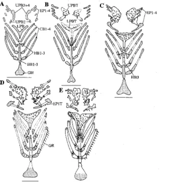
In 5.54 mm SL specimen (O day), the branchial arch elements were cartilaginously formed (Fig. 6). The upper pharyngobranchial bones (UPB2 and UPB3+4) started ossifying fibrously with the appearance of pharyngeal teeth in 7.36 mm SL specimen (8 days) and became fu11y ossified in 9.43 mm SL specimen (13 days) (Figs. 1 and 6B-C). The epibranchials (EPI-4) were chondrified in 5.54 mm SL specimen (O day). They became partially osshied in 11.43 mm SL specimen (16 days) and fu11y ossified in 15.80 mm SL specimens (22 days) (Figs. 1 and 6D-E). The first epibranchial (EPI) developed teeth on it at 11.43 mm SL (16 days) specimens
A HH IH
D
EH.
CHB
BR),'. rl/•,,t ]•k-'•...l, iL-- l•i( '•- Ess••
DHHc
/-UH 'K. CH vHH}<illlllili xFig. 5. Development of hyoid arch in C)IPselurus heternrus doederleini. Stippled area, cartilage. Broken 1ine, fibrously ossified Open area, ossified portion. A) 5.54 mm; B) 7.36 mm; C) 9.43 mm; D)
11.43 mm; E) 15.80 mm. BR -branchiostegal rays; CH-ceratohyal; CH-EH-cerato-epihyal; DHHrrdorsal hypohyal; EH-epihyal; Hll-hypohyal; IHminterhyal; VHH-ventral hypohyal;
UH-urohyal. Bars = O.5 mm.
A
twS-P-4F•.
CBI-4 HBI-3 3\:/,tlll,llg:;•i"D"
la tgXX"s:fng.62
,N,M!tss2' EatNe,ss,p`;b e' Ats :t
...i. ,ij,-r-EPiT K .,•"'v,gf'`" i:iiii/1/i'S/ji., e estr •ii///11i'i'iiiii" .. iei, vt,t hi=} o', 2 C VK$ti••"l
'
:lli!Ilirgi'i/t2
x,xZ i.,
HB3 t:,,.t,..,. -, l'l•.' ' "i ' t EPI-4Fig. 6. Development of upper and lower branchial arches in CyPselurus heterurus doederleini. Stippled
area, cartilage. Open area, ossified portion. A) 5.54 mm; B) 7.36 mm; C) 9.43 mm; D) 11.43 mm; E) 15.80 mm. BBIL3-basibranchial 1-3; CBI-4-ceratobranchial 1'4; hial 1-4; EPIT-epibranchial 1 teeth; GH-glossohyal; GR -gil1 raker; HBI-3-hypobranchial 1-3; LPB -lower pharyngeal bone; LPBT-lower pharyngeal bone teeth; UPB2-upper ngeal bone 2; UPB3+4-upper pharyngeal bone 3+4; UPBT-upper pharyngeal teeth. Bars =
The lower branchial elements were cartilaginously formed in 5.54 mm SL specirnen (O-day) (Fig. 6A). The glossohyal started to be osshied in 11.43 mm SL specimen (16 days) and be-came fu11y ossdied in 15.80 mm SL specimen (22 days) (Figs. 1 and 6D-E). The basibranchials (BBI+2+3) were cartilaginously fused as one element in 5.54 mm SL specimen (O day) (Figs. 6A). in 9.43 mm SL specimen (13 days), the third basibranchial (BB3) became partially ossfied and separated from the first and second basibranchials (BBI and 2) (Figs. 1 and 6C). It became fully ossbied in 11.43 mm SL specimen (16 days) with simultaneous separation and fu11 ossifica-tion of the first and second basibranchjals (BBI and 2) (Fig. 6D). The hypobranchials became partially ossified in 11.43 mm SL specimen (16 days) and fully ossified in 15.80 mm SL speci-men (22 days) (Figs. 1 and 6D-E). The ceratobranchials became partially ossfied in 9.43 mm SL (13 days) specimens and fu11y ossfied in 15.80 mm SL specimen (22 days) (Figs. 1 and 6C-E). The gil1 rakers were developed on the ceratobranchials (CB) in 11.43 mm SL specirnen (16 days) and on the hypobranchials (HB) in 15.80 mm SL specimen (22 days) (Figs. 1 and 6D-E). The lower pharyngeal bones became fibrously ossified with the appearance of teeth in 7.36 mm SL specimen (8 days) and fully ossified in 9.43 mm SL specimen (13 days) (Figs. 1 and 6B-C).
Vertebral column
In 5.54 mm SL specimen (O day), the notochord and the vertebral column (centra) were fib-rously ossfied, whereas the neural and haemal spines were fully chondrified (Figs. 7A and 8A). The centra became partially visible in 9.43 mm SL (13 days) and fully osshied in 11.43 mm SL specimen (16 days) (Figs. 1, 7B-C and 8C-E). The corresponding neural and haemal spines started ossifying fibrously in 7.36 mm SL specimen (8 days) and became fu11y ossified in 11.43
A
Cffl!/1/l//f
'D
iilil'li' EPRB
9
7Ltr? ..,1..,rr...L....;,.. i/e,liiiii••111-1,Ill.1(,1••i'tl•i.11••i"l///li-i,t}iii kpltil/ >x x`fi li s, 'Y<, PARAP-C
NPU NAp xxlE
NAP poscz J-. ". , PU / l.e t- '{)' Aet tt i NS PREZFig. 7. Development of few anterior vertebral colunm in CyPselurus hetentrus doederleini. Stippled area, cartilage. Dotted area, fibrously ossified. Open area, ossified portion. A) 5.54 mrn; B) 9.43 mm;
C) 11.43 mm; D) 15.80 mm; E) 30.04 mm. C-cranium; EPR-epipreural rib; NAP-neural arch plate; NPU-neural plate of preural centrum; NS -neural spine; PA-i?A-P -parapophysis; POSTZ-postzygapophysis; PRR-preural rib; PREZ-prezygapophysis; PU-preural cenmm. Bars = 1 mm.
PR
A ti4'blfptsfff.eif.. Ns
twll!if.!Zf!naztwaA "••',en•i Hy3+4+s
t t.
HP'kglllltt'NNittNN.,,..N.ll)FXNX,$.51i"ng.,l',,,Li•Hyi42'
'
DR
Bf22kig2lkimleSf2f2le;ek t.fEPit3
...n f.. .g z.,g f...f-.! z. ff zau. ..ffztz.tt-, •,•,..i..i . ..tt..ttt ...tt] ' 'e 'E a 'N'N'N'"'N'xNNXXNXXtlljillli .ll ecÅétts>puptgeaj•fupt DFRctwk,i,gk•g.',k'gst<Sxge
?'iXfva/r/f//1$(1.,n,,<ZlZs,//<fgg(ag,k PFR CIptil,l:l`GA'w"Z-: .,
Sli'itr ss
N\tt.
icXk'la{{pt-(ttt/ZiZi2ti2gtajag.,Npec XY/Nillixiliil{iliiillllllllil;IS:iisislili;iltL us /ti "{•[• CIHPUH
PR DR
Gt,st'i "//////.,i '' y,,.•/'`'t' Zi,.,p i Ni ..?.. X• ,. N.. L..tt. t,.Ut ,..Z.. tt't.. ""/"'ttttlxl'3top.'"Z'gx.e
NPU FZ
11 -t a' NAP t:' et-lrv" SNFig. 8. Development of vertebral colurmi, dorsal, anal and caudal tiins and supports in Cl)tPselurns uras doederleini. Stippled area, cartilage. Dotted area, fibrously ossified. Open area, osshied tion. A) 5.54 mm; B) 7.36 mm; C) 9.43 mm; D) 11.43 mm; E) 15.80 mm; F) 30.04 mm; G) 17.72 mm; H) 30.04 mm. AFR anal fin ray; CFR caudal fin ray; DFR dorsal fin ray; DR
distal radial or median segment; CIHPU-interhaemal spine cartilage; CIEP -interepural age; EPI-3 - epural 1-3; HP - haemapophysis; HS - haemal spine; HSI - first haemal spine;
HYI +2-hypural 1+2; HY3+4+5-hypural 3+4+5; NAP-neural arch plate; IVPU-neural
plate of preural centrum; NS -neural spine; PFR -procurrent fin ray; PH-parhypural;
preural centrum; PR-proximal radial or pterygiophore; TV-terminal vertebra; UN-uroneural; US-urostyle. Bars = 1 mm
mm SL specimen (16 days) (Figs. 1, 7B-C and 8C-E). Three neural plates of the preural cen-tnm of the precaudal vertebrae and four neural plates of the preural centrum of the caudal vertebrae started to be developed in 11.43 mm SL specimen (16 days) (Figs. 1, 7C and 8E). The neural arch plates started forming on the first few anterior preural centra (trunk region) and on the last few posterior preurai centra (caudai region) of the vertebrai coium at 11.43
mm SL specirnen (16 days) (Figs. 7C-E and 8E, F, H). The preural ribs appeared fibrously in 9.43 mm SL specimen (13 days) and became fu11y ossified in 11.43 mm SL specimen (16 days) (Figs. 1 and 7B-C). The epipreural ribs appeared to be fully ossified in 15.80 mm SL specimen
(22 days) (Figs. 1 and 7D).
Dorsat and anal fins and supports
Twelve to 14 dorsal pterygiophores or proximal radials and 10 to 11 anal proximal radials were present as cartilaginous elements in 5.54 mm SL (O day) (Figs. 8A). The distal radials and the dorsal and anal Iin rays were absent. The proximal radials became partially ossfied in 11.43 mm SL specimen (16 days) and were fully ossified in 15.80 mm SL specimen (22 days) (Figs. 1 and 8E-F). In 6.71 mm SL specimen (5 days), the distal radials were cartilagirtously formed on the respective pterygiophores posteriorly (except on the last element). They be-came partially ossified in 17.72 mm SL specimen (30 days) and were fu11y ossified in 30.04 mm SL specirnen (44 days) (Figs. 1 and 8G-H). The dorsal and anal fin rays became fibrously visi-ble in 7.36 mm SL specimen (8 days) and were fu11y ossified in 11.43 mm SL (16 days) (Figs. 1 and 8C-E).
Caudal fin and supports
All elements supporting the caudal fm such as epurals (EPI-3), hypurals (HYI+2, HY3+4 + 5), PHI-4) and parhypurals were cartilaginous in 5.54 mm SL specimen (O day) (Fig. 8). The hypurals and parhypurals started ossifying partially together with the fibrous appearance of the uroneural in 9.43 mm SL specimen (13 days) (Figs. 1 and 8D). They became fully•osshied with the uroneural in 15.80 mm SL specimen (22 days) (Fig. 8F). In 5.54 mm SL specimen (O day), the frrst epural (EPI) was absent (Fig. 8A). It appeared in 6.71 mm SL specimen (5 days) (Fig. 8B) and became fu11y ossified with the second and third epurals in 15.80 mm SL specimen (22 days) (Figs. 1 and 8F). The fibrous terminal vertebra started to be formed in 7.36 mm SL (8 days), followed by the fibrous forrnation of the urostyle in 9.43 mm SL men (13 days) (Figs. 1 and 8C-D). Both elements became fully ossified in 11.43 mm SL speci-men (16 days) (Fig. 8E). The caudal fm rays became fibrously visible in 7.36 mm SL specispeci-men (8 days) and fully ossfied in 9.43 mm SL specimen (13 days) (Figs. 1 and 8C-D).
Pectorat girdle and fin supports
In 5.54 mm SL specimen (O day), the pectoral girdle was preforrned by cartilaginous ele-ments except the cleithrum.which was formed as a thread-like fibrous ossified element (Figs. 9A). The cleithrum became fully ossified in 11.43 mm SL specimen (16 days) (Figs. 1 and 9D). The coracoid, scapula and radials were fused cartilaginously as one element in 5.54 mm SL specimen (O day) (Figs. 1 and 9A). The coracoid and scapula became separated and both were panially ossified in 11.43 mm SL (16 days) (Figs. 1 and 9D). They became fully ossified in 13.82 mm SL specimen (20 days) (Fig. 9E). The radials started to be separated from coracoid-scapula in 7.36 mm SL specimen (8 days) (Fig. 9B). They became partially ossified in 11.43 mm SL (16 days) and fu11y ossified in 13.82 mrn SL specimen (20 days) (Figs. 1 and 9D-E). Several pectoral fm rays started forrning fibrously in 7.36 mm SL specirnen (8 days) (Fig. 9B) and 15 to 16 fin rays becarne fully ossified in 9.43 mm SL specimen (l3 days) (Figs. 1 and 9C). The glenoid foramen staned to be formed in the scapula in 7.36 mm SL (8 days) specimens and became fully developed in 9.43 mm SL (13 days) (Figs. 1 and 9B-C). The ventral fenestra was panially formed in the coracoid in 9.43 mm SL specimen (13 days) and became fully de-veloped in 13.82 mrn SL specimen (20 days) (Fig. 9C-E). The postcleithrurn started forming fibrously in 9.43 mm SL specimen (13 days) and becarne fully osshied in 11.43 mm SL specimen
A
CL]t/.111iil.t.ii'..Il.'i..llll,.IX"",Ii-[1111'11111,111-B
eFH g. ik, ,i,//il,/,lll',' 'iPFR '
rt.t.t .t t
i ,,.•••ii[•.11illlil,illill/illl,11•lll.. "C ' 1111illlllllllllllllillllllillllli-1 ./t.t.tt/. t/..1//tt.t././.t.t/t. .1'iliii'i,/,tt•i,1'i,iilill'-"N,R
GF //,1 ., l,ISIIil,.,,•iiii•i•••iii•i•,•l•i•il••.Itst"•us-•,{,-.."li • 'V-PCL "i 'HbtL.-... pt' ;:/:D
SCL syvraa
-0•
/i.i',.1""t' '"/'//lt'/'1' '' 'f/'tj ''
't:•-i' f'-COR E :tlllllStwvF0
"N=:==:N
Fig. 9. Development of pectoral fin and support in CyPselurus heterurus doederleini. Stippled area, cartil-age. Open area, ossified portion. A) 5.54 mm; B) 7.36 mm; C) 9.43 mm; D) 11.43 mm; E) 15.80
mm; F) 30.04 mm. CL-cleithrum; CORrmcoracoid; GFrglenoid foramen; PCLmpostcleithrum;
PFR pectoral fin ray; PTposttemporal; R radial; SCL supracleithrum; S scapula; VF -ventral fenestra. Bars = 1 mm.
A
?tw
B
C bgl)1 ;L/fRE
F cp EVW Niii•ii,-i, 111i. ,111,/,;11•i.•• LP KQ,tsit ./tJ [ N tJ , wo Fig. 10.enW
Development of pelvic liin and support in C)tPselurus heterurus doederleini. Stippled area, carti-age. Open area, ossified ponion. A) 5.54 mm; B) 6.71 mm; C) 7.36 mm; D) 9.43 mm; E) 11.43
mm; F) 14.83 mm. CP-central process; EDVV-external dorsal wing; EVPV-external ventral wing; LP-lateral process; MEPmmetapterygium; R-ray. Bars = 1 mm.
(16 days) (Figs. 1 and 9C-D). ln 11.43 mm SL (16 days) specimens, the-posttemporal and supracleithrum started forming fibrously and were fully ossified in 13.82 mm SL specimen (20 days) (Figs. 1 and 9D-E).
Pelvic girdle and fin supports
The pelvic girdle was preformed as a cartilaginous element in 5.54 mm SL specimen (O-day) (Fig. 10A). It was fibrously osshied through the central process with the appearance of fibrous external ventral wing, external dorsal wing and lateral process in 11.43 mn SL specimen (16 days) (Figs. 1 and 10E). The pelvic girdle became fu11y ossdied in 13.82 mm SL (20 days) (Fig. 10F). The pelvic external ventral and dorsal wings started appearing fibrously in 11.43 mm SL specimen (16 days) and became fu11y osshied in 13.82 mm SL (20 days) (Figs. 1 and 10E-F). The cartilaginous metapterygium and the fibrous fin rays started to be formed in 7.36 mm SL specimen (8 days) (Figs. 1 and 10C). The pelvic fin rays became fu11y ossfied in 9.43 mm SL specimen (13 days) (Figs. 1 and 10D).
DISCUSSION
Although it was not easy to trace the developmental type and origin of individual cranial bones in Cypselurus heterurus doederleini they could be recognized by the patterns of their
de-velopment. First, an individual cranial bone was primarily preforrned as a cartilaginous element (cartilage bone) which was later osshied as a bone. Second, it arose directly as a result of
fib-rous ossification of dermal (derrnal and membrane bones) connective tissue (Harder, 1975). As a complex organ, every element in the skull of fish eXhibited individual growth pattern and
tm-ing•
In the cranium of flyingfish CyPselurus heterarus doederleini, the parasphenoid that bridged the vomerine and basioccipital region was the first element to be fully ossdied at 7.36 mm SL, followed by the basioccipital and prootic at 9.43 mm. Early successive ossMcation of these three connecting elements provides the neurocranium with strength, thus tolerating the press-ures coming from the weight of the developing brain, vertebral column and the trunk during
movement (Harder, 1975). The skull of flyingfish, C)Åë}selurus heterurus doederleini is light but strong (Parin, 1961b). During the ontogeny, the sphenotic, pterotic, supraoccipital, epiotic and
occipital showed the same growth pattern, becoming fully ossdied at 11.43 rnm SL. The ossi-fication of these elements may protect the brain from the pressures and forces arising from other developing elements such as the visceral elements and the developing muscle mass. Other elements that were ossified fully in the later stage, particularly in the nasal region (ethmoid, lateral etihmoid, prevomer and nasal) and sphenoid region foasisphenoid) may also
support the cranium. Although flyingfishes started darting at an early larval stage (Tsukahara et al., 1957), the darting ability can be traced in the early ossfication of the skull. This
ossifica-tion gave strength in the cranium, so that it would resist water pressure during swimming and
pre-dight motion.
The most intriguing point in the development of the cranium was the early ossincation of the feeding apparatus in the visceral region such as the maxilla, premaxi1la, dentary and the jaw teeth which started to be formed upon hatching. Following this development was the appear-ance of the pharyngeal teeth, followed by the osshication of the branchial arches and other visceral elements. These were the very reasons why C)IPselurus heteruras doederleini and probably most of the fiyingiishes could feed actively on zooplanktons (artemia) just after hatch-ing (Tsukahara et al., 1957; unpublished data). in fact, durhatch-ing the developmental process of the
mov-ing (unpublished data).
Upon hatching, larvae of C. heterurus doe(lerleini swam actively and darted occasionally few millimeters above the water surface (unpublished data). Although the vertebral column was not fLdly ossified at that time, the rudder-1ike structure of the hypurals had provided enough space for the attachment of muscle mass, resulting in getting a sufficient force to move the body dur-ing flexion (Dasilao et al., 1998). These were further augmented by the pectoral and the ven-tral fin rudiments which were formed earlier during the embryonic stage. While it is true that C. heterurus doederleini exhibited darting behaviour upon hatching, however, gliding was initi-ated in the postlarval stage (between 9.00 mm to 11.00 mm SL; 12 day-old) (unpublished data). Most probably, the initiation of the gliding ability was attributable to the appearance of
the radials and the fibrous pectoral im rays at 7.36 mm SL (8 day) (Fig. 9B) or possibly during
the fu11 ossification of pectoral fin rays at 9.43 mm SL (13 day-old) (Fig. 9C). Further, the
glid-ing effeciency was augmented by the same osshication timglid-ing of the pelvic fm rays which was fully ossified at 9.43 mm SL specimen (Fig. 10D). Stability was then augmented during the appearance of the lateral process of the pelvic girdle at 11.43 mm SL that provides more strength during the gliding (16 day-old) (Fig. 10E). We could not fmd any clear reason for the
later development of the dorsal fin supports such as the distal radials in a later stage.
The broadening of the neural arch of the preural centrum is one of the most important ontogenetic character changes in flyingfishes (Dasilao et al., 1998). The enlargement of the neural arches and the formation of the neural plates (Figs. 7C and 8E) at 11.43 mm SL (16 days) had provided stable insert]ion sites for ligaments, muscles and other connective tissues linking the vertebral column and the cranium. Although the larvae were observed darting over the water surface after hatching, darting effeciency may be started at the appearance of the neural plates and the neural arch plates, and the ossification of the vertebral colurrm at 11.43 mm SL specimen (16 days).
Generally, the ossification process in flyingfish, Cl)Åë,selurus heterurus doederleini started upon
hatching and were almost fully ossified at 15.80 mm SL (22 days). The early development of feeding mechanism in C. heterurzts doederleini had enabled them to feed just after hatching, and their ability to swim, dart and glide to evade predators enabled them to survive in a higher rate.
ACKNOWLEDGMENTS
We are grateful to Mr. Susumu Umeda of Usa Marine biological Institute for his valuable
help duimg the period of this study.
REFERENCES
BRuuN, A.F., 1935. Flying-fishes (Exocoetidae) of the AtlaT}tic. Dana Rep., 6: 1-106.
BREDER, C.M., 1938. A contribution to the life histories of Atlantic Ocean flyingfishes. Bull. Bingham Oceanogr. Coll. 6(5): 1-126.
DINGERKus, G. and L.D. UHLER, 1977. Enzyrne clearing of alcian blue stained whole smab vertebrates for stration of cartilage. Stain Technol., 52: 229-232.
DAsaAo, Jr, J.D. and K. YAMAoKA, 1998. Development of vertebral column and caudal complex in a flyingfish, Parexocoetus mento mento (Teleostei: Exocoetidae). IchthyoL Res., 45; 303-308.
F{llITA, K., l990. The caudal skeleton of teleostean fishes. Tokai Univ. Press, Tokyo. xili+897 pp. (in Japanese with English smmary.)
107-109.
Fu]ITA, K. and Y. OozEKI, 1994. Development of the caudal skeleton in the saury, Cololabt's saira. Japan. J. Ichthyol., 41: 334-337.
HARDER, W., 1975. Anatomy of Fi,shes Part 1. Hans Richarz, Publikations-Service, 5205 Sankt Augustin, many.
HuBBs, C.L. and E.M. KAMpA, 1946. The eariy stages (eggs, prelarva and juvenile) and classfication of the fomia flying fishes. Copeia, N4.
IMAi, S., 1959. Studies on the ure history of the Flying-fishes found in the adjacent Waters of Japan-I. Mem. Fac. Fish. Kagoshima Univ., 7: 1-85.
NAyuDu, M.R., 1923. A note on the eggs and early embryonic develpment of ClvÅë,silurus. Madras Fish. Bull., v. 15.
PARIN, N.V., 1961b. The bases for the classhication of the flying-fishes (Families Oxyporhamphidae and dae). Trudi. Inst. Okean. 43: 92L286.
RosEN, D.E., 1964. The relationships and taxonomic position of halfbeaks, killifishes, silversides and their tives. Bul1. Am Mus. Nat. Hist. 127: 217-286.
'
TsuKAHARA, H. and T. SHioKAwA, 1957. Studies on the fiying-fishes of the Arriakusa Islands. Part 2. The 1ife tory and habits ofParexocoetus mento (Cuvier et Valenciennes). Sci. Bul1. Fac. Agriculture, Kyushu Univ., 16:
275-286. (In Japanese with English r6sum6.) .
WEITzMAN, S.H., 1962. The osteology ofB2o,con meeki, a generalized characid fish, with an osteological deimition