In Vivo Mutagenicity of Ethyl Methanesulfonate Detected by Pig-a and PIGRET Assays
5
0
0
全文
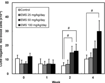
(2) EMS Mutagenesis Detected by Pig-a & PIGRET Assays. HIS49 positive red blood cells or one million CD71 and HIS49 double positive reticulocytes was calculated in the Pig-a or PIGRET assays, respectively. Incidence of reticulocytes: A mixture of acridine orange and peripheral blood was dropped onto a glass slide and a cover glass was placed over the drop. The incidence of reticulocytes in the erythrocytes was calculated by counting 111 erythrocytes within one-ninth of a split square in an objective adaptor glass under a fluorescence microscope (BX60F5, Olympus, Tokyo) and multiplying the number of reticulocytes and erythrocytes by 9 to obtain the number of reticulocytes among approximately 1000 erythrocytes. Statistical analyses: The following statistical analyses were performed by EXSUS Ver. 7.7 (CAC EXICARE Corporation, Tokyo, Japan) with a significance level of 5z. The incidence of CD59 negative red blood cells or reticulocytes was analyzed by Bartlett test to evaluate the homogeneity of variance. The parameter was further analyzed by a parametric Dunnett test when the variance was homogeneous or by a Steel (nonparametric Dunnett) test when it was not.. 6 weeks of age. Certified pellet food and tap water were provided ad libitum. The dose levels of EMS were selected based on the previous findings. Namely, the maximum tolerated dose of EMS for 28 days treatment in rats was reported to be 100 mg/kg/day (5), therefore, 25, 50 and 100 mg/kg/ day of EMS were set for the 28-day repeated dose study. In a single dose study, 360 mg/kg of EMS induced statistically significant increase in Pig-a mutant frequency 4 weeks after the single dosing (6); this dose level was set as a low dose level and twice that dose was set as a high dose level in the present study. EMS was administered by oral gavage at a dose volume of 10 mL/kg to 5–6 animals per group. Approximately 250 mL of blood was collected from the tail vein using a wing-type needle (Terumo Corporation, Tokyo, Japan) and blood sampling tube containing EDTA 2-potassium to prevent coagulation (Microtainer, Nippon Becton Dickinson Co., Ltd., Tokyo, Japan). This study was conducted in compliance with the following law and guidelines; ``Law Concerning the Protection and Control of Animals'', Japanese Law No. 105, October 1, 1973, revised on June 22, 2005, ``Standards Relating to the Care and Management of Laboratory Animals and Relief of Pain'', Notification No. 88 issued by the Japanese Ministry of the Environment, Japan, April 28, 2006 and ``Guidelines for Animal Experimentation'', issued by the Japanese Association for Laboratory Animal Science, May 22, 1987. Pig-a and PIGRET assays: Sample preparation in the present study was performed according to the method described in a previous report (4). Namely, a suspension of blood and EDTA mixture in PBS was labeled with FITC-conjugated anti-rat CD59 and biotinylated HIS49 antibodies (BD Biosciences, Tokyo, Japan) for the Pig-a assay. After incubation, the samples were suspended in PBS and incubated with APCconjugated streptavidin (BD Biosciences). The resultant cell samples were resuspended in PBS. For the PIGRET assay, the suspension of blood and EDTA mixture in PBS was incubated with PE-conjugated anti-rat CD71 (BD Biosciences). Labeled cells were washed with IMag Buffer (BD Biosciences) and the cells were mixed with PE Particles Plus-DM (BD Biosciences). After incubation, the CD71 positive cell fraction was enriched using a device, IMagnet (BD Biosciences). Enriched samples were labeled with anti-CD59 and HIS49 antibodies as described for the Pig-a assay. Unlike the method in the previous report, depletion of leukocytes was not performed. Flow cytometric analysis on CD59 negative red blood cells and reticulocytes was conducted on a FACSCanto flow cytometer equipped with 488 nm and 635 nm lasers and FACSDiva (ver. 6.0) software (BD Biosciences). The incidence of CD59 negative cells in one million. Results Pig-a assay in 28-day repeated dose study with EMS: The results obtained from the Pig-a assay in the 28-day repeated dose study with EMS are shown in Fig. 1. A statistically significant increase in Pig-a mutant (CD59 negative red blood cells) frequency was observed after 2 weeks of EMS treatment in the groups given 50 and 100 mg/kg/day, and after 4 weeks of that given 100. Fig. 1. Time-course for induction of Pig-a mutant (CD59 negative red blood cells) in rats treated orally for 28 days with ethyl methanesulfonate by the Pig-a assay. Mean of 6 animals. The x-axis represents the sampling time after first dosing. The vertical bars represent SD. Statistical analysis was performed on the incidence of CD59 negative red blood cells by Bartlett-Dunnett test (#) with significance level at 5z. 175.
(3) Satoru Itoh et al.. mg/kg/day. The Pig-a mutant frequencies in the control groups and pre-dosing values of the EMS treatment groups were 5.7–12.2×10⁻⁶, and were comparable to those obtained in the previous study (3). PIGRET assay in single dose study with EMS: The results obtained from the PIGRET assay in the single dose study with EMS are shown in Fig. 2, and the incidence of reticulocytes is summarized in Table 1. A statistically significant increase in Pig-a mutant (CD59. negative reticulocytes) frequency was observed after 1 week of EMS treatment in groups given 360 and 720 mg/kg, and after 2 weeks of that given 720 mg/kg. Induction in Pig-a mutant frequency on week 1 was stronger than that on week 2. No change in the percentage of reticulocytes was observed at either sampling point after treatment with EMS.. Discussion Repeated dosing with EMS for 28 days resulted in a statistically significant increase in Pig-a mutant frequency on weeks 2 and 4 at a dose level of 100 mg/kg/day. A similar result has been reported (7), which confirms that the Pig-a assay can detect mutagenesis induced by EMS on the Pig-a gene in erythroid cells. In general, genotoxicants require relatively high exposure to express their genotoxicity in each target tissue in vivo. Therefore, setting the dose level of the test compound is an important factor for detection of in vivo genotoxicity, especially in the repeated dose toxicity study. Frequently the highest dose level of a genotoxicant is lower in the repeated dose toxicity study than in the single dose toxicity study due to its general toxicity. As an illustration, the clastogenicity of mitomycin C, a strong micronucleus inducer in vivo, was not detected in the 14-day repeated dose toxicity study because adequate dose levels could not be used due to its general toxicity (8). In the present case, although we used 100 mg/kg/day of EMS, which was reported to be the maximum tolerated dose in a 28-day repeated dose toxicity study (5), no deaths or serious toxicity was observed in. Fig. 2. Time-course for induction of Pig-a mutant (CD59 negative reticulocytes) in rats treated orally with single dose of ethyl methanesulfonate by the PIGRET assay. Mean of 5 animals. The xaxis represents the sampling time after single dosing. The vertical bars represent SD. Statistical analysis was performed on the incidence of CD59 negative reticulocytes by Bartlett-Steel test (*) or Bartlett-Dunnett test (#) with significance level at 5z.. Table 1. Compound Water for injection. Ethyl methanesulfonate. Ethyl methanesulfonate. Incidence of reticulocytes in rats treated orally with single dose of ethyl methanesulfonate Dose (mg/kg). Animal No.. 0. Week 1. Week 2. —. 10101 10102 10103 10104 10105. 13.49 13.74 13.79 13.54 10.04. 7.17 7.16 6.81 6.34 6.40. 7.30 6.06 7.62 6.29 5.94. Mean±SD. 12.92±1.62. 6.78±0.40. 6.64±0.77. 10201 10202 10203 10204 10205. 8.72 12.77 10.19 11.40 9.53. 6.77 5.13 6.34 7.75 7.22. 6.88 6.18 5.33 4.52 5.57. Mean±SD. 10.52±1.59. 6.64±0.99. 5.70±0.89. 10301 10302 10303 10304 10305. 11.75 10.71 9.68 13.82 9.82. 7.29 8.07 9.44 6.93 7.58. 4.82 6.07 5.08 4.48 6.23. Mean±SD. 11.16±1.70. 7.86±0.98. 5.34±0.78. 360. 720. unit: z 176.
(4) EMS Mutagenesis Detected by Pig-a & PIGRET Assays. our study and induction in Pig-a mutant frequency by EMS was seen. These results support the possibility of integration of the Pig-a assay into repeated dose general toxicity studies. The integration of the Pig-a assay provides us with some benefits, compared with performing each study as a stand-alone, not only by reducing the amount of test chemical but also by reducing the number of experimental animals, which is in accord with a recent movement and an unavoidable issue in genotoxicity testing (9). Further studies are necessary to evaluate whether the integration of this assay into the repeated dose general toxicity study is acceptable. The increase in Pig-a mutant frequency was observed at weeks 2 and 4 during the 28-day treatment with EMS, and higher Pig-a mutant frequency was obtained at week 4. The values at 50 and 100 mg/kg/day of week 2 are statistically significant, but are comparable to the statistically insignificant value obtained at 50 mg/kg/ day of week 4. Therefore, it seems that a 4-week dosing period is required for detecting the mutagenicity of EMS with certainty in the Pig-a assay. In the single dose study with the PIGRET assay, the Pig-a mutant frequency was significantly increased 1 week after the single dose of EMS at both dose levels (360 and 720 mg/kg) and the effect was reduced at week 2. Briefly, the induction of Pig-a mutations was detected later by Pig-a assay than by PIGRET assay. The same situation was reported in the bone marrow and peripheral blood micronucleus tests (10). The peak of micronucleus induction occurred later in peripheral blood than in bone marrow. This is caused by the transition time from micronucleated immature erythrocytes in bone marrow to micronucleated reticulocytes in peripheral blood (11,12). The target cell population in the PIGRET assay is the reticulocytes and that in the Pig-a assay is the more mature red blood cells in peripheral blood. Therefore, the PIGRET assay could detect Pig-a mutants earlier than the original Pig-a assay, which is similar to the situation with the bone marrow micronucleus test. This is the one of the strengths of the PIGRET assay over the Pig-a assay. Another strength of the PIGRET assay is an improvement in the sensitivity of the assay to in vivo mutagens. The target cell population of the PIGRET assay is reticulocytes and it is expected to concentrate the target cell population. Actually, the maximum increase in Pig-a mutant frequency in the EMS group compared with the concurrent control group was 13.5 fold in the PIGRET assay, and 2.9 fold in the Pig-a assay, although it is difficult to directly compare them because the dose levels and dosing frequency were different in the two studies. The weakness of the PIGRET assay, on the other hand, is that if the test compound suppresses bone marrow, it would be difficult to analyze the Pig-a mutants because the target cell population might be reduced. In the case of EMS,. this was not a problem because no change in the incidence of reticulocytes occurred during EMS treatment. Further comparison studies between the PIGRET and Pig-a assays using genotoxicants including myelotoxic compounds should be performed. In conclusion, the results obtained in the present study indicate that the PIGRET assay can detect Pig-a mutants much earlier than the original Pig-a assay can by focusing on reticulocytes rather than red blood cells as the target cell population.. Acknowledgements: This work was partly supported by the Japan Health Sciences Foundation, Grant number KHB1209. Conflict of interest statement: The authors declare that there are no conflicts of interest.. References 1. 2. 3. 4. 5. 6. 7. 8. 9. 177. Dobrovolsky VN, Miura D, Heflich RH, Dertinger SD, The in vivo Pig-a gene mutation assay, a potential tool for regulatory safety assessment. Environ Mol Mutagen. 2010; 51: 825–35. OECD Guidelines for the Testing of Chemicals. Test No. 488: Transgenic Rodent Somatic and Germ Cell Gene Mutation Assays. 28 July 2011. Kimoto T, Horibata K, Chikura S, Hashimoto K, Itoh S, Sanada H, et al. Interlaboratory trial of the rat Pig-a mutation assay using an erythroid maker HIS49 antibody. Mutat Res. 2013; 755: 126–34. Kimoto T, Chikura S, Suzuki K, Kobayashi X-m, Itano Y, Horibata K, et al. Further development of the rat Piga mutation assay: measuring rat Pig-a mutant bone marrow erythroids and a high throughput assay for mutant peripheral blood reticulocytes. Environ Mol Mutagen. 2011; 52: 774–83. Pfister T, Eichinger-Chapelon A. General 4-week toxicity study with EMS in the rat. Toxicol Lett. 2009; 190: 271–85. Kimoto T, Chikura S, Suzuki-Okada K, Kobayashi X, Itano Y, Miura D, et al. Effective use of the Pig-a gene mutation assay for mutagenicity screening: measuring CD59-deficient red blood cells in rats treated with genotoxic chemicals. J Toxicol Sci. 2012; 37: 943–55. Dobo KL, Fiedler RD, Gunther WC, Thiffeault CJ, Cammerer Z, Coffing SL, et al. Defining EMS and ENU dose-response relationships using the Pig-a mutation assay in rats. Mutat Res. 2011; 725: 13–21. Hamada S, Sutou S, Morita T, Wakata A, Asanami S, Hosoya S, et al. Evaluation of the rodent micronucleus assay by a 28-day treatment protocol: summary of the 13th collaborative study by the Collaborative Study Group for the Micronucleus Test (CSGMT)/Environmental Mutagen Society of Japan (JEMS)-Mammalian Mutagenicity Study Group (MMS). Environ Mol Mutagen. 2001; 37: 93–110. Pfuhler S, Kirkland D, Kasper P, Hayashi M,Vanparys P, Carmichael P, et al. Reduction of use of animals in.
(5) Satoru Itoh et al.. 10. 11. regulatory genotoxicity testing: Identification and implementation opportunities—Report from an ECVAM workshop. Mutat Res. 2009; 680: 31–42. The Collaborative Study Group for the Micronucleus Test. Micronucleus test with mouse peripheral blood erythrocytes by acridine orange supravital staining: the summary report of the 5th collaborative study by CSGMT/JEM・MMS. The Collaborative Study Group for the Micronucleus Test. Mutat Res. 1992; 278: 83–98.. 12. 178. Hayashi M, Kodama Y, Awogi T, Suzuki T, Asita AO, Sofuni T. The micronucleus assay using peripheral blood reticulocytes from mitomycin C- and cyclophosphamidetreated rats. Mutat Res. 1992; 278: 209–13. Iwakura K, Tamura H, Matsumoto A, Ajimi S, Ogura S, Kakimoto K, et al. The micronucleus assay with peripheral blood reticulocytes by acridine orange supravital staining with 1-b-d-arabinofuranosylcytosine. Mutat Res. 1992; 278: 131–7..
(6)
図

関連したドキュメント
We present sufficient conditions for the existence of solutions to Neu- mann and periodic boundary-value problems for some class of quasilinear ordinary differential equations.. We
Analogs of this theorem were proved by Roitberg for nonregular elliptic boundary- value problems and for general elliptic systems of differential equations, the mod- ified scale of
Then it follows immediately from a suitable version of “Hensel’s Lemma” [cf., e.g., the argument of [4], Lemma 2.1] that S may be obtained, as the notation suggests, as the m A
Correspondingly, the limiting sequence of metric spaces has a surpris- ingly simple description as a collection of random real trees (given below) in which certain pairs of
[Mag3] , Painlev´ e-type differential equations for the recurrence coefficients of semi- classical orthogonal polynomials, J. Zaslavsky , Asymptotic expansions of ratios of
This problem becomes more interesting in the case of a fractional differential equation where it closely resembles a boundary value problem, in the sense that the initial value
In this article we prove the following result: if two 2-dimensional 2-homogeneous rational vector fields commute, then either both vector fields can be explicitly integrated to
While early experiments with algebraic multigrid solvers have shown promising results [2], herein we focus on a domain decomposition approach based on the finite element tearing
