Suppression of hypersynchronous network
activity in cultured cortical neurons using an
ultrasoft silicone scaffold
著者
Takuma Sumi, Hideaki Yamamoto, Ayumi
Hirano-Iwata
journal or
publication title
Soft Matter
volume
16
number
13
page range
3195-3202
year
2020-02-17
URL
http://hdl.handle.net/10097/00131034
doi: 10.1039/C9SM02432H1
Suppression of hypersynchronous network activity in cultured
1
cortical neurons using an ultrasoft silicone scaffold
2 3
Takuma Sumi
a, Hideaki Yamamoto*
ab, and Ayumi Hirano-Iwata
ab4 5
aResearch Institute of Electrical Communication, Tohoku University, 2-1-1 Katahira, Aoba-ku, 6
Sendai 980-8577, Japan. E-mail: [email protected] 7
bWPI-Advanced Institute for Materials Research (WPI-AIMR), Tohoku University, 2-1-1 8
Katahira, Aoba-ku, Sendai 980-8577, Japan 9
2
Abstract
11
The spontaneous activity pattern of cortical neurons in dissociated culture is characterized by
12
burst firing that is highly synchronized among a wide population of cells. The degree of
13
synchrony, however, is excessively higher than that in cortical tissues. Here, we employed
14
polydimethylsiloxane (PDMS) elastomers to establish a novel system for culturing neurons on a
15
scaffold with an elastic modulus resembling brain tissue, and investigated the effect of the
16
scaffold’s elasticity on network activity patterns in cultured rat cortical neurons. Using
17
whole-cell patch clamp to assess the scaffold effect on the development of synaptic connections,
18
we found that the amplitude of excitatory postsynaptic current, as well as the frequency of
19
spontaneous transmissions, was reduced in neuronal networks grown on an ultrasoft PDMS with
20
an elastic modulus of 0.5 kPa. Furthermore, the ultrasoft scaffold was found to suppress neural
21
correlations in the spontaneous activity of the cultured neuronal network. The dose of GsMTx-4,
22
an antagonist of stretch-activated cation channels (SACs), required to reduce the generation of
23
the events below 1.0 event/min on PDMS substrates was lower than that for neurons on a glass
24
substrate. This suggests that the difference in the baseline level of SAC activation is a molecular
25
mechanism underlying the alteration in neuronal network activity depending on scaffold
26
stiffness. Our results demonstrate the potential application of PDMS with biomimetic elasticity
27
as cell-culture scaffold for bridging the in vivo-in vitro gap in neuronal systems.
3
Main text
29
1. Introduction
30
In vitro modelling of in vivo multicellular functions is essential in biology and medicine not 31
only for basic studies but also for applied research, such as the screening of candidate molecules
32
in drug development.1,2 In fields such as cardiology and oncology, cultured-cell models have 33
been established and are used in disease modelling and toxicity assays.1,3 However, in 34
neuroscience, cortical and hippocampal neurons in dissociated culture generate a
35
non-physiological activity characterized by globally synchronized burst firing, often referred to
36
as ‘network bursts’.4-7 This activity pattern is significantly different from that observed in an 37
animals’ cortex or hippocampus, which is highly complex both spatially and temporally.8,9 Such 38
complexity in neural activity is important, as it underlies the computational capacity of the
39
neuronal networks.10,11 40
Several approaches have been taken to suppress the globally synchronized bursting in
41
cultured neuronal networks. For instance, it has been shown that the synchronized bursts are
42
inhibited and the complexity in the spontaneous activity is upregulated by growing cultured
43
neurons on micropatterned surfaces to induce a network architecture such as those observed in
44
the in vivo networks.12 The role of external inputs in shaping the spontaneous dynamics of the 45
cultured neural networks has also been investigated both experimentally and computationally,
46
showing that chronic application of external stimulus that resembles thalamic input decorrelates
47
cortical neuronal network activity.13-15 Furthermore, pharmacological blockade of an 48
AMPA-type glutamate receptor with CNQX at a dose below its IC50 reduces the spatial extent 49
of the burst spreading,5 possibly through a reduction in the excitatory synaptic strength that is 50
excessively strong in cultured neurons as compared to the in vivo cortex.16-18 51
4
Another major difference between the in vitro and in vivo neuronal networks is the
52
mechanical property of their scaffolds. Cultured neurons are usually grown on a polystyrene or
53
glass substrate, whose elastic moduli, E, are in the order of GPa.19,20 In contrast, the brain is the 54
softest tissue in an animals’ body, with an E below 1 kPa.21 Several studies on non-neuronal 55
cells have pointed to the importance of culturing cells on a scaffold with biomimetic elasticity.
56
For instance, mesenchymal stem cells commit to the lineage specified by scaffold elasticity.22 57
Furthermore, the expression of chondrocyte phenotype is stabilized when cultured on a scaffold
58
with an E of 5.4 kPa, similar to that of the in vivo environment.23 Based on these observations, 59
we hypothesized that the non-physiological synchronized bursting in cultured neuronal
60
networks could be suppressed by growing neurons on a biomimetic scaffold.
61
In this work, we established a biomimetic culture platform using polydimethylsiloxane
62
(PDMS) that is as soft as brain tissue (i.e. E ~ 0.5 kPa). PDMS is a well-established
63
biocompatible material, whose elasticity can be tuned in a wide range, from ~0.1 kPa to tens of
64
MPa by choosing the precursors and changing their mixing ratio.24,25 It also offers several 65
advantages over more commonly used materials (e.g. polyacrylamide), such as being
66
compatible with surface modification techniques, being electrically insulating, and having a
67
long shelf life.26 Primary rat cortical neurons, one of the most well-established systems in 68
dissociated culture of neuronal cells, were cultured on the PDMS substrate, and the effect of the
69
scaffold’s stiffness on synaptic strength and the complexity of the neuronal network activity was
70
assessed using whole-cell patch-clamp recording and fluorescent calcium imaging, respectively.
71
We show that the excitatory synapses are weakened on the softer substrates and that the
72
neuronal correlation in spontaneous network activity is significantly reduced on the PDMS
73
substrate with an E ~ 0.5 kPa. The underlying molecular mechanism responsible for the
5
stiffness-dependent modulation on spontaneous network activity is pharmacologically explored
75
by blocking stretch-activated cation channels (SACs).
76 77 78
2. Experimental
79
2.1 Mechanical characterization of the PDMS
80
PDMS was prepared using Sylgard 184 (Dow Corning; mixing ratio = 50:1) and Sylgard 527
81
(Dow Corning; mixing ratio = 5:4). For each PDMS, 200 g of the mixtures were poured in a
82
glass petri dish (diameter, 90 mm; height, 60 mm), degassed in a vacuum chamber, and cured in
83
an oven (AS-ONE SONW-450S) for two days at 80 oC. 84
The elastic modulus of the PDMS was determined by the spherical indentation method
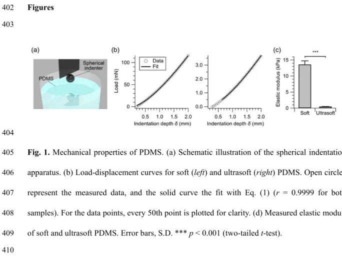
85
(Fig. 1a) following Zhang et al.27,28 Briefly, a chromium steel ball of 3.175-mm radius (R) was 86
attached onto the load cell of the Instron 5943 Universal Testing System. The depth
87
(δ)-indentation load (P) curves were measured (Fig. 1b), and the elastic moduli, E, were
88
determined by fitting the load curves to the following equation:
89
√ δ 1 0.15δ . (1)
90
2.2 PDMS substrates for neuronal culture
91
Glass coverslips (Matsunami C018001; diameter, 18 mm; thickness 0.17 mm) were first cleaned
92
by sonication in 99.5% ethanol and rinsed two times in Milli-Q grade water. After a thorough
93
mixing of the two PDMS components and subsequent degassing, 100 μL of the mixture was
94
drop casted on the coverslip. PDMS was then cured in an oven for 11 h at 80 oC. 95
96
2.3 Contact angle measurement
6
The hydrophilicity of the surfaces was characterized by measuring the water contact angle.
98
Using the LSE-B100 equipment (NiCK Corporation, Japan), a 0.5-μL water droplet was
99
dropped onto the substrate and was imaged from the side. The contact angle of the droplet was
100
measured using the i2win software (NiCK Corporation, Japan). Three samples were prepared
101
for each condition, and measurements were performed at three different positions for each
102 sample. 103 104 2.4 Cell culture 105
For cell culturing, the PDMS substrate was first treated in air plasma (Yamato PM-100) for 10 s
106
and was sterilized under UV light (Toshiba GL-15; wavelength, ~253.7 nm) for 60 min. The
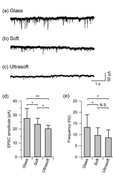
107
exposure to UV light itself did not affect the surface properties, as confirmed by water contact
108
angle measurements (data not shown). In order to promote the adhesion of neuronal cells, the
109
surface of the PDMS was then coated with poly-D-lysine (PDL; Sigma P-0899) by floating the
110
sample upside-down on a phosphate-buffered saline (Gibco 14190-144) containing 50 μg/mL
111
PDL overnight. The sample was then rinsed two times in sterilized water and dried in air inside
112
a laminar flow hood. One day prior to cell plating, the sample was immersed in the plating
113
medium [minimum essential medium (Gibco 11095-080) + 5% foetal bovine serum + 0.6%
114
D-glucose] and stored in a CO2 incubator (37 ºC). Glass coverslips without the PDMS layer 115
were used in control experiments. These were prepared by cleaning coverslips in ethanol and
116
water, treating the surface with air plasma (60 s), UV-sterilization (60 min), and subsequent
117
coating with PDL (overnight).
118
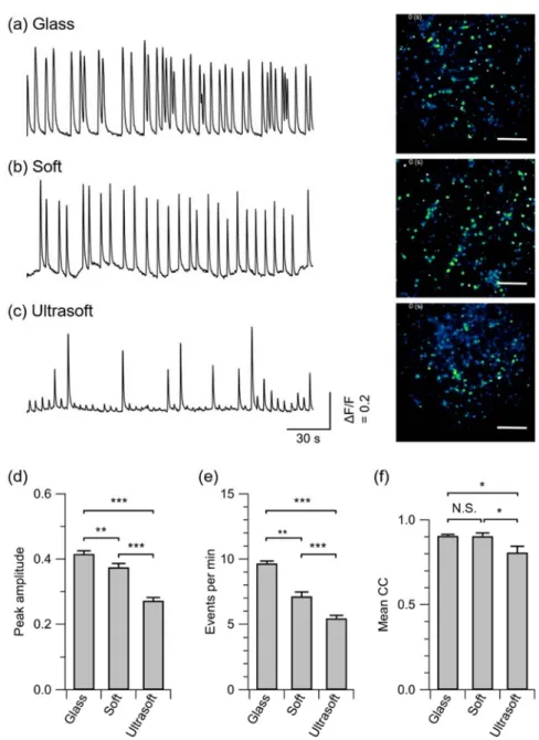
Rat cortical neurons from 18-d old embryos were used in our experiments. All
119
procedures comply with the Regulations for Animal Experiments and Related Activities at
120
Tohoku University and were approved by the Center for Laboratory Animal Research, Tohoku
7
University (approval number: 2017AmA-001-1). After dissection of the cortical tissues and cell
122
dispersion, the cells were plated on the samples immersed in the plating medium. After a 3 h
123
incubation, the medium was changed to Neurobasal medium [Neurobasal (Gibco 21103-049) +
124
2% B-27 supplement (Gibco 17504-044) + 1% GlutaMAX-I (Gibco 3505-061)]. Half of the
125
medium was replaced with fresh Neurobasal medium at 4 and 8 days of the culture.
126 127
2.5 Electrophysiology
128
Whole-cell patch-clamp recordings (HEKA EPC-10) were performed on neurons at 14−18 DIV
129
under the voltage-clamp mode (holding potential, -70 mV). Signals were sampled at 20 kHz and
130
filtered with 10 kHz and 2.9 kHz Bessel filters. Recordings were performed at room temperature.
131
The intracellular solution contained: 146.3 mM KCl, 0.6 mM MgCl2, 4 mM ATP-Mg, 0.3 mM 132
GTP-Na, 5 U/mL creatine phosphokinase, 12 mM phosphocreatine, 1 mM EGTA, and 17.8 mM
133
HEPES (pH 7.4). The extracellular solution for the recording contained: 140 mM NaCl, 2.4 mM
134
KCl, 10 mM HEPES, 10 mM glucose, 2 mM CaCl2, and 1 mM MgCl2 (pH 7.4).18 The GABAA 135
receptor antagonist, bicuculline (Sigma 14343; 10 μM), was added to the extracellular solution
136
to block inhibitory synaptic transmission. The membrane resistance was ~30 MΩ, and the
137
synaptic currents with amplitude of 10−150 pA were analysed using a custom code written in
138
MATLAB (Mathworks).
139 140
2.6 Fluorescent calcium imaging
141
Cultured neurons were loaded with a fluorescence calcium indicator Cal-520 AM (AAT
142
Bioquest).12 The cells were first rinsed in HEPES-buffered saline (HBS) containing 128 mM 143
NaCl, 4 mM KCl, 1 mM CaCl2, 1 mM MgCl2, 10 mM D-glucose, 10 mM HEPES, and 45 mM 144
sucrose, and subsequently incubating in HBS containing 2 μM Cal-520 AM for 30 min at 37 °C.
8
The cells were then rinsed in fresh HBS and were imaged on an inverted microscope (IX83,
146
Olympus) equipped with a 20× objective lens (numerical aperture, 0.70), a light-emitting diode
147
light source (Lambda HPX, Sutter Instrument), a scientific complementary metal-oxide
148
semiconductor camera (Zyla 4.2, Andor), and an incubation chamber (Tokai Hit). All recordings
149
were performed at 14−18 DIV, while incubating in HBS at 37 °C. In some experiments,
150
GsMTx-4 (Peptide Institute 4393-s) was added to the HBS to inhibit SACs.29 Each recording 151
was performed for 10 min at a frame rate of 10 Hz.
152 153
2.7 Statistical analysis
154
The results are presented as mean ± S.D. unless otherwise as stated in the main text. Samples
155
sizes (n) are also presented at each section in the text. Statistical significance of the mean values
156
between two groups were compared using Student’s t-tests.
157 158 159
3. Results and discussion
160
3.1 Material properties of silicone scaffolds
161
The elastic scaffolds for neuronal culture were prepared with two types of PDMS, i.e. Sylgard
162
184 mixed at a ratio of 50:1 (hereafter referred to as ‘soft’) and Sylgard 527 mixed at a ratio of
163
5:4 (hereafter referred to as ‘ultrasoft’). We first prepared the PDMS in glass petri dishes and
164
determined their elastic moduli by the spherical indentation method27,28 (Fig. 1). The elastic 165
moduli of soft and ultrasoft PDMS were determined to be 13.6 ± 1.1 kPa (n = 4) and 0.5 ± 0.03
166
kPa (n = 5), respectively (Fig. 1c). The values are in good agreement with previous studies,24,27 167
and the elastic modulus of the ultrasoft PDMS was nearly equal to that of brain tissue.21 168
9
We next evaluated the wettability of the PDMS surface by measuring water contact
169
angles. Neurons require the scaffold surface to be coated with cationic molecules, such as PDL.
170
However, the strong hydrophobicity of as-prepared PDMS prevents the molecules from stably
171
adsorbing on the surface.30 Therefore, the samples were exposed to air plasma for a designated 172
amount of time, which hydrophilizes the PDMS surface by substituting methyl groups with
173
hydroxyl groups.31 The changes in water contact angle θ of the soft and ultrasoft PDMS upon 174
the plasma treatment are shown in Fig. 2a. Prior to the plasma treatment, the PDMS surface was
175
hydrophobic, and θ were measured to be 127.6 ± 6.6 and 123.9 ± 5.1 (n = 40) for the soft and
176
ultrasoft PDMS, respectively. The hydrophilicities of samples increased with the plasma
177
exposure time. For the cell-culture experiment, samples exposed to the plasma for 10 s were
178
used in order to minimize the effect of surface vitrification and cracking.31,32 It has also been 179
previously studied by MacNearney et al.32 that the elastic modulus of Sylgard 527 did not 180
change upon a plasma treatment for less than 10 s, although a plasma treatment for more than
181
30 s resulted in a significant increase in the elastic modulus.
182
The hydrophilized surface was finally coated with PDL, and rat cortical neurons were
183
cultured on the substrates. As shown in Fig. 2a, θ for the soft and ultrasoft PDMS immediately
184
after the 10 s plasma treatment were significantly different. However, the values of θ for the two
185
scaffolds were found to converge after the PDL and the subsequent immersion in the neuronal
186
plating medium (Fig. 2b). This suggests that the surfaces were chemically consistent between
187
the two substrates and validates the comparison of the two substrates focusing solely on their
188
mechanical properties. Representative micrographs of the rat cortical neurons cultured on the
189
soft and ultrasoft PDMS are shown in Figs. 2c–e. Plain glass coverslips coated with PDL were
190
used as controls. The cell bodies of the neurons were well spread, and the neurites uniformly
191
covered the entire surface. In order to compensate for the difference in cell affinity between
10
glass and PDMS, initial plating density was increased 1.5-fold for the two PDMS scaffolds to
193
achieve a constant attachment density of ~950 cells/mm2 (Fig. 2f). 194
195
3.2 Reduction of excitatory synaptic currents on ultrasoft scaffolds
196
Previous work has shown that the amplitude of excitatory postsynaptic current (EPSC) in
197
hippocampal neurons cultured on Sylgard 184 with E = 457 kPa was significantly higher than
198
that of neurons on Sylgard 184 with E = 46 kPa.27 To investigate whether a further reduction of 199
substrate stiffness to mimic that of the brain tissue (E ~ 0.5 kPa) influences the synaptic
200
strengths, we compared the amplitude and frequency of spontaneous EPSC (sEPSC) in neuronal
201
networks grown on the soft (E = 14 kPa) and ultrasoft (E = 0.5 kPa) PDMS. sEPSC was
202
recorded from cultured cortical neurons at 14−18 DIV under whole-cell patch clamp. To inhibit
203
spontaneous inhibitory transmissions, a GABAA receptor blocker, bicuculline (10 μM), was 204
added to the extracellular solution during recording
205
Representative traces from neurons cultured on glass, soft PDMS, and ultrasoft PDMS
206
are shown in Figs. 3a–c, respectively. The amplitude of sEPSC observed in the neurons on soft
207
substrates was 15% lower than those on glass substrates [soft: 23.5 ± 4.1 pA (n = 13), glass:
208
27.8 ± 7.0 pA (n = 11)]. sEPSC amplitude in neurons on ultrasoft substrates was further reduced
209
from those on soft substrates and was approximately 30% lower than those on glass substrates
210
[ultrasoft: 20.4 ± 2.3 pA (n = 12)]. In addition, the frequency of sEPSC from the neurons on soft
211
and ultrasoft substrates was significantly lower than that on glass substrates (ultrasoft: 8.7 ± 3.3
212
Hz, soft: 9.7 ± 3.3 Hz, glass: 13.3 ± 5.6 Hz). These data are summarized in Figs. 3d and 3e.
213
These results indicate that ultrasoft substrates that resemble the elastic moduli of brain tissues
214
suppress the excitatory synaptic strength in cultured cortical neurons. The molecular mechanisms 215
underlying the observations are further investigated and discussed in section 3.4. 216
11 217
3.3 Suppression of neural synchrony on ultrasoft scaffolds
218
Next, fluorescence calcium imaging was used to quantify the difference in the spontaneous
219
firing patterns of neuronal networks on respective substrates. Representative traces of relative
220
fluorescence intensity (ΔF/Fo) from single neurons are shown in Figs. 4a–c. On the glass surface,
221
the peak amplitude of the calcium transients was 0.42 ± 0.01, and the rate was 9.7 ± 0.2
222
events/min (mean ± S.E.M.; n = 500). Both the peak amplitude and the event rate were
223
significantly reduced on the soft PDMS (0.37 ± 0.01 and 7.1 ± 0.3 events/min, respectively; n =
224
500). On the ultrasoft substrates, both the amplitude and rate were further reduced as compared
225
to the soft substrate and the control (0.27 ± 0.01 and 5.5 ± 0.2 events/min, respectively; n = 500).
226
The reduction is likely to be caused by the reduction in the excitatory synaptic strength. These
227
data are summarized in Figs. 4d and 4e.
228
In order to analyse the degree of neural correlations in the spontaneous activity, we
229
evaluated the correlation coefficient, rij, between neurons i and j, as:
230
∑
∑ ∑
, (2)
where fi(t) is the relative fluorescence intensity of cell i at time t, and the overline represents
231
time average. Then, we compared their mean, ̅ ∑, / , where N (= 50) is the total
232
number of analysed neurons on respective substrates. Although no significant difference in ̅
233
was observed between glass and soft substrates, the value was significantly lower in the
234
neuronal network grown on the ultrasoft scaffold (Fig. 4f). These results show that excessive
235
neural synchronization was suppressed by reducing the scaffold stiffness to 0.5 kPa.
236
The results obtained in this work are in agreement with the previous study, which
237
showed that a stiff PDMS substrate with E = 457 kPa increased hippocampal neuronal network
238
activity as compared to a PDMS substrate with E = 46 kPa.27 However, no discernible change in 239
12
network synchrony was observed within the range of the elasticities investigated by the previous
240
study. In the present study, we found that the non-physiological bursting activity is suppressed,
241
and the mean correlation coefficient significantly decreases when the elastic modulus of the
242
scaffold is further reduced to 0.5 kPa. Thus, Sylgard 527 is a promising scaffold for suppressing
243
the hypersynchrony in neuronal culture.
244 245
3.4 Molecular mechanism of the scaffold effect
246
The above results show that the ultrasoft scaffold weakens the excitatory synaptic strength and
247
reduces the synchrony in the neuronal network activity. We hypothesized that SACs, whose
248
activity is downregulated on softer substrates,33 would be the underlying molecular mechanism 249
and investigated the effect of its pharmacological blockade on the neuronal network activity.
250
GsMTx-4 is a selective antagonist for SACs with an equilibrium constant of
251
approximately 500 nM.29,34 We first investigated the effect of reducing SAC activity in neurons 252
on glass substrates. Bath application of GsMTx-4 at a concentration of 250 nM was found to
253
reduce the peak amplitude and the rate of spontaneous calcium transients [0.30 ± 0.01 and 4.2 ±
254
0.1 events/min (mean ± S.E.M.), respectively; Fig. 5]. When GsMTx-4 was applied at a higher
255
concentration of 500 nM, the rate was further reduced to 0.24 ± 0.01 events/min (Fig. 5b), while
256
the peak amplitude did not significantly vary from the value observed at 250 nM (Fig. 5a).
257
These results indicate that the fraction of active SACs in the neuronal plasma membrane plays a
258
key role in the generation of spontaneous bursting events and the size of individual events.
259
We next examined the impact of GsMTx-4 application on cortical neurons grown on
260
the PDMS substrates. Application of GsMTx-4 at a concentration of 250 nM reduced the rate of
261
spontaneous calcium transients down to 0.62 ± 0.06 and 0.42 ± 0.02 events/min on the soft and
262
ultrasoft substrates, respectively (Fig. 5b). Therefore, the dose of GsMTx-4 required to reduce
13
the spontaneous occurrence of the calcium transients below 1.0 event/min was lower than that
264
for the neurons on the glass substrate. This suggests that the difference in the baseline level of
265
SAC activation is a molecular mechanism that contributes to the alteration in neuronal network
266
activity depending on scaffold stiffness.
267
Penn et al.35 previously showed that synchronized network activity in cultured 268
hippocampal neurons decreased with extracellular calcium concentration, which was discussed
269
to be caused by a reduction in presynaptic vesicle release probability. Considering that SACs
270
permeate calcium ions,36 the decrease in SAC activation could underlie the reduction in sEPSC 271
amplitude and frequency, and neuronal synchrony on ultrasoft substrates.35,37 Another 272
possibility is that the influx of sodium ions through SACs36 could directly enhance neuronal 273
excitability independent of the modulation of synaptic strength (e.g. through facilitation of
274
action potential generation). Finally, a mechanism independent of SACs could also have a role.
275
A recent study reported that stiff substrates increase the number of synapses and reduce
276
voltage-dependent Mg2+ blockade in N-methyl-D-aspartate receptors, which lead to higher 277
postsynaptic activity in cultured hippocampal neurons.38 Figure 6 summarizes the above 278
discussion concerning the underlying molecular mechanisms for the suppression of
279
hypersynchrony on the ultrasoft substrate.
280 281
3.5 Mechanobiology of neuronal cells
282
Understanding of cellular mechanosensitivity has advanced rapidly since Engler et al.22 found in 283
2006 that mesenchymal stem cells commit to the lineage specified by scaffold elasticity. With
284
neuronal cells, studies during the last decade have shown that the stiffness of scaffolds affects
285
multiple properties of cultured neurons, including neuritogenesis, neurite outgrowth, branching,
286
and axon pathfinding.39-41 For instance, Sur et al.39 has used mouse hippocampal neurons 287
14
cultured on peptide amphiphile gels to show that the growth rate of neurites in immature
288
neurons significantly increased on scaffolds with lower elastic modulus. The neurite outgrowth
289
of rat spinal cord neurons was also found to be accelerated on softer substrates.40 290
Although the molecular mechanism behind the mechanosensitive responses yet remain
291
to be fully elucidated, more recent works have identified that SACs, including the Piezo1
292
channels, are primarily responsible for the effects.41 This was shown, for example, in the 293
pathfinding and branching of axons in Xenopus retinal ganglion cells41, as wells as in the 294
determination of cell fate in human neural stem cells.33 In the current work, we showed that the 295
SAC activity also affects the spontaneous network activity of cultured cortical neurons,
296
providing novel insights into the mechanobiology of neuronal cells and the role of SACs
297 therein. 298 299 4. Conclusions 300
We established a protocol for culturing primary cortical neurons on an ultrasoft PDMS gel that
301
mimics the elasticity of brain tissues and investigated the impact of the biomimetic scaffold on
302
synaptic strength and spontaneous activity patterns. Our study showed that the ultrasoft
303
substrate reduces the amplitude of sEPSCs (Fig. 3) that are excessively strong in the in vitro
304
cultures. This led to significant reduction in the peak fluorescence amplitude and event rate of
305
spontaneous network bursts on the ultrasoft substrate as compared to the glass substrate (Fig. 4).
306
No significant difference in the correlation of neuronal network activity was observed on the
307
scaffolds with E > 13.5 kPa. In contrast, this value was significantly lower for the neuronal
308
network grown on the scaffold with E = 0.5 kPa (Fig. 4f), a stiffness similar to that of brain
309
tissue. This is the first evidence that the ultrasoft scaffold with biomimetic elasticity effectively
310
suppresses the hypersynchrony in the spontaneous network activity. A difference in the baseline
15
activation of SACs underlie these stiffness-dependent changes in synaptic transmission and
312
neuronal network activity.
313
The ultrasoft PDMS scaffold offers a mechanically biomimetic culture platform that is
314
beneficial in suppressing the synchronous bursting in neuronal cultures. Moreover, it is a useful
315
platform to study the influence of mechanical cues on neuronal network development. Further
316
work is necessary to fully suppress the synchronized bursting in neuronal cultures. This could
317
be accomplished by integrating cell micropatterning technology with ultrasoft scaffolds or by
318
adding external noise to fill in for functional interactions between brain regions.12,13 319
320
Conflicts of interest
321
There are no conflicts to declare.
322 323
Acknowledgements
324
We acknowledge Prof. Hisashi Kino and Prof. Tetsu Tanaka of Tohoku University for the
325
mechanical analysis of PDMS. This work was supported by the Japan Society for the Promotion
326
of Science (Kakenhi Grant No. 18H03325) and by the Japan Science and Technology Agency
327
(PRESTO: JPMJPR18MB and CREST: JPMJCR14F3).
328 329
16
References
330
[1] K. H. Benam, S. Dauth, B. Hassell, A. Herland, A. Jain, K.-J. Jang, K. Karalis, H. J. Kim, L.
331
MacQueen, R. Mahmoodian, S. Musah, Y. Torisawa, A. D. van der Meer, R. Villenave, M.
332
Yadid, K. K. Parker and D. E. Ingber, Annu. Rev. Pathol. Mech. Dis., 2015, 10, 195-262.
333
[2] G. Quadranto, J. Brown and P. Arlotta, Nat. Med., 2016, 22, 1220-1228.
334
[3] A. Skardal, S. V. Murphy, M. Devarasetty, I. Mead, H-W. Kang, Y-J. Seol, Y. S. Zhang,
335
S.-R. Shin, L. Zhao, J. Aleman, A. R. Hall, T. D. Shupe, A. Kleensang, M. R. Dokmeci, S. J.
336
Lee, J. D. Jackson, J. J. Yoo, T. Hartung, A. Khademhosseini, S. Soker, C. E. Bishop and A.
337
Atala, Sci. Rep., 2017, 7, 8837.
338
[4] D. A. Wagenaar, J. Pine and S. M. Potter, BMC Neurosci., 2006, 7:11, 1271-2202.
339
[5] J. Soriano, M. Rodríguez Martínez, T. Tlusty and E. Moses, Proc. Natl. Acad. Sci. U.S.A.,
340
2008, 105, 13758-13763.
341
[6] J. G. Orlandi, J. Soriano, E. Alvarez-Lacalle, S. Teller and J. Casademunt, Nat. Phys., 2013
342
9, 582-590.
343
[7] H. Yamamoto, S. Kubota, Y. Chida, M. Morita, S. Moriya, H. Akima, S. Sato, A.
344
Hirano-Iwata, T. Tanii and M. Niwano, Phys. Rev. E, 2016, 94, 012407.
345
[8] P. Golshani, J. T. Goncalves, S. Khosahkhoo, R. Monstany, S. Smirnakis and C.
346
Portera-Cailliau, J. Neurosci., 2009, 29, 10890-10899.
347
[9] J. K. Miller, I. Ayzenshtat, L. Carrillo-Reid and R. Yuste, Proc. Natl. Acad. Sci. U.S.A., 8,
348
E4053-E4061 (2014).
349
[10] E. M. Izhikevich, Neural Comput., 2006, 18, 245-282.
350
[11] H. Ju, M. R. Dranias, G. Banumurthy and A. M. J. VanDongen, J. Neurosci., 2015, 35,
351
4040-4051.
352
[12] H. Yamamoto, S. Moriya, K. Ide, T. Hayakawa, H. Akima, S. Sato, S. Kubota, T. Tanii, M.
17
Niwano, S. Teller, J. Soriano and A. Hirano-Iwata, Sci. Adv., 2018, 4, eaau4914.
354
[13] K. P. Dockendorf, I. Park, P. He, J. C. Princípe and T. B. DeMarse, BioSystems, 2009, 95,
355
90-97.
356
[14] E. M. Izhikevich, J. A. Gally and G. M. Edelman, Cereb. Cortex, 2004, 14, 933-944.
357
[15] J. Zierenberg, J. Wilting and V. Priesemann, Phys. Rev. X, 2018, 8, 031018.
358
[16] A. K. Vogt, L. Lauer, W. Knoll and A. Offenhäusser, Biotechnol. Prog., 2003, 19,
359
1562-1568.
360
[17] S. Song, P. J. Sjöström, M. Reigl, S. Nelson and D. B. Chklovskii, PLoS Biol., 2005, 3,
361
e68.
362
[18] H. Yamamoto, R. Matsumura, H. Takaoki, S. Katsurabayashi, A. Hirano-Iwata and M.
363
Niwano, Appl. Phys. Lett., 2016, 109, 043703.
364
[19] G. V. Lubarsky, M. R. Davidson and R. H. Bradley, Surf. Sci., 2004, 558, 135-144.
365
[20] N. Soga, J. Non-Cryst. Solids, 1985, 73, 305-313.
366
[21] N. D. Leipzig and M. S. Shoichet, Biomaterials, 2009, 30, 6867-6878.
367
[22] A. J. Engler, S. Sen, H. L. Sweeney and D. E. Discher, Cell, 2006, 126, 677-689.
368
[23] T. Zhang, T. Gong, J. Xie, S. Lin, Y. Liu, T. Zhou and Y. Lin, ACS Appl. Mater. Interfaces,
369
2016, 8, 22884-22891.
370
[24] C. Moraes, J. M. Labuz, Y. Shao, J. Fu and S. Takayama, Lab Chip, 2015, 15, 3760-3765.
371
[25] M. P. Wolf, G. B. Salieb-Beugelaar and P. Hunziker, Prog. Polym. Sci., 2018, 83, 97-134.
372
[26] H. Yamamoto, L. Grob, T. Sumi, K. Oiwa, A. Hirano-Iwata and B. Wolfrum, Adv. Biosys.,
373
2019, 3, 1900130.
374
[27] Q.-Y. Zhang, Y.-Y. Zhang, J. Xie, C.-X. Li, W.-Y. Chen, B.-L. Liu, X.-a. Wu, X.-N. Li, B.
375
Huo, L.-H. Jiang and H.-C. Zhao, Sci. Rep., 2014, 4, 6215.
376
[28] M. G. Zhang, Y.-P. Cao, G.-Y. Li and X.-Q. Feng, Biomech. Model. Mechanobiol., 2014, 13,
18
1-11.
378
[29] T. M. Suchyna, J. H. Johnson, K. Hamer, J. F. Leykam, D. A. Gage, H. F. Clemo, C. M.
379
Baumgarten and F. Sachs, J. Gen. Physlol., 2000, 115, 583-598.
380
[30] K. Kang, I. S. Choi and Y. Nam, Biomaterials, 2011, 32, 6374-6380.
381
[31] N. Y. Adly, H. Hassani, A.Q. Tran, M. Balski, A. Yakuschenko, A. Offenhäusser, D. Mayer
382
and B. Wolfrum, Soft Matter, 2017 13, 6297-6303.
383
[32] D. MacNearney, B. Mak, G. Ongo, T. E. Kennedy and D. Juncker, Langmuir, 2016, 32,
384
13525-13533.
385
[33] M. M. Pathak, J. L. Nourse, T. Tran, J. Hwe, J. Arulmoli, D. T. T. Le, E. Bernardis, L. A.
386
Flanagan and F. Tombola, Proc. Natl. Acad. Sci. U.S.A., 2014, 111, 16148-16153.
387
[34] C. L. Bowman, P. A. Gottlieb, T. M. Suchyna, Y. K. Murphy and F. Sachs, Toxicon, 2007,
388
49, 249-270.
389
[35] Y. Penn, M. Segal and E. Moses, Proc. Natl. Acad. Sci. U.S.A., 2016, 113, 3341-3346.
390
[36] F. B. Kalapesi, J. C. H. Tan and M. T. Coroneo, Clin. Exp. Optom., 2005, 33, 210-217.
391
[37] N. R. Hardingham, N. J. Bannister, J. C. A. Read, K. D. Fox, G. E. Hardingham and J. J. B.
392
Jack, J. Neurosci., 2006, 26, 6337-6345.
393
[38] Y. Yu, S. Liu, X. Wu, Z. Yu, Y. Xu, W. Zhao, I. Zavodnik, J. Zheng, C. Li and H. Zhao, ACS
394
Biomater. Sci. Eng, 2019, 5, 3475-3482. 395
[39] S. Sur, C. J. Newcomb, M. J. Webber and S. I. Stupp, Biomaterials, 2013, 34, 4749-4757.
396
[40] F. X. Jiang, B. Yurke, R. S. Schloss, B. L. Firestein and N. A. Langrana, Tissue Eng. Part A,
397
2010, 16, 1873-1889.
398
[41] D. E. Koser, A. J. Thompson, S. K. Foster, A. Dwivedy, E. K. Pillai, G. K. Sheridan, H.
399
Svoboda, M. Viana, L. F. Costa, J. Guck, C. E. Holt and K. Franze, Nat. Neurosci., 2016,
400
19, 1592-1598.
19
Figures
402 403
404
Fig. 1. Mechanical properties of PDMS. (a) Schematic illustration of the spherical indentation
405
apparatus. (b) Load-displacement curves for soft (left) and ultrasoft (right) PDMS. Open circles
406
represent the measured data, and the solid curve the fit with Eq. (1) (r = 0.9999 for both
407
samples). For the data points, every 50th point is plotted for clarity. (d) Measured elastic moduli
408
of soft and ultrasoft PDMS. Error bars, S.D. *** p < 0.001 (two-tailed t-test).
409 410
20 411
412
Fig. 2 Culturing primary neurons on PDMS. (a) Change in water contact angles of soft and
413
ultrasoft PDMS upon exposure to air plasma. (b) Water contact angles measured after plasma
414
irradiation for 10 s, after coating with PDL, and after immersion in the plating medium
415
overnight. The surfaces of both samples were superhydrophilic after the immersion in the
416
plating medium, and thus the data are plotted as 0o. No significant difference was found between 417
the soft and ultrasoft substrates for the datapoints not marked with asterisks. (c–e) Primary
418
cortical neurons cultured on (c) glass, (d) soft, and (e) ultrasoft scaffolds. Scale bars, 50 μm. (f)
419
Average cell densities on the glass, soft, and ultrasoft substrates. Error bars, S.D. * p < 0.05; **
420
p < 0.01; *** p < 0.001 (two-tailed t-test). 421
21 423
424
Fig. 3. Effects of elastic modulus on sEPSC. (a–c) Representative recordings of spontaneous
425
EPSCs on (a) glass, (b) soft, and (c) ultrasoft scaffolds. (d and e) The mean values of the
426
amplitude (d) and frequency (e) of sEPSCs on respective surfaces. Error bars, S.D. * p < 0.05;
427
** p < 0.01 (one-tailed t-test).
428 429
22 430
431
Fig. 4. Impact of substrate stiffness on network activity of cultured cortical neurons. (a–c)
432
Fluorescence intensity traces of representative neurons on (a) glass, (b) soft, and (c) ultrasoft
433
scaffolds. Fluorescence micrographs are shown on the right. Scale bars, 100 μm. (d and e)
434
Average peak amplitudes (d) and frequency of bursting events (e) on respective substrates. (f)
435
Mean correlation coefficient (mean CC) of neural activity on respective substrates. Error bars,
436
S.E.M. * p < 0.05; ** p < 0.01; *** p < 0.001 (two-tailed t-test).
437 438
23 439
440
Fig. 5. Impact of the pharmacological blockade of SAC on neuronal network activity. (a and b)
441
Average peak amplitudes (a) and rate of bursting events (b) at various concentrations of
442
GsMTx-4 on respective substrates. Error bars, S.E.M. * p < 0.05; ** p < 0.01; *** p < 0.001
443
(two-tailed t-test).
444 445 446
24 447
448
Fig. 6. Diagram summarizing the present findings and the mechanisms underlying the
449
suppression of hypersynchronous neuronal network activity on soft scaffolds. Abbreviations:
450
SAC, stretch-activated cation channel; NMDAR, N-methyl-D-aspartate receptor; sEPSC,
451
spontaneous excitatory postsynaptic current.