REVIEWS
The DNA-damage response in human
biology and disease
Stephen P. Jackson
1& Jiri Bartek
2The prime objective for every life form is to deliver its genetic material, intact and unchanged, to the next generation. This must be achieved despite constant assaults by endogenous and environmental agents on the DNA. To counter this threat, life has evolved several systems to detect DNA damage, signal its presence and mediate its repair. Such responses, which have an impact on a wide range of cellular events, are biologically significant because they prevent diverse human diseases. Our improving understanding of DNA-damage responses is providing new avenues for disease management.
E
ach of the ,1013cells in the human body receives tens of thousands of DNA lesions per day1. These lesions can block genome replication and transcription, and if they are not repaired or are repaired incorrectly, they lead to mutations or wider-scale genome aberrations that threaten cell or organism viability. Some DNA aberrations arise via physiological processes, such as DNA mismatches occasionally introduced during DNA rep- lication and DNA strand breaks caused by abortive topoisomerase I and topoisomerase II activity. In addition, hydrolytic reactions and non-enzymatic methylations generate thousands of DNA-base lesions per cell per day. DNA damage is also produced by reactive- oxygen compounds arising as by-products from oxidative respira- tion or through redox-cycling events involving environmental toxic agents and Fenton reactions mediated by heavy metals2. Reactive oxygen and nitrogen compounds are also produced by macrophages and neutrophils at sites of inflammation and infections3. Such chemicals can attack DNA, leading to adducts that impair base pairing and/or block DNA replication and transcription, base loss, or DNA single-strand breaks (SSBs). Furthermore, when two SSBs arise in close proximity, or when the DNA-replication apparatus encounters a SSB or certain other lesions, double-strand breaks (DSBs) are formed. Although DSBs do not occur as frequently as the other lesions listed above, they are difficult to repair and extremely toxic4.The most pervasive environmental DNA-damaging agent is ultra- violet light. Although the ozone layer absorbs the most dangerous part of the solar ultraviolet spectrum (ultraviolet C), residual ultra- violet A and ultraviolet B in strong sunlight can induce ,100,000 lesions per exposed cell per hour. Ionizing radiation also generates various forms of DNA damage, the most toxic of these being DSBs5. Some ionizing radiation results from radioactive decay of naturally occurring radioactive compounds. For example, uranium decay pro- duces radioactive radon gas that accumulates in some homes and contributes to lung-cancer incidence. Exposure to natural or man- made radioisotopes also occurs during cancer radiotherapy, whereas the radioactive compounds iodine-131 and technetium-99m are exploited to diagnose and treat benign and malignant thyroid diseases. Lessons about the health consequences of excessive radi- ation exposure are provided by the aftermaths of the Chernobyl nuclear-reactor disaster and nuclear detonations over Japan in the Second World War.
Today, probably the most prevalent environmental cancer-causing chemicals are those produced by tobacco products, which trigger
various cancers, most notably those of the lung, oral cavity and adja- cent tissues6,7. Cancer-causing DNA-damaging chemicals can also contaminate foods, such as aflatoxins found in contaminated pea- nuts and heterocyclic amines in over-cooked meats7. DNA-damaging chemicals have also been used in warfare, and on a more positive note, are widely used to treat cancer8and ailments such as psoriasis9. Here, we describe how DNA lesions are dealt with at the molecular level. We then explain how such responses affect many cellular pro- cesses, their biological significance and their roles in preventing human diseases. Finally, we illustrate how our increasing knowledge of DNA-damage responses is providing opportunities for improving disease detection and management.
An integrated signalling and genome-maintenance network To combat threats posed by DNA damage, cells have evolved mechanisms—collectively termed the DNA-damage response (DDR)—to detect DNA lesions, signal their presence and promote their repair10–12. Cells defective in these mechanisms generally display heightened sensitivity towards DNA-damaging agents and, as described below, many such defects cause human disease. Although responses differ for different classes of DNA lesions, they usually occur by a common general programme (Fig. 1). Although we focus on DNA repair and DNA-damage signalling separately, we stress that these operate collectively and share many components.
DNA-repair pathways. The wide diversity of DNA-lesion types necessitates multiple, largely distinct DNA-repair mechanisms (Table 1). Whereas some lesions are subject to direct protein- mediated reversal, most are repaired by a sequence of catalytic events mediated by multiple proteins. In mismatch repair, detection of mis- matches and insertion/deletion loops triggers a single-strand incision that is then acted upon by nuclease, polymerase and ligase enzymes13. In base-excision repair, a damaged base is often recognized by a DNA glycosylase enzyme that mediates base removal before nuclease, poly- merase and ligase proteins complete the repair14in processes overlap- ping with those used in SSB repair15,16. The nucleotide excision repair (NER) system, which recognizes helix-distorting base lesions, operates via two sub-pathways that differ in the mechanism of lesion recog- nition: transcription-coupled NER, which specifically targets lesions that block transcription, and global-genome NER15. A key aspect of NER is that the damage is excised as a 22–30-base oligonucleotide, producing single-stranded DNA (ssDNA) that is acted upon by DNA polymerases and associated factors before ligation ensues15.
1The Gurdon Institute and Department of Biochemistry, University of Cambridge, Tennis Court Road, Cambridge CB2 1QN, UK.2Danish Cancer Society, Centre for Genotoxic Stress Research, Strandboulevarden 49, DK-2100 Copenhagen, Denmark, and Institute of Molecular Genetics, CZ-14220 Prague, Czech Republic.
1071
Notably, some DNA lesions are not repaired but are instead bypassed during DNA replication by polymerases with less stringent base-pairing requirements than replicative polymerases17.
For DSB repair, two principal mechanisms are used: non- homologous end-joining (NHEJ)18and homologous recombination (HR)19. In NHEJ, DSBs are recognized by the Ku protein that then binds and activates the protein kinase DNA-PKcs, leading to recruit- ment and activation of end-processing enzymes, polymerases and DNA ligase IV. A less-well-characterized Ku-independent NHEJ pathway, called microhomology-mediated end-joining (MMEJ) or alternative end-joining, also exists; it always results in sequence dele- tions20. Although both NHEJ and MMEJ are error-prone, they can operate in any phase of the cell cycle. By contrast, HR is generally restricted to S and G2 because it uses sister-chromatid sequences as the template to mediate faithful repair. Although there are several HR sub-pathways19, HR is always initiated by ssDNA generation, which is promoted by various proteins including the MRE11–RAD50–NBS1
(MRN) complex. In events catalysed by RAD51 and the breast-cancer susceptibility proteins BRCA1 and BRCA2, the ssDNA then invades the undamaged template and, following the actions of polymerases, nucleases, helicases and other components, DNA ligation and sub- strate resolution occur. HR is also used to restart stalled replication forks and to repair interstrand DNA crosslinks, the repair of which also involves the Fanconi anaemia protein complex21.
DNA-damage signalling and cell-cycle checkpoints. Key DDR- signalling components in mammalian cells are the protein kinases ATM and ATR, which are recruited to and activated by DSBs and replication protein A (RPA)-coated ssDNA, respectively (Table 1)22–24. Two of the best studied ATM/ATR targets are the protein kinases CHK1 and CHK2 which, together with ATM and ATR, act to reduce cyclin-dependent kinase (CDK) activity by various mechanisms, some of which are mediated by activation of the p53 transcription factor23,25,26. Inhibition of CDKs slows down or arrests cell-cycle progression at the G1–S, intra-S and G2–M ‘cell-cycle check- points’, which is thought to increase the time available for DNA repair before replication or mitosis ensues. In parallel, ATM/ATR signalling enhances repair by inducing DNA-repair proteins transcriptionally or post-transcriptionally; by recruiting repair factors to the damage; and by activating DNA-repair proteins by modulating their phosphoryla- tion, acetylation, ubiquitylation or SUMOylation27. Proteomics studies have recently identified a great many as-yet uncharacterized ATM/ATR-mediated phosphorylation sites, suggesting that the DDR modulates additional cellular processes28. If the above events allow effective DNA repair, DDR inactivation ensues, allowing the resump- tion of normal cell functioning. Alternatively, if the damage cannot be removed, chronic DDR signalling triggers cell death by apoptosis or cellular senescence (that is, permanent cell-cycle withdrawal), both of which have potential antitumour functions29,30. As discussed in later sections, another important SSB- and DSB-signalling protein is the enzyme poly(ADP-ribose) polymerase (PARP).
It is becoming increasingly clear that chromatin structure has an impact on the DDR and is modulated in response to DNA damage23,31. The best characterized example of this is ATM/ATR/DNA-PK- mediated phosphorylation of serine 139 of the histone H2A variant, H2AX, on chromatin flanking DSB sites. This brings about ubiquitin- adduct formation in such regions and the recruitment of DDR factors
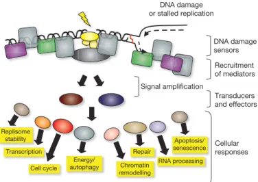
DNA damage sensors
Cellular responses Transducers and effectors Recruitment of mediators
Apoptosis/ senescence
Cell cycle
Repair Transcription
Chromatin remodelling
DNA damage or stalled replication
Signal ampliication
Replisome stability
RNA processing Energy/
autophagy
Figure 1|Model for the DDR. The presence of a lesion in the DNA, which can lead to replication stalling, is recognized by various sensor proteins. These sensors initiate signalling pathways that have an impact on a wide variety of cellular processes. See text for details.
Table 1|DDR mechanisms and components
DDR mechanism Prime lesions acted upon Key protein components
Direct lesion reversal O6alkylguanine O6-methylguanine methyltransferase Mismatch repair DNA mismatches and insertion/deletion
loops arising from DNA replication
Sensors MSH2–MSH6 and MSH2–MSH3 plus MLH1–PMS2, MLH1–PMS1, PLH1–MLH3, EXO1, polymerases d and e, PCNA, RFC, RPA, ligase I Base excision repair and SSB repair Abnormal DNA bases, simple base adducts,
SSBs generated as base-excision repair intermediates by oxidative damage or by abortive topoisomerase I activity
DNA glycosylases (sensors), APE1 endonuclease, DNA polymerases (b, d, e) and associated factors, flap endonuclease FEN1, ligase I or ligase III. SSB repair can also involve polymerase b lyase activity, XRCC1, PARP1, PARP2, polynucleotide kinase and aprataxin.
Nucleotide excision repair (NER) Lesions that disrupt the DNA double helix, such as bulky base adducts and ultraviolet photo-products
Sensors elongating RNA polymerase, XPC-HR23B and DDB1/2, plus XPA, XPE, XPF/ERCC1, XPG, CSA, CSB, TFIIH (containing helicases XPB and XPD), DNA polymerases including polymerase k and associated factors, PCNA, RPA, ligase I and III
Trans-lesion bypass mechanisms Base damage blocking replication-fork progression
‘Error-prone’ DNA polymerases, including polymerases g, i, k, REV3 and REV1; plus associated factors
Non-homologous end-joining (NHEJ) Radiation- or chemically-induced DSBs plus V(D)J and class-switch recombination intermediates
Sensors Ku and DNA-PKcs plus XRCC4, XLF/Cernunnos and ligase IV. Can also use the MRE11–RAD50–NBS1 complex, Artemis nuclease, polynucleotide kinase, aprataxin and polymerases m and l.
Homologous recombination (HR) DSBs, stalled replication forks, inter-strand DNA crosslinks and sites of meiotic recombination and abortive topoisomerase II action
RAD51, RAD51-related proteins (XRCC2, XRCC3, RAD51B, RAD51C, RAD51D, DMC1), RAD52, RAD54, BRCA2, RPA, FEN1, DNA polymerase and associated factors. Promoted by MRN, CtIP, BRCA1 and the ATM signalling pathway.
Fanconi anaemia (FANC) pathway Inter-strand DNA crosslinks FANCA, FANCC, FANCD1/BRCA2, FANCD2, FANCE, FANCF, FANCG, FANCI, FANCJ, FANCL, FANCM, FANCN plus factors including PALB2 and HR factors
ATM-mediated DDR signalling DSBs ATM, MRN and CHK2. Promoted by mediator proteins such as MDC1, 53BP1 MCPH1/BRIT1, and by ubiquitin ligases RNF8, RNF168/RIDDLIN and BRCA1. ATR-mediated DDR signalling ssDNA, resected DSBs Sensors ATR, ATRIP and RPA plus the RAD9–RAD1–HUS1 (9-1-1) complex, RAD17 (RFC1-like) and CHK1. Promoted by MRN, CtIP and mediator proteins such as TOPBP1, Claspin, MCPH1/BRIT1 and BRCA1.
See text for details. 1072
plus other chromatin-modifying components which, together, are thought to promote DSB repair and amplify DSB signalling27. Notably, ATM activation leads to chromatin relaxation at sites of DSBs32, and H2AX tyrosine 142 phosphorylation was recently shown to function in the DDR33,34. It therefore seems likely that further DDR- induced chromatin modifications await discovery.
DDR events operate in diverse biological settings
Generating immune-receptor diversity. The only known pro- grammed genome alterations in vertebrates are V(D)J recombination, class-switch recombination and somatic hyper-mutation35,36. These occur in developing B and T lymphocytes to generate immuno- globulin and T-cell receptor (TCR) diversity, thus allowing effective recognition of diverse pathogens and antigens. Immunoglobulin and TCR proteins comprise variable regions that specify antigen binding, and constant regions that endow specific properties to the TCR or the various immunoglobulin classes. Exons encoding the antigen-binding portions of these molecules are composed of V, D and J segments that are combined in various ways to generate mature immunoglobulin and TCR genes. Each segment is flanked by recombination-signal sequences that are recognized by the RAG1–RAG2 protein complex, which generates a blunt DSB at the signal sequence and a covalently- closed DNA hairpin at the coding end. These structures are then processed and ligated by the NHEJ apparatus35. Consequently, besides causing ionizing radiation hypersensitivity, NHEJ defects yield severe- combined immune-deficiency.
A rearranged immunoglobulin heavy-chain variable domain is initially expressed fused to an Igm constant region but, during antigen-stimulated B-cell differentiation, class-switch recombina- tion can juxtapose a V region to any of several constant regions that bestow distinct properties on the encoded immunoglobulin. B cells undergoing antigen stimulation also activate somatic hyper-mutation to increase mutation rates in the heavy- and light-chain V regions, thus expanding the repertoire of variable segments and allowing selec- tion of B cells expressing immunoglobulin molecules with heightened antigen affinity. Unlike V(D)J recombination, class-switch recom- bination and somatic hyper-mutation require activation-induced deaminase (AID). AID targeting to variable-region exons and IgH switch regions is believed to trigger deamination of cytosine to uracil, resulting in UNG mismatches that are processed by mismatch repair and/or base-excision repair to yield SSBs. In somatic hyper-mutation, error-prone repair of these SSBs is thought to yield mutations within the variable exon; whereas in class-switch recombination, SSBs are converted into DSBs that are acted upon by NHEJ to juxtapose the immunoglobulin variable exon to a constant-region exon35. Production of gametes for sexual reproduction. The DDR also has a key role in generating genetic diversity via sexual reproduction, a stage in which is meiosis, the cell-division pathway that generates haploid gametes. After DNA replication, meiosis proceeds by two successive cell divisions: MI that reductionally segregates the two copies of individual chromosomes; and MII that separates resulting sister-chromatid pairs. Before MI, homologous chromosomes align and exchange genetic information by HR37. In species ranging from yeast to human, meiotic
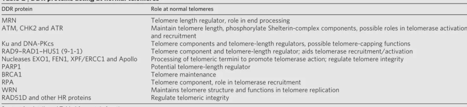
HR is triggered by the topoisomerase-II-related enzyme, Spo11, which generates Spo11-bound DSBs. Spo11 removal and DSB resection then ensue by mechanisms requiring the MRN complex, resulting in ssDNA that promotes HR with the homologous chromosome. These events require all mitotic HR components together with the meiosis-specific RAD51-like protein DMC137. Consequently, mice deficient in Spo11 or Dmc1 are healthy but infertile. In addition, DDR factors such as ATM, MRN and H2AX monitor and coordinate meiotic HR progression. Telomere homeostasis and ageing. In most organisms, the ends of chromosomes are organized into telomeres that comprise stretches of short-tandem-DNA repeats terminating in a 3’ protruding ssDNA over- hang. These repeats are normally generated by the ribonucleoprotein complex telomerase, but in some cancer cells they are maintained by HR-based ‘alternative lengthening of telomeres’ mechanisms38. Although telomeres possess DNA ends, their sequestration into a com- plex termed Shelterin prevents them from engaging in NHEJ-mediated fusions or activating ATM/ATR signalling39. Nevertheless, various DDR proteins have important roles at normal telomeres (Table 2); conse- quently, their defects cause telomere shortening and/or telomere dys- function that trigger chromosome fusions and ensuing chromosomal instability38,40. Furthermore, mammalian telomeres are recognized by MRN and ATM during G2, possibly to trigger a localized DDR that promotes telomere end-processing and Shelterin-complex formation. HR proteins such as RAD51D are also required for telomere integrity, which might reflect their involvement in establishing T-loop structures wherein the telomeric 3’ overhang invades the DNA duplex of internal telomeric sequences38–41.
Excepting specialized cells such as stem cells, human cells generally do not express sufficient telomerase to counteract telomere shortening caused by the inability of the DNA replication machinery to fully replicate chromosomal ends. Thus, human telomeres generally shorten with each cell division until some retain hardly any terminal telomeric repeats. These then fail to act as telomeres and are instead recognized as DSBs, triggering chromosomal fusions and ensuing breakage–fusion–breakage cycles. Under such situations of chronic DDR activation, cells enter into apoptosis or senescence40,41. There is evidence that such processes occur during natural ageing and under circumstances of high cell turnover, such as chronic inflammation or persistent infections. Consistent with ageing in part reflecting the accumulation of shortened telomeres and/or DNA damage, markers of unrepaired DSBs accumulate with age in human and mouse cells, and certain DDR-defective mice strains display hallmarks of acceler- ated ageing42–44. Furthermore, senescent cells occur at sites of age- related pathologies in man, including atherosclerotic lesions, skin ulcers and arthritic joints29,44.
Physiological control of the DDR. The differing physiologies of various cells presumably impose different DDR requirements. Indeed, some DNA-repair pathways are downregulated upon cell differentiation, possibly reflecting rapid DNA repair being less imperative for non-dividing cells. For example, work on terminally differentiated neurons and macrophages has indicated the existence of a new type of NER, termed differentiation-associated repair, in which both transcribed and non-transcribed DNA strands are
Table 2|DDR proteins acting at normal telomeres
DDR protein Role at normal telomeres
MRN Telomere length regulator, role in end processing
ATM, CHK2 and ATR Maintain telomere length, phosphorylate Shelterin-complex components, possible roles in telomerase activation and recruitment
Ku and DNA-PKcs Telomere components and telomere-length regulators, possible telomere-capping functions RAD9–RAD1–HUS1 (9-1-1) Telomere component and telomere-length regulator; aids telomerase recruitment/activation Nucleases EXO1, FEN1, XPF/ERCC1 and Apollo Processing of telomeric termini to promote telomerase action; regulate telomere integrity
PARP1 Potential telomere-length regulator
BRCA1 Telomere maintenance
RPA Telomere component, role in telomerase recruitment
WRN Maintains telomere structure and functions in telomere replication
RAD51D and other HR proteins Regulate telomeric integrity See text for details and Table 1 for protein functions.
1073
repaired effectively but non-transcribed loci are repaired poorly or not at all45. DSB-repair requirements also change during mammalian nervous-system development, with HR being crucial during neuron proliferation, whereas NHEJ becomes critical as neurons terminally differentiate46. This could reflect NHEJ being the prime DSB mech- anism available to post-mitotic neurons, whereas dividing neural precursors also use HR. Because of their importance for tissue home- ostasis and renewal, it has been speculated that stem cells will rely heavily on the DDR. Indeed, defects in base-excision repair, NER, mismatch repair, HR or the Fanconi anaemia complex impair stem- cell function47, and NER or NHEJ defects trigger age-related haema- topoietic stem-cell failure in mice48,49.
Many organisms regulate physiological processes in synchrony with the light–dark cycle via the circadian rhythm/biological clock that is controlled by light stimuli. Recent work has established molecular linkages between the biological clock and DDR events50. For instance, the Caenorhabditis elegans biological-clock gene clk-2 affects radiation sensitivity, and CLK-2 and its mammalian counter- part control the S-phase checkpoint in response to replication stress51. Also, it was recently reported that NER is regulated by the circadian clock52. Perhaps such linkages allow cells to enhance DDR proficiency at times when physiological or environmentally-induced DNA lesions are most prevalent.
Life cycles of pathogens. The cells of pathogens also possess DDR proteins to mitigate the effects of DNA damage. Furthermore, muta- tional repair and recombination occasionally occur in viruses, thus fuelling evolution of pathogens such as avian influenza (H5N1) and swine-origin influenza (H1N1) viruses53. In addition, certain patho- gens use DDR mechanisms to promote virulence. For example, African trypanosomes—unicellular eukaryotic parasites that infect mammals, including humans—evade immune surveillance by using HR to periodically alter their protective variant-surface-glycoprotein coat54. Furthermore, acquisition of drug or pesticide resistance in certain bacteria, plants and unicellular eukaryotic pathogens often involves integration of resistance genes into the organism’s genome via DSB-repair mechanisms. DDR activation is also triggered when cells are infected by viruses, including retroviruses (such as HIV-1), adenoviruses, herpes simplex viruses 1 and 2, hepatitis B virus, Epstein–Barr virus, cytomegalovirus, Kaposi’s sarcoma virus, simian virus 40 and polyomavirus55. Indeed, DDR factors often provide a line of defence against these pathogens, and in many cases viruses have evolved ways to evade such responses. For example, the E6 protein of human papilloma virus types 16 and 18 targets the p53 tumour suppressor for proteolytic degradation to prevent apoptosis of infected cells56. Furthermore, the MRN complex and NHEJ com- ponents curtail adenovirus infectivity by concatemerizing the viral genome; the virus circumvents this by impairing DNA-PK activity, disrupting complexes containing MRN, and targeting MRN for degradation. Conversely, host cell DDR activities sometimes facili- tate viral infectivity. For instance, NHEJ-mediated conversion of linear viral double-stranded (ds) DNA into circles seems to be important for herpes simplex virus replication57. Furthermore, CHK2 deficiency or ATM inhibition impairs HSV-1 growth58. Retroviruses have dsRNA genomes that are converted into dsDNA, which must then integrate into the host genome to produce new retroviruses. Notably, ATM, MRN and NHEJ proteins are required for efficient retrovirus infection, probably by promoting repair of viral integration intermediates55,59.
The DDR and human disease
Cancer and DNA damage: an intimate relationship. A fundamental feature of cancer is genome instability60. For example, genomic instability in lymphoid tumours frequently corresponds to chromo- somal translocations, wherein proto-oncogene loci are fused to those of antigen receptors, apparently by aberrant antigen-receptor recom- bination35,36. In addition, mismatch repair defects cause microsatel- lite instability that predisposes to colorectal and endometrial
carcinomas13. Furthermore, chromosomal instability is seen in most sporadic solid tumours61. It is likely that transient chromosomal instability arises when telomeres in a nascent tumour become critically short and prone to chromosomal fusions62, whereas activated onco- genes and ensuing DNA-replication stress with DSB formation fuel chromosomal instability continuously30. At later stages of cancer pro- gression, chronic hypoxia and/or cycles of hypoxia and re-oxygenation might also contribute to genomic instability and deregulate DDR pathways63.
Most carcinogens operate by generating DNA damage and causing mutations15,26. Furthermore, inherited DDR defects commonly pre- dispose to cancer, contribute to the ‘mutator phenotype’ of many malignancies, and may allow tumour-cell survival and proliferation despite enhanced mutation rates and genome instability (Sup- plementary Table 1). Notably, aberrant cell proliferation, caused by oncogene activation or inactivation of certain tumour suppressors, elicits DNA-replication stress and ongoing DNA-damage formation. Such damage activates ATR/ATM-mediated signalling, causing cell death or senescence in cell-culture models and during tumorigenesis in vivo29,30,64,65. Indeed, the DDR is commonly activated in early neo- plastic lesions and probably protects against malignancy64,65. It has been suggested that breaches to this barrier, arising through muta- tional or epigenetic inactivation of DDR components, are subse- quently selected for during tumour development, thus allowing malignant progression. This model for the DDR as an anticancer barrier helps to explain the high frequency of DDR defects in human cancers30.
Neurodegenerative disorders. Accumulation of DNA lesions in neurons is associated with neurodegenerative disorders, including ataxias together with Alzheimer’s, Huntington’s and Parkinson’s diseases (Supplementary Table 1)66,67. One reason for this may be that neurons generally exhibit high mitochondrial respiration and associated reactive-oxygen-species production that can damage mitochondrial and nuclear DNA68. Consistent with a role for base- excision repair and SSB repair in repairing such lesions, defects in these pathways trigger neuronal dysfunction and degeneration66,69. Another reason why the nervous system is particularly vulnerable to DNA damage is its limited capacity for cell replacement in adulthood, potentially leading to accumulation of damaged but irreplaceable terminally differentiated neurons. Furthermore, being in G0, such cells do not repair DSBs by HR but must use error-prone NHEJ66. It is also noteworthy that neurons rely heavily on transcription and that oxidative DNA damage can block this. Thus, accumulation of DNA lesions in repair-defective patients—and possibly in ageing normal individuals—might progressively deprive neurons of vital tran- scripts, leading to cell dysfunction or apoptosis70. Such processes presumably contribute to the neurodegeneration observed in ataxias and in Cockayne syndrome, which are caused by defects in DNA strand-break repair and transcription-coupled NER, respectively66,67. Genome instability in other heritable human diseases. DNA-repeat instability causes some 40 known diseases that result from expansions or contractions of genetically unstable DNA repeat sequences, usually a trinucleotide motif, within a specific locus for each disease. This instability is thought to arise through the repetitive nature of these regions allowing aberrant DNA-secondary-structure formation during DNA replication or DNA-repair processes71,72. These neuro- muscular and neurodegenerative diseases include fragile X syndromes, Friedrich’s ataxia, spinocerebellar ataxias, diabetes mellitus type 2, Creutzfeldt–Jakob disease, myotonic dystrophy and Huntington’s disease (see Supplementary Table 1 for examples). Analogously, muta- tions or rearrangements of mitochondrial DNA can lead to impaired mitochondrial function as found in amyotrophic lateral sclerosis, mitochondrial encephalomyopathy, Leigh syndrome, myoclonic epi- lepsy, Leber’s hereditary optic neuropathy, and additional neuropa- thies and myopathies73.
Immune deficiencies and infertility. Genome rearrangements involving DDR factors occur during immune-system development, 1074
meaning that DDR defects can cause immune deficiency. For instance, mutations in NHEJ factors yield B- and T-cell immune deficiency, whereas ataxia telangiectasia (AT) and Nijmegen break- age syndrome (NBS) patients (defective in ATM and NBS1, respec- tively) are prone to sometimes-fatal infections, partly due to impaired immunity (class-switch recombination is particularly affected in AT patients). Furthermore, many cancers arising in such conditions are lymphomas and leukaemias of B- and T-cell origin that can result from impaired V(D)J recombination. Human infer- tility is a significant issue, with ,20% of males in western countries being affected74. As meiotic recombination involves DSB generation, it seems likely that certain DDR defects would cause human infertility. Indeed, DDR signalling is readily detectable during human spermatogenesis75, and various inherited DDR deficiencies are characterized by infertility or sub-fertility74. A significant proportion of human infertility might therefore be caused by DDR deficiencies.
Ageing, stem-cell dysfunction, cardiovascular disease and meta- bolic syndrome. There is evidence that ageing is in part caused by accumulated DNA damage76. First, various endogenously arising DNA lesions accumulate with age in the nuclear and mitochondrial genomes of healthy mammals, including humans42,76,77. This may reflect not only ongoing DNA-damage induction but also declining DNA-repair capacity over time68,73. Second, patients with inherited DDR defects often display features of premature ageing (Supplemen- tary Table 1). Third, work in various organisms has implicated growth hormone and insulin-like growth factor 1 signalling in regu- lating longevity, and notably, such signalling is downregulated in response to DNA damage76. The evolutionary honing of longevity- regulating pathways may serve as an example of antagonistic pleio- tropy, where processes selected as advantageous early in life (such as active DDR signalling) because they promote reproduction and pre- vent tumorigenesis are detrimental later in life because they lead to stem-cell depletion, hence contributing to ageing.
Cell senescence and apoptosis are suspected causes of ageing under conditions where attempted tissue regeneration causes stem-cell exhaustion44,78. Indicative of DNA damage contributing to such epi- sodes, impaired stem-cell function is exhibited in mice with defects in the Fanconi anaemia, NER, mismatch repair or NHEJ path- ways48,49,79,80. Furthermore, whereas p53-induced cell death protects against tumorigenesis, pro-apoptotic p53 activity is harmful in set- tings such as stroke or heart attack81,82. Induction of p53 by oxidative stress and other sources of DNA damage can also affect the develop- ment of atherosclerosis, thus providing a link between the DDR and
cardiovascular disease83. Indeed, growing evidence points to human atherosclerosis being characterized by enhanced DNA damage and DDR signalling, leading to senescence of vascular smooth muscle cells and death of other cells to yield atherosclerotic lesions. Modulating ROS production and the DDR therefore represent potential therapeutic opportunities for atherosclerosis.
Metabolic syndrome is a relatively common condition characterized by aberrant glucose metabolism, insulin resistance and atherosclerosis. Interestingly, ATM-defective patients commonly exhibit insulin resi- stance and glucose intolerance, whereas mice heterozygous or homo- zygous for ATM mutations display features associated with metabolic syndrome and atherosclerosis84,85. Furthermore, DDR-regulated kinases target multiple substrates involved in glucose metabolism and the insulin–AKT kinase signalling network28,84,85. Thus, although some linkages between the DDR and metabolic syndrome might be indirect, it is possible that the DDR directly modulates certain aspects of energy metabolism and vascular physiology of relevance to meta- bolic syndrome.
Harnessing DDR knowledge for treating disease
Cancer. Other than surgery, the most prevalent cancer treatments are radiotherapy and chemotherapies that function by generating DNA damage (Table 3). Although such therapies generate dose-limiting toxicities in normal tissues, they are often efficacious. In part, this reflects most cancer cells being DDR-impaired and them prolif- erating more rapidly than most normal cells (S phase is a particularly vulnerable time for DNA-damage exposure). Nevertheless, DNA repair provides a common mechanism for cancer-therapy resistance. For instance, it has been reported that glioma stem cells display a heightened DDR and are refractory to radiation treatment86, thus potentially helping to explain why glioblastoma is difficult to cure (radiation- and chemotherapy-resistance of cancer stem cells might more generally reflect unique properties of their DDR machinery). It has therefore been speculated that DDR inhibition might enhance the effectiveness of radiotherapy and DNA-damaging chemotherapies; and indeed, various DDR-inhibitory drugs are in pre-clinical and clinical development to test this premise87,88. Another possible application for DDR inhibitors is to block apoptotic events, such as those mediated by CHK2 and p53, thus alleviating toxicities to normal tissues.
Many, and possibly all, cancer cells lack one or other aspect of the DDR due to selective pressures operating during tumour evolution (see above). Indeed, reduced or absence of DDR factors correlates, usually positively, with therapeutic outcome (exceptions are defects in p53 and
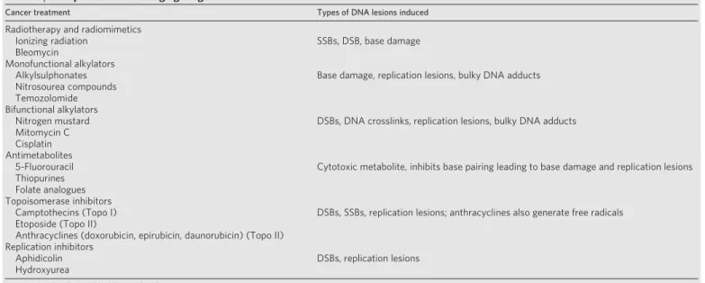
Table 3|Examples of DNA-damaging drugs used to treat cancer
Cancer treatment Types of DNA lesions induced
Radiotherapy and radiomimetics
Ionizing radiation SSBs, DSB, base damage
Bleomycin
Monofunctional alkylators
Alkylsulphonates Base damage, replication lesions, bulky DNA adducts
Nitrosourea compounds Temozolomide Bifunctional alkylators
Nitrogen mustard DSBs, DNA crosslinks, replication lesions, bulky DNA adducts
Mitomycin C Cisplatin Antimetabolites
5-Fluorouracil Cytotoxic metabolite, inhibits base pairing leading to base damage and replication lesions Thiopurines
Folate analogues Topoisomerase inhibitors
Camptothecins (Topo I) DSBs, SSBs, replication lesions; anthracyclines also generate free radicals Etoposide (Topo II)
Anthracyclines (doxorubicin, epirubicin, daunorubicin) (Topo II) Replication inhibitors
Aphidicolin DSBs, replication lesions
Hydroxyurea
See text for details (modified from ref. 87).
1075
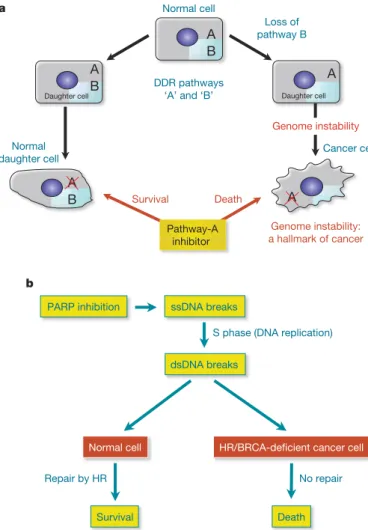
other pro-apoptotic proteins, which commonly yield therapy resist- ance82,89). Because different DNA-repair pathways can overlap in func- tion, and as one pathway can sometimes ‘back-up’ for defects in another, inhibition of pathways present in a cancer cell should in some cases have a greater impact on the cancer than on normal tissues (Fig. 2a). A paradigm for this is provided by drugs targeting the enzyme PARP1, which binds SSBs and base-excision repair intermediates to facilitate these repair processes. Notably, PARP inhibitors are rela- tively non-toxic to normal cells but are strikingly cytotoxic towards HR-defective cells, particularly those impaired in BRCA1 or BRCA2 (Fig. 2b)90,91. On the basis of promising phase 1 data, phase 2 trials are currently underway to test PARP1 inhibitors in BRCA-defective breast cancer and ovarian cancer patients (http://www.cancer.gov/ search/ResultsClinicalTrialsAdvanced.aspx?protocolsearchid55678174). Significantly, some sporadic breast, ovarian, prostate, pancreatic and other tumours also possess HR defects due to mutation or epigenetic inactivation of HR components, suggesting that PARP inhibitors might be more broadly applicable. Furthermore, as other DDR pathways are frequently impaired in cancers, there may be addit- ional situations where DDR inhibitors would display selective antitu- mour effects. Consistent with this idea, CHK1 inhibition reportedly
sensitizes p53-deficient cells to DNA-damaging agents more than p53- proficient cells92. The development of diagnostic procedures to identify DDR differences between cancer and normal cells therefore holds great promise for intelligent tailoring of DNA-damaging therapies and DDR-inhibitor therapies for the individual patient. Furthermore, as DDR activation is prevalent during oncogenesis, screening for DDR markers could enhance the reliability and sensiti- vity of cancer detection, and might allow effective detection of pre- malignant disease. In the longer term, it might be possible to develop drugs that enhance DDR events, thus reducing cancer incidence. In this regard, it is noteworthy that mice engineered to exhibit enhanced p53-dependent DNA-damage responses are less tumour prone than wild-type mice93.
Ischaemia-reperfusion injury, inflammatory diseases and ageing. Although DDR mechanisms generally protect against disease, their hyper-activation can contribute to pathology. A prime example of this is in ischaemia-reperfusion episodes associated with stroke and myo- cardial infarction, where PARP1 can become hyper-activated through DNA damage caused by re-oxygenation and nitric-oxide production. This depletes intracellular pools of nicotinamide adenine dinucleo- tide, resulting in impaired ATP production and cell death, often by necrosis. Notably, genetic inactivation or pharmacological inhibition of PARP1 in rodents provides considerable protection towards such cell death, consequently diminishing ensuing organ dysfunction. Similarly, animal models have shown PARP1 inhibition to protect against traumatic brain injury, endotoxic shock, tissue damage caused by chronic inflammation and drug-induced diabetes (for example see refs 94, 95). Thus, PARP1 inhibitors might find utility in treating such conditions in people. It is also noteworthy that p53 dysfunction is associated with inflammatory diseases and atherosclerosis96,97, indi- cating that pharmacological modulation of p53, and its upstream activator ATM, might ameliorate such pathologies85. With our grow- ing realization that DNA damage and sub-optimal DDR events are associated with neurodegenerative disease and various other age- related degenerative pathologies, it is also tempting to speculate that DDR-modulatory drugs will one day be used to slow down or prevent such conditions; perhaps even certain aspects of the normal ageing process.
Viral, parasitic and other diseases. DDR proteins function in the life cycles of human parasites and pathogens, suggesting that DDR inhi- bitors could be used to treat their associated pathologies. For instance, the reliance of HIV on host-cell DDR factors suggests a potential for DDR inhibitors in AIDS therapy98,99. Although such treatments would need to be evaluated for potential side effects, a possible advantage over conventional treatments that target the pathogen itself is that they would not be easily subject to evolution of resistance. Furthermore, antibacterial agents could be developed that target aspects of bacterial DDR mechanisms that are distinct from those of host cells.
Gene therapy. Correcting gene dysfunction is a long-sought-after treatment for many human maladies, including immune deficiencies, cystic fibrosis, muscular dystrophy and hereditary blindness. Although some success has been achieved, such approaches have been plagued by safety issues, largely arising through unwanted NHEJ- mediated integration of the introduced gene into tumour-suppressor loci. Although it is difficult to imagine such obstacles being sur- mounted in the near future, the development of methods to interfere with NHEJ or promote gene integration into desired loci (for example, see ref. 100) offers exciting prospects for gene-therapy optimization.
Future challenges
Great progress has been made towards understanding the DDR but much remains to be learned. One major future challenge is to under- stand in more detail how the activities of DDR proteins are con- trolled. Other challenges are to determine precisely how and why the DDR impacts on myriad cellular functions and how such com- plex programmes are orchestrated. Additional important issues to be
Normal cell a
A B
Loss of pathway B
A
Daughter cell
A
Daughter cell B
DDR pathways
‘A’ and ‘B’
Normal daughter cell
A B
Cancer cell
A
Genome instability
Genome instability: a hallmark of cancer Pathway-A
inhibitor Death Survival
b
ssDNA breaks PARP inhibition
dsDNA breaks
HR/BRCA-deicient cancer cell S phase (DNA replication)
Death Survival
Normal cell
No repair Repair by HR
Figure 2|Exploitation of DDR pathways to enhance therapeutic responses. a, Model: normal cells have two DDR pathways: A and B. If one pathway (B) is eliminated, genome instability results, which can foster the evolution of a cancer cell. Addition of an inhibitor targeting the second pathway (A) leads to cell death. Normal cells that still retain an active B pathway, however, survive.b, Treatment with a PARP inhibitor selectively kills HR/BRCA-deficient cells. PARP inhibition impairs the repair of SSBs, which are converted to DSBs in S phase. Such DSBs are effectively repaired by HR in non-cancerous cells but not in BRCA-deficient cancer cells. Adapted with permission from ref. 101.
1076
addressed are how the DDR can be shaped and fine-tuned by other pathways and events, and how the same DDR stimulus can yield markedly different responses in different cells and tissues, including cancer cells and stem cells. Such knowledge will not only enhance our appreciation of DDR functions but will undoubtedly present exciting opportunities for better understanding and managing human health and disease.
1. Lindahl, T. & Barnes, D. E. Repair of endogenous DNA damage. Cold Spring Harb. Symp. Quant. Biol.65, 127–134 (2000).
An excellent overview of the extent of endogenous DNA damage, the types of DNA lesions arising from cell autonomous sources, and the pathways that repair such lesions.
2. Valko, M., Rhodes, C. J., Moncol, J., Izakovic, M. & Mazur, M. Free radicals, metals and antioxidants in oxidative stress-induced cancer. Chem. Biol. Interact. 160, 1–40 (2006).
3. Kawanishi, S., Hiraku, Y., Pinlaor, S. & Ma, N. Oxidative and nitrative DNA damage in animals and patients with inflammatory diseases in relation to inflammation- related carcinogenesis. Biol. Chem. 387, 365–372 (2006).
4. Khanna, K. K. & Jackson, S. P. DNA double-strand breaks: signalling, repair and the cancer connection. Nature Genet. 27, 247–254 (2001).
5. Ward, J. F. DNA damage produced by ionizing radiation in mammalian cells: identities, mechanisms of formation, and reparability. Prog. Nucleic Acid Res. Mol. Biol.35, 95–125 (1988).
6. Doll, R. & Peto, R. The causes of cancer: quantitative estimates of avoidable risks of cancer in the United States today. J. Natl Cancer Inst. 66, 1191–1308 (1981). Classical overview of epidemiological evidence for DNA-damaging
environmental insults implicated as carcinogens, and suggestions for measures to prevent such tumours.
7. Wogan, G. N., Hecht, S. S., Felton, J. S., Conney, A. H. & Loeb, L. A. Environmental and chemical carcinogenesis. Semin. Cancer Biol. 14, 473–486 (2004). 8. Espinosa, E., Zamora, P., Feliu, J. & Gonzalez Baron, M. Classification of anticancer
drugs—a new system based on therapeutic targets. Cancer Treat. Rev. 29, 515–523 (2003).
9. Lebwohl, M., Ting, P. T. & Koo, J. Y. Psoriasis treatment: traditional therapy. Ann. Rheum. Dis.64 (suppl. 2), 83–86 (2005).
10. Harper, J. W. & Elledge, S. J. The DNA damage response: ten years after. Mol. Cell 28, 739–745 (2007).
11. Rouse, J. & Jackson, S. P. Interfaces between the detection, signalling, and repair of DNA damage. Science 297, 547–551 (2002).
12. Harrison, J. C. & Haber, J. E. Surviving the Breakup: The DNA damage checkpoint. Annu. Rev. Genet.40, 209–235 (2006).
13. Jiricny, J. The multifaceted mismatch-repair system. Nature Rev. Mol. Cell Biol. 7, 335–346 (2006).
14. David, S. S., O’Shea, V. L. & Kundu, S. Base-excision repair of oxidative DNA damage. Nature 447, 941–950 (2007).
15. Hoeijmakers, J. H. J. Genome maintenance mechanisms for preventing cancer. Nature411, 366–374 (2001).
A highly informative review of the links between DNA damage, DNA-repair pathways and their defects contributing to tumorigenesis.
16. Friedberg, E. C. et al. DNA Repair and Mutagenesis 2nd edn (ASM Press, 2006). An excellent, comprehensive multi-author book covering essentially the entire field of DNA repair, from basic mechanisms in diverse organisms, to human diseases associated with defective DNA repair.
17. Loeb, L. A. & Monnat, R. J. Jr. DNA polymerases and human disease. Nature Rev. Genet.9, 594–604 (2008).
18. Lieber, M. R. The mechanism of human nonhomologous DNA end joining. J. Biol. Chem.283, 1–5 (2008).
19. San Filippo, J., Sung, P. & Klein, H. Mechanism of eukaryotic homologous recombination. Annu. Rev. Biochem. 77, 229–257 (2008).
20. McVey, M. & Lee, S. E. MMEJ repair of double-strand breaks (director’s cut): deleted sequences and alternative endings. Trends Genet. 24, 529–538 (2008). 21. Kennedy, R. D. & D’Andrea, A. D. The Fanconi Anemia/BRCA pathway: new faces
in the crowd. Genes Dev. 19, 2925–2940 (2005).
22. Cimprich, K. A. & Cortez, D. ATR: an essential regulator of genome integrity. Nature Rev. Mol. Cell Biol.9, 616–627 (2008).
23. Bartek, J. & Lukas, J. DNA damage checkpoints: from initiation to recovery or adaptation. Curr. Opin. Cell Biol. 19, 238–245 (2007).
24. Shiloh, Y. ATM and related protein kinases: safeguarding genome integrity. Nature Rev. Cancer3, 155–168 (2003).
Describes the key DDR kinases ATM, ATR and DNA-PK, provides an overview of their substrates, and outlines the cellular pathways affected by DNA-damage signalling.
25. Riley, T., Sontag, E., Chen, P. & Levine, A. Transcriptional control of human p53- regulated genes. Nature Rev. Mol. Cell Biol. 9, 402–412 (2008).
26. Kastan, M. B. & Bartek, J. Cell-cycle checkpoints and cancer. Nature 432, 316–323 (2004).
27. Huen, M. S. & Chen, J. The DNA damage response pathways: at the crossroad of protein modifications. Cell Res. 18, 8–16 (2008).
28. Matsuoka, S. et al. ATM and ATR substrate analysis reveals extensive protein networks responsive to DNA damage. Science 316, 1160–1166 (2007).
Milestone report on the proteomic identification of ATM/ATR substrates and their assignment to various cellular functions, including RNA processing and other protein networks not previously recognized as DDR targets.
29. Campisi, J. & d’Adda di Fagagna, F. Cellular senescence: when bad things happen to good cells. Nature Rev. Mol. Cell Biol. 8, 729–740 (2007).
30. Halazonetis, T. D., Gorgoulis, V. G. & Bartek, J. An oncogene-induced DNA damage model for cancer development. Science 319, 1352–1355 (2008). 31. Misteli, T. & Soutoglou, E. The emerging role of nuclear architecture in DNA repair
and genome maintenance. Nature Rev. Mol. Cell Biol. 10, 243–254 (2009). 32. Ziv, Y. et al. Chromatin relaxation in response to DNA double-strand breaks is
modulated by a novel ATM- and KAP-1 dependent pathway. Nature Cell Biol. 8, 870–876 (2006).
33. Xiao, A. et al. WSTF regulates the H2A.X DNA damage response via a novel tyrosine kinase activity. Nature 457, 57–62 (2009).
34. Cook, P. J. et al. Tyrosine dephosphorylation of H2AX modulates apoptosis and survival decisions. Nature 458, 591–596 (2009).
35. Bassing, C. H. & Alt, F. W. The cellular response to general and programmed DNA double-strand breaks. DNA Repair 3, 781–796 (2004).
36. Schlissel, M. S., Kaffer, C. R. & Curry, J. D. Leukemia and lymphoma: a cost of doing business for adaptive immunity. Genes Dev. 20, 1539–1544 (2006).
37. Richardson, C., Horikoshi, N. & Pandita, T. K. The role of the DNA double-strand break response network in meiosis. DNA Repair 3, 1149–1164 (2004). 38. Verdun, R. E. & Karlseder, J. Replication and protection of telomeres. Nature 447,
924–931 (2007).
39. de Lange, T. Shelterin: the protein complex that shapes and safeguards human telomeres. Genes Dev. 19, 2100–2110 (2005).
40. d’Adda di Fagagna, F., Teo, S. H. & Jackson, S. P. Functional links between telomeres and proteins of the DNA-damage response. Genes Dev. 18, 1781–1799 (2004).
References 39 and 40 illustrate the intimate links between the telomere maintenance and DDR machineries.
41. Longhese, M. P. DNA damage response at functional and dysfunctional telomeres. Genes Dev. 22, 125–140 (2008).
42. Sedelnikova, O. A. et al. Senescing human cells and ageing mice accumulate DNA lesions with unrepairable double-strand breaks. Nature Cell Biol. 6, 168–170 (2004).
43. Niedernhofer, L. J. et al. A new progeroid syndrome reveals that genotoxic stress suppresses the somatotroph axis. Nature 444, 1038–1043 (2006).
Reports a powerful mouse model of multifaceted premature ageing, based on engineered deficiency in the Xpf gene involved in transcription-coupled NER. 44. Jeyapalan, J. C. & Sedivy, J. M. Cellular senescence and organismal aging. Mech.
Ageing Dev.129, 467–474 (2008).
45. Nouspikel, T. P., Hyka-Nouspikel, N. & Hanawalt, P. C. Transcription domain- associated repair in human cells. Mol. Cell. Biol. 26, 8722–8730 (2006). 46. Orii, K. E., Lee, Y., Kondo, N. & McKinnon, P. J. Selective utilization of
nonhomologous end-joining and homologous recombination DNA repair pathways during nervous system development. Proc. Natl Acad. Sci. USA 103, 10017–10022 (2006).
47. Park, Y. & Gerson, S. L. DNA repair defects in stem cell function and aging. Annu. Rev. Med.56, 495–508 (2005).
48. Nijnik, A. et al. DNA repair is limiting for haematopoietic stem cells during ageing. Nature447, 686–690 (2007).
49. Rossi, D. J. et al. Deficiencies in DNA damage repair limit the function of haematopoietic stem cells with age. Nature 447, 725–729 (2007).
50. Collis, S. J. & Boulton, S. J. Emerging links between the biological clock and the DNA damage response. Chromosoma 116, 331–339 (2007).
51. Collis, S. J. et al. HCLK2 is essential for the mammalian S-phase checkpoint and impacts on Chk1 stability. Nature Cell Biol. 9, 391–401 (2007).
52. Kang, T. H., Reardon, J. T., Kemp, M. & Sancar, A. Circadian oscillation of nucleotide excision repair in mammalian brain. Proc. Natl Acad. Sci. USA 106, 2864–2867 (2009).
53. Writing Committee of the Second World Health Organization Consultation on Clinical Aspects of Human Infection with Avian Influenza A (H5N1) Virus.. Update on avian influenza A (H5N1) virus infection in humans. N. Engl. J. Med. 358, 261–273 (2008).
54. McCulloch, R. & Barry, J. D. A role for RAD51 and homologous recombination in Trypanosoma bruceiantigenic variation. Genes Dev. 13, 2875–2888 (1999). 55. Lilley, C. E., Schwartz, R. A. & Weitzman, M. D. Using or abusing: viruses and the
cellular DNA damage response. Trends Microbiol. 15, 119–126 (2007). 56. Narisawa-Saito, M. & Kiyono, T. Basic mechanisms of high-risk human
papillomavirus-induced carcinogenesis: roles of E6 and E7 proteins. Cancer Sci. 98, 1505–1511 (2007).
57. Muylaert, I. & Elias, P. Knock-down of DNA ligase IV/ XRCC4 by RNAi inhibits herpes simplex virus type I DNA replication. J. Biol. Chem. 282, 10865–10872 (2007).
58. Li, H. et al. Chk2 is required for HSV-1 ICP0-mediated G2/M arrest and enhancement of virus growth. Virology 375, 13–23 (2008).
59. Smith, J. & Daniel, R. Following the path of the virus: the exploitation of host DNA repair mechanisms by retroviruses. ACS Chem. Biol. 1, 217–226 (2006). 60. Stratton, M. R., Campbell, P. J. & Futreal, P. A. The cancer genome. Nature 458,
719–724 (2009).
A comprehensive overview of cancer-predisposing mutations and advances in cancer genetics.
1077
61. Lengauer, C., Kinzler, K. W. & Vogelstein, B. Genetic instabilities in human cancers. Nature 396, 643–649 (1998).
62. Maser, R. S. & DePinho, R. A. Connecting chromosomes, crisis, and cancer. Science 297, 565–569 (2002).
63. Bristow, R. G. & Hill, R. P. Hypoxia and metabolism. Hypoxia, DNA repair and genetic instability. Nature Rev. Cancer 8, 180–192 (2008).
64. Bartkova, J. et al. DNA damage response as a candidate anti-cancer barrier in early human tumorigenesis. Nature 434, 864–870 (2005).
65. Gorgoulis, V. G. et al. Activation of the DNA damage checkpoint and genomic instability in human precancerous lesions. Nature 434, 907–913 (2005). References 64 and 65 provide evidence for activation of the DDR machinery in early human oncogenic lesions and models of oncogenic transformation, and propose that the DNA-damage checkpoint activated by oncogene-evoked replication stress and DNA breakage provides an inducible barrier against tumour progression.
66. Rass, U., Ahel, I. & West, S. C. Defective DNA repair and neurodegenerative disease. Cell 130, 991–1004 (2007).
67. Kulkarni, A. & Wilson, D. M. III. The involvement of DNA-damage and -repair defects in neurological dysfunction. Am. J. Hum. Genet. 82, 539–566 (2008). 68. Weissman, L., de Souza-Pinto, N. C., Stevnsner, T. & Bohr, V. A. DNA repair,
mitochondria, and neurodegeneration. Neuroscience 145, 1318–1329 (2007). 69. Caldecott, K. W. Single-strand break repair and genetic disease. Nature Rev. Genet.
9, 619–631 (2008).
70. Ljungman, M. & Lane, D. P. Transcription—guarding the genome by sensing DNA damage. Nature Rev. Cancer 4, 727–737 (2004).
71. Mirkin, S. M. Expandable DNA repeats and human disease. Nature 447, 932–940 (2007).
72. Kovtun, I. V. & McMurray, C. T. Features of trinucleotide repeat instability in vivo. Cell Res.18, 198–213 (2008).
73. Yang, J. L., Weissman, L., Bohr, V. A. & Mattson, M. P. Mitochondrial DNA damage and repair in neurodegenerative disorders. DNA Repair 7, 1110–1120 (2008).
74. Matzuk, M. M. & Lamb, D. J. The biology of infertility: research advances and clinical challenges. Nature Med. 14, 1197–1213 (2008).
75. Bartkova, J., Rajpert-De Meyts, E., Skakkebaek, N. E., Lukas, J. & Bartek, J. DNA damage response in human testes and testicular germ cell tumours: biology and implications for therapy. Int. J. Androl. 30, 282–291 (2007).
76. Schumacher, B., Garinis, G. A. & Hoeijmakers, J. H. Age to survive: DNA damage and aging. Trends Genet. 24, 77–85 (2008).
A thought-provoking review of the evidence for causative links between DNA- damage accumulation and organismal ageing, which proposes the concept of a survival response that allows the organism’s resources to be shifted from emphasis on growth, to survival of DNA damage and other stresses. 77. Herbig, U., Ferreira, M., Condel, L., Carey, D. & Sedivy, J. M. Cellular senescence in
aging primates. Science 311, 1257 (2006).
78. Sharpless, N. E. & DePinho, R. A. How stem cells age and why this makes us grow old. Nature Rev. Mol. Cell Biol. 8, 703–713 (2007).
79. Navarro, S. et al. Hematopoietic dysfunction in a mouse model for Fanconi anemia group D1. Mol. Ther. 14, 525–535 (2006).
80. Reese, J. S., Liu, L. & Gerson, S. L. Repopulating defect of mismatch repair-deficient hematopoietic stem cells. Blood 102, 1626–1633 (2003).
81. Mocanu, M. M. & Yellon, D. M. p53 down-regulation: a new molecular mechanism involved in ischaemic preconditioning. FEBS Lett. 555, 302–306 (2003).
82. Vousden, K. H. & Lane, D. P. p53 in health and disease. Nature Rev. Mol. Cell Biol. 8, 275–283 (2007).
83. Mercer, J., Mahmoudi, M. & Bennett, M. DNA damage, p53, apoptosis and vascular disease. Mutat. Res. 621, 75–86 (2007).
84. Schneider, J. G. et al. ATM-dependent suppression of stress signaling reduces vascular disease in metabolic syndrome. Cell Metab. 4, 377–389 (2006).
85. Kastan, M. B. DNA damage responses: mechanisms and roles in human disease. 2007 G.H.A. Clowes Memorial Award Lecture. Mol. Cancer Res. 6, 517–524 (2008).
86. Bao, S. et al. Glioma stem cells promote radioresistance by preferential activation of the DNA damage response. Nature 444, 756–760 (2006).
87. Helleday, T., Petermann, E., Lundin, C., Hodgson, B. & Sharma, R. A. DNA repair pathways as targets for cancer therapy. Nature Rev. Cancer 8, 193–204 (2008). 88. Martin, S. A., Lord, C. J. & Ashworth, A. DNA repair deficiency as a therapeutic
target in cancer. Curr. Opin. Genet. Dev. 18, 80–86 (2008).
89. Jiang, H. et al. The combined status of ATM and p53 link tumor development with therapeutic response. Genes Dev. 23, 1895–1909 (2009).
90. Farmer, H. et al. Targeting the DNA repair defect in BRCA mutant cells as a therapeutic strategy. Nature 434, 917–921 (2005).
91. Bryant, H. E. et al. Specific killing of BRCA2-deficient tumours with inhibitors of poly(ADP-ribose) polymerase. Nature 434, 913–917 (2005).
References 90 and 91 document the potential of personalized cancer treatment, based on the exceptional sensitivity of tumour cells defective in BRCA1/BRCA2- dependent HR towards small molecule inhibitors of PARP1; these studies support the principle of synthetic-lethal relationships between complementary DDR pathways.
92. Chen, Z. et al. Selective Chk1 inhibitors differentially sensitize p53-deficient cancer cells to cancer therapeutics. Int. J. Cancer 119, 2784–2794 (2006). 93. Garcı´a-Cao, I. et al. ‘Super p53’ mice exhibit enhanced DNA damage response, are
tumor resistant and age normally. EMBO J. 21, 6225–6235 (2002). 94. Pacher, P. & Szabo, C. Role of poly(ADP-ribose) polymerase 1 (PARP-1) in
cardiovascular diseases: the therapeutic potential of PARP inhibitors. Cardiovasc. Drug Rev.25, 235–260 (2007).
95. Moroni, F. Poly(ADP-ribose)polymerase 1 (PARP-1) and postischemic brain damage. Curr. Opin. Pharmacol. 8, 96–103 (2008).
96. Guevara, N. V., Kim, H. S., Antonova, E. I. & Chan, L. The absence of p53 accelerates atherosclerosis by increasing cell proliferation in vivo. Nature Med. 5, 335–339 (1999).
97. Andreassi, M. G. DNA damage, vascular senescence and atherosclerosis. J. Mol. Med.86, 1033–1043 (2008).
98. Lau, A. et al. Suppression of HIV-1 infection by a small molecule inhibitor of the ATM kinase. Nature Cell Biol. 7, 493–500 (2005).
99. Smith, J. A. et al. Evidence that the Nijmegen breakage syndrome protein, an early sensor of double-strand DNA breaks (DSB), is involved in HIV-1 post-integration repair by recruiting the ataxia telangiectasia-mutated kinase in a process similar to, but distinct from, cellular DSB repair. Virol. J. 5, 11 (2008).
100. Moehle, E. A. et al. Targeted gene addition into a specified location in the human genome using designed zinc finger nucleases. Proc. Natl Acad. Sci. USA 104, 3055–3060 (2007).
101. Jackson, S. P. The DNA-damage response: new molecular insights and new approaches to cancer therapy. Biochem. Soc. Trans. 37, 483–494 (2009). Supplementary Information is linked to the online version of the paper at www.nature.com/nature.
Acknowledgements We thank S. Polo and P. Huertas for advice, and K. Dry for expert help with the text and figures. The S.P.J. laboratory is supported by grants from Cancer Research UK, the European Commission (projects GENICA and DNA Repair), the Wellcome Trust and the Biotechnology and Biological Sciences Research Council. The J.B. laboratory is supported by grants from the Danish Cancer Society, the Danish National Research Foundation and the European Commission (projects GENICA, Active p53, TRIREME and DNA Repair). Author Contributions S.P.J. and J.B. conceived of and wrote all aspects of this article.
Author Information Reprints and permissions information is available at www.nature.com/reprints. Correspondence should be addressed to S.P.J. ([email protected]).
1078