日本血管外科学会雑誌 2017; 26: 9–12
9
重症下肢虚血に対する下肢動脈バイパス術後,
2D Perfusion Angiography
を用いて末梢血流評価を行った
1
例
米山
文弥
1,*
,佐藤
藤夫
1,大坂
基男
2,坂本
裕昭
2,軸屋
智昭
1,平松
祐司
2要 旨: 2D perfusionは手術室ハイブリッド装置付属のアプリケーションであり,可視的かつ定量的な血流評価
が可能である.今回,下肢動脈バイパス術後の血流評価に2D perfusion angiographyを用い,その有効性を検討し
た.症例は49歳男性.右足趾の潰瘍を主訴に来院,重症下肢虚血の診断で入院となった.下肢血管造影検査では
右前脛骨動脈・腓骨動脈が近位部で閉塞し,外科的血行再建術の方針となった.大伏在静脈をグラフトとして右
膝窩–右足背動脈バイパス術,同時にショパール関節離断術を施行した.2D perfusion angiographyではカラーマッ
ピングされた血流と各ファンクショナルパラメーターはいずれも改善を認め,術後2カ月で植皮術まで到達し踵
部温存を得た.2D perfusion angiographyは下肢動脈バイパス術後の末梢血流を評価する検査として有用と考えられ
た.
(日血外会誌2017; 26: 9–12)
索引用語:重症下肢虚血,下肢動脈バイパス術,2D perfusion angiography
背
景
下肢血行再建術後の血流評価や予後予測の手段は,足 関節上腕血圧比(Ankle-Brachial Index; ABI),皮膚灌流 圧測定(Skin perfusion pressure; SPP),動脈造影検査,造 影CT検査等多岐に渡る1).2D perfusion angiographyは血
管内治療(Endovascular therapy; EVT)後の末梢血流評価
に有用とされているが2, 3),外科的血行再建術の術後評
価のために使用された報告は今までにない.今回われわ れは,重症下肢虚血(Critical limb ischemia; CLI)に対す
る下肢動脈バイパス術の術後評価に2D perfusion
angiog-raphyを用いて,可視的かつ定量的な末梢血流評価に有
効であったので報告する.
症
例
症 例:49歳,男性
主 訴:右足趾壊死(第I趾∼足底)
現病歴:約1カ月前に右足第I趾先端に潰瘍が出現し,
その後急速に壊死が進行し近医を受診した.抗生剤とプ ロスタグランジン製剤を投与したが改善せず,当院に紹 介された.
既往歴:高血圧,高脂血症,2型糖尿病,慢性腎不全
(維持透析中),狭心症(#7 75%,#10 50%)
手術歴:2年前に左CLI病変に対して左膝上部膝窩–足
背動脈バイパス術を施行した(術後は器具を用いて歩行 可能であった).
身体所見:右足の第I趾から足底にかけて広範囲に悪
臭を伴う潰瘍・壊死を認めた(Rutherford 6)(Fig. 1A).
入院後よりベッドサイドでデブリドマンを施行した,右
第I趾の腐骨と足底筋腱膜の融解を認めた(Fig. 1B).
血 液 検 査 所 見:白 血 球24000/µL, Hb 12.5 g/dL, CRP
17.7 mg/dL
術前ABI:左1.17,右0.90
術前SPP(右):足背I 39 mmHg,足背V 39 mmHg,足底
I 29 mmHg,足底V 48 mmHg
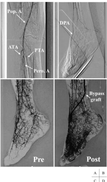
術前下肢血管造影(Fig. 2A, B):右膝窩動脈は開存し,
右前脛骨動脈と右腓骨動脈は近位部で閉塞していた.右 後脛骨動脈の近位部は開存していたが,遠位側でびまん 性の狭窄と閉塞を認めた.足部では側副血行路を介して 右足背動脈が描出された.
手術所見:全身麻酔,仰臥位で手術を開始した.右膝
上部膝窩動脈と右足背動脈を露出し皮下トンネルを作成
した.全身ヘパリン化(ACT 200秒以上)して,右大伏
在静脈をnon-reversed vein graftとして用い,中枢側を7-0
1筑波メディカルセンター病院心臓血管外科
〒305–0005 茨城県つくば市天久保1–3–1
2筑波大学附属病院心臓血管外科
* E-mail: fumiya.y@hotmail.co.jp
受付:2016年10月31日 受理:2016年12月12日 doi: 10.11401/jsvs.16-00074
10
日本血管外科学会雑誌 第26巻 第1号(2017)
ネスピレン1針連続で吻合した.グラフトをトンネルに
通し,右下腿部をターニケット・エスマルヒ法で血流遮
断し,遠位側を7-0ネスピレン1針連続で吻合した.術
中グラフト血流計測では45 mL/minであった.2D
perfu-sion angiography撮像後にショパール関節離断術を施行し
た.
術中下肢血管造影(2D perfusion angiography):イオ
パミロン注300シリンジ(50 mL)(イオパミドール,バ
イエル薬品,東京)をバイパス近位側吻合の浅大腿動
脈から3 mL/秒でインジェクターを用いて注入し造影し
た.DSAでは,術前と比べてバイパスグラフトを介し
てより早期に末梢まで血流を確認した(Fig. 2C, D).ま
たカラーマッピング画像でも同様に末梢血流の改善が 認 め ら れ た(Fig. 3A, B).Time density curveで 得 ら れ
た各ファンクショナルパラメーターを術前と術後で比
較 す る と, ① Arrival Time: 8.2→5.9秒(術 前→ 術 後), ② Time to Peak: 9.4→6.9秒, ③ Wash-in Rate: 37.9→56.7, ④ Mean Transit Time: 11.3→8.1秒,⑤ Area Under Curve:
23140→27800といずれも改善値を示した(Fig. 3C, D).
術後経過:第1病日からベッドサイドで創部のデブ
リドマンと,V.A.C.システム(KCI株式会社,米国)を
用 い た 持 続 陰 圧 療 法 を 開 始 し た(Fig. 1C). 術 後7日 目 の 造 影CTで は 下 肢 バ イ パ ス は 開 存 し て お り, 右 ABIは1.24(術 前1.12), 右 足 背SPPは69 mmHg(術 前
34 mmHg)に改善し.術後2カ月で切離断面への植皮術
まで到達した(Fig. 1D).踵部温存に成功し補助器具を
Fig. 1 A: Right foot gangrene on admission. B: Performing debride-ment preoperatively to control local infection. C: Performing debridement postoperatively followed by negative pressure wound therapy with vacuum assisted closure (V.A.C.) system. D: Skin grafting in postoperative 2 month.
11
米山ほか:2D perfusionでのバイパス評価
用いた歩行が可能になり,リハビリテーションを目的に
第82病日に転院した.現在,術後1年を経過したが,潰
瘍の再発を認めず外来で経過観察している.
考
察
2D perfusion angiographyは,ハイブリッド手術室に設
置 す るX線 血 管 造 影 装 置Allura Clarity FD20(FHILIPS 社)で撮像したdigital subtraction angiography(DSA)を,
Xtravision搭載の2D Perfusionソフトウェアを用いて画像
変換することで得られる,組織灌流量を定量的に表示し た血管造影画像である.造影剤が組織に流入する速度に 応じて血流をカラーマッピングしており,いわば造影剤 の速度,分布に関するデータを通して血流を可視化した ものといえる.また造影剤を一定の時間・量で投与し横 軸に時間,縦軸に造影剤濃度をプロットし,Time density
curveを作成することで,定量的に血流を評価できる.
Time density curveからは各ファンクショナルパラメー
ターが得られ,血行再建前後での比較はそれぞれ以下の
ように解釈される.① Arrival Timeは 最初のフレーム
から造影剤が検出されたフレームまでの時間 とされ,
Bypassを介して組織灌流が始まる時間を示し,inflowの
増加量を表している.② Time to Peakは Arrival Timeか ら造影剤濃度が最大になるまでの時間 とされ,最大
血流量に到達するまでの時間を示している.③ Wash-in
Rateは 造影剤流入速度 であり,outflowの血流の抵抗
を示している.④ Mean Transit Timeは 造影剤が組織内 を通過するのに要する平均時間の目安 であり,その時 間が短い程,全体の組織灌流が早いと考えられる.⑤
Area Under Curveは 総血流量の目安 であり,バイパス
遠位側吻合部以遠の組織灌流量を示している.
既に,頸動脈狭窄症に対する頸動脈ステント留置術の
術後の過灌流障害の評価,CLI病変に対する血管内治療
(Endovascular therapy; EVT)後の評価として2D perfusion
angiographyの使用は報告されている1, 2, 4).造影剤使用
量や放射線被爆量については従来の血管造影検査と変わ
りなく,患者の負担は少ない1).また
JensらはCLI病変
に対してEVTを施行した症例に対して2D perfusionを用
いて血行再建後の血流評価を試みており,Time-density
curveが改善した症例については下肢潰瘍が早期に改善
したと報告している2).従来の
DSAがグラフトの開存と
吻合部形態,末梢のrun offに関する情報を与えてくれる
のに対して,2D perfusion angiographyは更にグラフトが いかに末梢組織灌流を改善するのか,灌流速度と範囲に 関する情報まで外科医に直感的視覚的に呈示してくれ る.
一方,本検査の限界は,現在のところSPPのように創
12
日本血管外科学会雑誌 第26巻 第1号(2017)
のでもない.更に,局所に微小な動静脈シャントが存在 する場合に過大評価する可能性や,局所の動脈の攣縮時 と弛緩時で結果が異なる可能性も否定できない.また, 造影剤使用検査のため適応不可能な症例が存在するこ と,専用の設備とソフトウェアが必要なこと,患者の体 動により測定に正確性を失う可能性があることなどの欠 点も有する.体動の問題に関しては,われわれは全身麻 酔下の手術患者でこそ正確で有効な検査になり得ると考 えている.
また糖尿病は中小血管のみならずとくに毛細血管レベ ルで微小循環が障害される(microangiopathy)5).2D
per-fusion angiographyでは通常のDSAに比べより鮮明な末梢
循環評価を行うことができるので,とくに本症例のよう
な糖尿病性足壊疽を基盤としたCLI病変の血流評価には
適していると考える.本症例では,2D perfusion
angiogra-phyでのDSAやカラーマッピング画像で明らかな改善を
認め,またTime density curveの全てのファンクショナル
パラメーターで改善値を得られた.Azumaらは透析症例
のCLI病変に対する下肢動脈バイパス術において,術後
から潰瘍・壊死が治癒に至るまでの期間は,Rutherford 5(ischemic ulceration not exceeding ulcer of the digits of the foot)で73日,Rutherford 6(severe ischemic ulcers or frank
gangrene)で237日を要したと報告している6).本症例
はRutherford 6の透析症例であったが,術後2カ月とい
う短期間で植皮術まで到達し,踵部温存に成功してお り,2D perfusion angiographyの結果を良く反映した症例
であった.一方,Time density curveから得られる各ファ
ンクショナルパラメーターと,ABI, SPPなどの従来の検
査や,グラフト開存率,潰瘍治癒率および期間などの臨 床指標との関係は未だ明らかではない.今後はそれらを 検討していくことにより,下肢動脈バイパス術術後の血 流評価検査としての有用性を確立させていきたい.
利益相反
共著者全員が利益相反はない.
文
献
1) Iida O, Nakamura M, Yamauchi Y, et al. 3-year outcomes of the OLIVE Registry, a prospective multicenter study of patients with critical limb ischemia: a prospective, multi-center, three-year fol-low-up study on endovascular treatment for infra-inguinal vessel in patients with critical limb ischemia. JACC Cardiovasc Interv 2015; 8: 1493–1502.
2) Manzi M, Van Den Berg J. 2D perfusion angiography: a useful tool for CLI treatment. Endovasc Today 2015; 76–79.
3) Jens S, Marquering HA, Koelemay MJ, et al. Perfusion angiogra-phy of the foot in patient with critical limb ischemia: description of the techinique. Cardiovasc Intervent Radiol 2015; 38: 201–205. 4) Canty JM Jr., Judd RM, Brody AS, et al. First-pass entry of non-ionic contrast agent into the myocardial extravascular space: ef-fects on radiographic estimates of transit time and blood volume. Circulation 1991; 84: 2071–2078.
5) Santema TB, Poyck PP, Ubbink DT. Skin grafting and tissue replacement for treating foot ulcers in people with diabetes. Cochrane Database Syst Rev 2016; doi: 10.1002/14651858. CD011255.pub2
6) Azuma N, Uchida H, Kokubo T, et al. Factors influencing wound healing of critical ischemic foot after bypass surgery: is the angio-some important in selecting bypass target artery? Eur J Vasc En-dovasc Surg 2012; 43: 322–328.
Evaluation of Bypass Surgery for CLI Using 2D Perfusion Angiography
Fumiya Yoneyama
1, Fujio Sato
1, Motoo Osaka
2, Hiroaki Sakamoto
2, Tomoaki Jikuya
1, and Yuji Hiramatsu
21 Department of Cardiovascular Surgery, Tsukuba Medical Center Hospital 2 Department of Cardiovascular Surgery, University of Tsukuba Hospital
Key words: critical limb ischemia, bypass surgery, 2D perfusion angiography
2D perfusion angiography is a useful tool in terms of visualization and quantitative evaluation of blood perfusion. It can be applied to the evaluation of the bypass surgery for critical limb ischemia (CLI). Herein, we described a 49-year-old man presented with CLI. On preoperative angiography, the right anterior tibial and peroneal arteries were obstructed at proximal site. We performed bypass procedure; right popliteal–dorsalis pedis artery bypass with a saphenous vein graft. During the bypass surgery and after anastomosis of bypass, we evaluated peripheral perfusion with 2D perfusion angiography. We could confirm improved peripheral perfusion with colored mapping image. In addition, all of the functional parameters were improved compared with preoperative values. At 2 month postoperatively, the patient underwent skin grafting surgery for preserving the ankle. This is the first case report regarding bypass surgery evaluated by 2D perfusion angiography.