New Insights into Selective Autophagy
in Yeast: Studies on Lap3
2008 (School Year)
Takuya Kageyama
Department of Basic Biology
School of Life Science
The Graduate University for Advanced Studies
DOCTOR OF PHYLOSOPHY
New Insights into Selective Autophagy
in Yeast: Studies on Lap3
March 24, 2009 by
Takuya Kageyama
DOCTRAL DISSERTATION
Department of Basic Biology School of Life Science
The Graduate University for Advanced Studies
ACKNOWLEDGEMENTS
I wish to gratefully express my sincere thanks to Dr. Yoshinori Ohsumi, Prof. of Molecular and Cellular Biology, National Institute for Basic Biology, for his generous guidance, supervision, valuable advises, stimulating discussion, valuable feedbacks throughout the writing process, and encouragement throughout this study. I wish to express my appreciation to Dr. Kuninori Suzuki for his constant and extensive advise, enormous support, encouragement, and critically reading my manuscript as well as for the valuable feedbacks in my occasions on my work. I am also thankful to Drs. Masahide Oku, Mamoru Oneda, and Koji Okamoto for help and technical assistance. Finally, I would like to express my deep gratitude to other past and current members of the Ohsumi laboratory for sharing with me their strains, thoughts, their constant friendship, helpful discussion, and encouragement. Thank you all!
March 24, 2009 Takuya Kageyama
National Institute The graduate University for Basic Biology for Advanced Studies
TABLE OF CONTENTS
ABSTRACT………..V
LIST OF FIGURES AND TABLE……….VIII
ABBREVIATIONS………...X
INTRODUCTIONS
1. The many routes to the yeast vacuole………...…...……....1
2. Degradation of short-lived and long-lived proteins………....1
3. The conceptual model of autophagy………..2
4. The ATG genes and the core autophagy machinery………….3
5. Selective type of Autophagy………...5
6. The aim of this study……….8
7. References………......9
MATERIALS AND METHODS 1. Cultivation and handling of yeast strains………...…………...………..20
2. Manipulation of yeast strains………...20
3. Antibodies……….......20
4. Immunoblotting of whole-cell lysates………21
5. Microscopy………...……….21
6. Alkaline Phosphatase (ALP) assay………...………...21
7. Vacuole isolation………..……….22
8. Peptide Mass Finger-printing (PMF)…………………...23
9. Immunoprecipitation……….………....23
10. Yeast two-hybrid analysis………24
11. World-Wide-Web databases………...………..24
12. References……….…24
RESULTS CHAPTER I LAP3 IS A SELECTIVE TARGET OF AUTOPHAGY DURING NITROGEN STARVATION IN THE YEAST Saccharomyces cerevisiae….……….28
I-A: Backgrounds I-A-1. Lap3 and Ape1 are self-compartmentalizing proteins………..29
I-A-2. Ape1 is selectively transported to the vacuole during nitrogen starvation…………...29
I-B-2. Lap3 is degraded in the vacuole during nitrogen starvation…………..……………...31
I-B-3. Lap3 localizes to the Cvt complex…………...32
I-B-4. Lap3 is selectively transported to the vacuole by a similar mechanism to Ape1…..32
I-C: Discussion………….33
CHAPTER II MACHINERY OF THE CVT PATHWAY ACTS FOR DEGRADATION DURING GLYCEROL GROWTH IN THE YEAST Saccharomyces cerevisiae………35
II-A: Backgrounds II-A-1. Yeast grows in a medium containing fermentable and non-fermentable carbon sources…..………....…..36
II-A-2. Studies on yeast autophagy under catabolite-repressed conditions…………36
II-B: Results in this chapter II-B-1. The Vacuolar transport pathways enhances during YPGly growth………..….37
II-B-2. Intravacuolar structures accumulate in pep4Δ cells during YPGly growth………..37
II-B-3. Non-selective autophagy is undetectable under YPGly growth conditions….….….38 II-B-4. Identification of enwrapped proteins in the intravacuolar structures…..…………...38
II-B-5. Behavior of Lap3 under the various growth conditions……….…………39
II-B-6. Atg19 acts as a receptor for Lap3 transport……….39
II-B-7. Lap3 is transported to the vacuole via the Cvt pathway……….40
II-B-8. Lap3 co-localizes with Ape1 at the extra- and intravacuolar dot………...41
II-B-9. Lap3 differs from the classical cargo in the transport mechanism……….41
II-B-10. Lap3 is unstable protein inside the vacuole…………....41
II-C: Discussion………..………42
GENERAL DISCUSSIONS 1. Selective transport of Lap3 into the vacuole………..44
2. Molecular mechanisms of Lap3 sequestration………...………...44
3. Accumulation at a punctate dot next to the vacuole………..45
4. Rate of Lap3 degradation inside the vacuole……….45
5. Role of the Cvt pathway in YPGly grown cells………...46
6. Perspective on this study -Physiological role of Lap3 degradation during YPGly growth-………...46
REFERENCES………...…..48
FIGURES………...52
ABSTRACT
Cellular activities require the maintenance of balance between the synthesis and degradation of proteins. Regulation of protein degradation is less understood compared to protein synthesis. The ubiquitin-proteasome system contributes to the selective degradation of short-lived protein. Since most of cellular proteins have long lifetimes, the turnover of long-lived proteins is important to the understanding of cell physiology. Macroautophagy (hereafter simply referred to as autophagy) is an intracellular non-selective degradation system, which is well conserved in eukaryotes; autophagy transports cytoplasmic constituents to the lysosomes/vacuoles for degradation. The autophagic degradation is a cellular response to starvation and plays a role in recycling of cytoplasmic components, which is important for cellular remodeling, development, and differentiation. Screens in the yeast Saccharomyces cerevisiae have led to the identification of 31 autophagy-related (ATG) genes involved in autophagy. Much progress has been made in the functional analysis of these genes.
Autophagy is initiated by the sequestration of cytoplasmic constituents in a double-membrane structure, termed the autophagosome. Fusion of an autophagosome membrane with the vacuole membrane results in the delivery of an inner vesicle (i.e., autophagic body). Eighteen Atg proteins comprise the core machinery essential for the biogenesis of the autophagosomes. Immuno-electron microscopy has revealed that ribosomes and typical cytosolic marker enzymes are present in the autophagosomes and autophagic bodies at the same densities as in the cytosol, indicating that autophagy executes a non-selective degradation. If degradation of long-lived proteins is exclusively mediated by autophagy, all proteins might be expected to have similar lifetimes. Long-lived proteins, however, have a variety of lifetimes; therefore, the autophagic process would have some selectivity.
Recently, different molecules or complexes are selectively recognized and delivered to the vacuoles via autophagy. Onodera and Ohsumi have reported (Onodera and Ohsumi, 2005) that Ald6 is preferentially sequestered in autophagosomes and is eliminated from cytoplasm during prolonged starvation; this mechanism, however, is not understood well. Mechanisms of such cargo selection have been well studied for aminopeptidase I (Ape1/Lap4), a vacuole-resident enzyme. Ape1 self-assembles and then forms an aggregate-like structure. Ape1 then is selectively
selectively degraded by yeast autophagy.
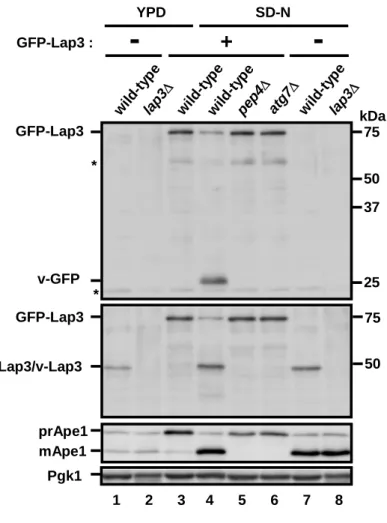
To address the issue of selective degradation via autophagy, I focused on leucine aminopeptidase III (Lap3). LAP3 was originally isolated, along with LAP1/APE2 and LAP4/APE1, in a genetic screen and is a widely conserved cytoplasmic cysteine protease among eukaryotes. Lap3 self-assembles in the cytosol, and Lap3 forms homohexameric complex. The first part of this study shows that Lap3 is a selective target of autophagy. When Lap3 tagged with GFP is overexpressed, it forms large aggregates next to the vacuole. Lap3 is transported to the vacuole in a manner dependent on autophagy during nitrogen starvation. Under these conditions, the rate of Lap3 transport is much higher than that of general cytosolic proteins; 27% of Pho8Δ60, an indicator of general cytosolic proteins, is transported to the vacuole within 6 h (Scot et al., 1996), whereas approximately 50% of the Lap3 is transported to the vacuole in 1.5 h. These results show that Lap3 is selectively transported to the vacuole. I also identified that ATG11 is involved in Lap3 transport. ATG11 is essential for selective types of autophagy: selective degradation of peroxisomes (pexophagy) and mitochondria (mitophagy).
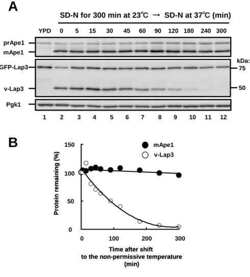
The amount of Lap3 in the vacuole is apparently reduced after nitrogen-starvation at 3 h. I hypothesized that the reduction in Lap3 was a result of degradation in the vacuole. To examine this possibility, the kinetics of Lap3 degradation via autophagic process was measured using temperature-sensitive (atg1ts) cell, which shows that most Lap3 is degraded in the vacuole within a couple of hours. Taken together, Lap3 is a novel target of selective degradation mediated by autophagy during nitrogen starvation. These results are described in Chapter I.
The yeast has a unique system, named the cytoplasm-to-vacuole-targeting (Cvt) pathway. This pathway utilizes common molecular machinery with autophagy under nutrient-rich and fermentable conditions, and constitutively delivers two vacuole-resident enzymes, Ape1 and α-mannosidase (Ams1), to the vacuole via a double-membrane structure (i.e., Cvt vesicle). The latter half of this study shows that the Cvt pathway is involved in not only a biosynthetic process but also protein degradation.
The Cvt pathway is enhanced during vegetative growth in a medium containing glycerol as non-fermentable carbon source (YPGly). Under the conditions, the amount of vacuole-resident enzymes is increased; the lytic function in the vacuole may be important during YPGly growth,
(pep4Δ) cells, single-membrane vesicles accumulate in the vacuole under the conditions. The intravacuolar vesicles are detected in ATG7-dependent manner, and ribosomes and membranes are apparently excluded from these vesicles.
To investigate the content of the vesicle, I isolated vacuoles from pep4Δ cells with or without ATG7 and subjected to proteomic analysis, leading to the identification of Lap3 and Ape1. When Lap3 tagged with GFP is endogenously expressed, it forms aggregates next to the vacuole during YPGly growth. Lap3 co-localizes with Ape1 and is transported into the vacuole. This transport requires Atg11 and Atg19, which are essential for the Cvt pathway. Atg19, which is a cargo receptor for Ape1, is immunoprecipitated with Lap3; Atg19 is likely to function as a receptor for Lap3 transport similar to Ape1 transport. These results show that Lap3 is constitutively transport to the vacuole during vegetative growth and is selectively sequestered in the Cvt-related vesicles.
I assumed that Lap3 would be unstable in the vacuole, since Lap3 is the target of selective autophagy (described in Chapter I). To test this assumption, the stability of Lap3 in the vacuole was examined in atg1ts cells. The amount of Lap3 in the vacuole decreased to 50% of its initial level within 1.5 h. I also isolated vacuoles from wild-type and pep4Δ cells, respectively and subjected to immunoblot, resulting that Lap3 is not detected in wild-type. This result indicates that Lap3 is degraded in the vacuole. Thus, I conclude that machinery of the Cvt pathway can function to eliminate certain proteins during vegetative growth. These results were described in Chapter II.
In this study, I revealed the following facts; 1) Lap3 is a novel cargo of selective autophagy during nitrogen starvation, and 2) the Cvt pathway is involved in protein degradation under growth conditions. Recently, protein degradation via autophagic process in mammalian cells was reported to play a crucial role in elimination of aberrant protein complexes, and the process would be performed constitutively and selectively. For instance, polyubiquitinated aggregates are recognized by p62, selectively sequestered by autophagosomes and are degraded in lysosomes. In contrast to p62, we do not know yet whether Lap3 are harmful or disadvantageous for yeast cells. As Lap3 is conserved widely in eukaryotes, it may serve as a model protein for analysis of selective autophagy in other organisms as well. Autophagy has traditionally been described as a non-selective degradation process. Several results shown in this thesis is the first study reporting that autophagy can be involved in selective and/or constitutive protein degradation in yeast cells. This thesis will allow researchers in this field to make new discoveries regarding the regulation
LIST OF FIGURES AND TABLE
Figure 1. Trafficking pathways to and from the vacuole……….…………13
Figure 2. Conceptual model for autophagy………….14
Figure 3. The classification of Atg proteins…………...………..15
Figure 4. Temporal order for packing cargo components in the Cvt pathway………...………..16
Figure 5. Schematic representation of autophagy and the Cvt pathway in yeast………...……..17
Figure 6. Selective types of autophagy……….18
Figure 7. Immunostaining image of alcohol dehydrogenase in yeast cells………...…...……….19
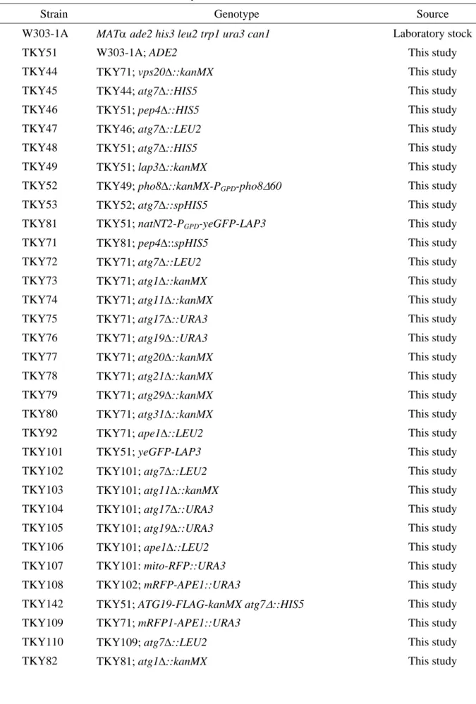
Table I. Yeast strains used in this study………...………....26
Figure 8. Crystal structures of Lap3 and Lap4/Ape1………..………52
Figure 9. Visualization of Lap3 transport to the vacuole during nitrogen starvation…………....53
Figure 10. Vacuolar transport of Lap3 during nitrogen starvation……………..54
Figure 11. Time course of GFP-Lap3 delivery during starvation………………….55
Figure 12. Lap3 degradation in the vacuole using an atg1 temperature-sensitive mutant………….56
Figure 13. Lap3 localizes to the Cvt complex……………….57
Figure 14. Machinery of the Cvt pathway is involved in Lap3 transport during nitrogen starvation……….58
Figure 15. Correlation of the mechanisms for Lap3 transport and Ape1 transport during vegetative growth………...59
Figure 16. Up-regulation of vacuolar hydrolase during YPG growing cells……...………....……60
Figure 17. Growth curve of yeast cells in YPD and YPGly media……………61
Figure 18. Intravacuolar structures accumulate in pep4Δ cells during YPGly growth………62
Figure 19. Accumulation of intravacuolar structures is ATG7-dependent manner during YPGly growth………63
Figure 20. Non-selective autophagy is undetectable in YPGly grown cells………64
Figure 21. Growth curve of wild-type and atg7Δ cells during YPGly growth…...…………..65
Figure 22. Identification of proteins in the intravacuolar structures………66
Figure 23. Illustration of procedures of peptide mass finger-printing (PMF)……………67
Figure 24. Quantification of Lap3 in YPD and YPGly growth………...68
Figure 25. The dynamic alterations of Lap3 localization under growing condition………69
Figure 26. Visualization of Lap3 transport to the vacuole during YPGly growth……...………...70
Figure 27. Vacuolar transport of Lap3 during YPGly growth……….71
Figure 29. Atg17 complex is not essential for Lap3 transport during YPGly growth…..……….73 Figure 30. The Lap3-Atg19 interaction is undetectable in yeast two-hybrid systems.…………..74 Figure 31. Co-immunoprecipitation of Lap3 and Atg19……….………..75 Figure 32. Enhancement of GFP-Lap3 signal at the punctate dot in atg mutants…………..76 Figure 33. Lap3 is localized to the Cvt complex during YPGly growth……………77 Figure 34. Lap3 degradation in the vacuole
using an atg1 temperature-sensitive mutant during YPGly growth………….……….78 Figure 35. Lap3 is degraded in the vacuole…………………...79 Figure 36. Model of the GAL regulation involved Autophagy
under YPGly growth conditions………...80
ABBREVIATIONS
A600 absorbance at 600 nm
Ape1 aminopeptidase I
Ams1 α-mannosidase I
CPY carboxypeptidase Cvt cytoplasm to vacuole targeting DIC differential interference contrast
DNA deoxyribonucleic acid
DTT ditiothreitol
EDTA ethyleediamine tetra-acetic acid GFP green fluorescent protein
KLH keyhole limpet homocyanin
Lap leucine aminopeptidase
MALDI-TOF matrix associated laser deionization-time of flight mRFP monomeric red fluorescence
ORF open reading frame
PAGE polyacrylamide gel electrophoresis
PAS pre-autophagosomal structure
PCR polymerase chain reaction
PrA PrB PrC PGK
proteinase A proteinase B
proteinase C (i.e., CPY) phosphoglycerate kinase
SD synthetic dextrose
SDS sodium dodecyl sulfate
TBS tris-buffered saline
INTRODUCTIONS
1. The Many Routes to the Vacuole -Correlation between vacuole and autophagy-
Already more than a half-century has passed since the lytic organelle, named lysosome, was discovered by de Duve using cell fractionation procedures (De Duve and Hooft, 1968). A vacuole in yeast continues to be a very informative model for mammalian lysosome. Multiple pathways can direct proteins to the vacuole in yeast (Figure 1). The “CPY pathway” is the best-studied pathway for newly synthesized vacuolar proteins and involves vesicular transport from the late Golgi through the multivesicular body (MVB) to the vacuole. A number of membrane proteins, including dipeptidyl aminopeptidase B (DPAP-B) and the V-ATPase also transit this pathway (Piper et al., 1995). Carboxypeptidase S (CPS) uses a variation in which it is transported to the MVB as a membrane protein, then inserted into intralumenal vesicles before transport to the vacuole and final processing to remove its membrane domain. The “ALP pathway”, named for its alkaline phosphatase cargo, is characterized by direct vesicular transport from the Golgi apparatus to the vacuole (Cowles et al., 1997; Piper et al., 1997; Stepp et al., 1997). In addition, aminopeptidase I (Ape1/Lap4) and α-mannosidase (Ams1) follows an unusual pathway to the vacuole, the Cvt pathway (see below), which is characterized by direct transport from the cytosol to the vacuole. In addition to these biosynthetic pathways, multiple proteins are delivered to the vacuole for degradation. Extracellular or cell surface proteins may enter the cells by endocytosis, first transiting to early endosomes and then intersecting the CPY pathway in MVBs before transport to the vacuole (Piper et al., 1995). Intracellular proteins are transported directly to the vacuole and digested in the vacuole under certain conditions. These proteins are generally targeted via the autophagy process (see below). In mammalian cells, since then many electron micrographs showing autophagy have been reported in lysosomes from different organs and cultured cells, it is now generally accepted that autophagy is ubiquitous intracellular activity of eukaryotic cells.
2. Degradation of Short-lived and Long-lived Proteins
Proper balance between protein synthesis and degradation (i.e., turnover) is required for cellular homeostasis. Almost proteins have its own lifetime of wide range, from a few minutes to more than ten days (Goldberg and Dice, 1974; Schimke and Doyle, 1970). We do not know yet the determinants of the lifetime of each protein and the exact meaning of protein turnover, but dynamic state of equilibrium itself must be crucial for maintenance of life. Recently, it was
ubiquitin-proteasome system in the cytosol, which is involved in degradation of short-lived, damaged, and/or misfolded proteins (Hershko and Ciechanover, 1998; Hochstrasser, 1991). Target protein to be degraded is a tagged with a ubiquitin and then digested by a huge proteinase complex, proteasome. Both ubiquitination and cleavage processes require ATP hydrolysis, and undergoes with strict recognition of target proteins by the sophisticated ubiquitin ligase system and proteasome. Short-lived proteins play crucial roles in important cellular events as transcriptional regulation and cell cycle model.
Long-lived proteins are believed to be degraded in a specific lytic compartment, lysosomes/vacuoles. So far, several transport routes to the compartment are proposed. General process of degradation of intracellular components in lysosomes/vacuoles is called “Autophagy” in contrast to heterophagy of extracellular materials (Mortimore and Poso, 1987). Macroautophagy is a principal pathway in autophagy, and initiates by enwrapping a portion of cytoplasm by membrane sac called isolation membrane, to form a double membrane structure, named autophagosome (Seglen and Bohley, 1992). Autophagosome then fuses with lysosomes and turns to be autolysosome and its inner membrane and contents are digested for reuse. Microautophagy is a process in which the lysosomes/vacuoles directly engulfs a portion of cytoplasm. Hereafter in this thesis, macroautophagy will be referred simply as autophagy.
Autophagy is characterized as non-selective and bulk degradation of intracellular proteins. More than 90% of intracellular proteins are long-lived proteins, thus turnover of long-lived proteins is important to understand the control of cell growth, because their degradation mainly contributes to the pools of amino acid. Bulk protein degradation is also play roles in the process of starvation response, remodeling, development, differentiation, and, some aspects of organelle homeostasis (Doelling et al., 2002; Levine and Klionsky, 2004; Tsukada and Ohsumi, 1993).
3. The Conceptual Model of (Macro)autophagy
These years, genetically and molecular biological approaches using the yeast Saccharomyces cerevisiae have begun to unravel a molecular dynamics of autophagy. The general mechanism of autophagy is the sequestration of cytoplasmic cargoes that have to be degraded into large double-membrane vesicle (i.e., autophagosome), which then fuse with the lysosomes/vacuoles liberating the internal vesicles, also called an autophagic body, into the interior of this organelle where, together with the cargo, it is degraded by vacuolar hydrolases (Figure 2). The biogenesis and consumption of autophagosome can be divided into four discrete morphological steps: (i) induction and expansion that is marked by the initiation of sequestration; (ii) completion during
outer vesicle membrane tethers to and fuses with the lysosomes/vacuoles; and (iv) breakdown that involves lysis of the inner vesicle and degradation of the cargo (Suzuki and Ohsumi, 2007). Autophagosome is formed from small initial cisternae also termed isolation membrane. It remains unknown from which organelle and how this compartment is generated. Studies in mammalian cells have indicated that several of these isolation membranes are present in the cytoplasm in resting situations. After autophagy induction, they simultaneously give rise to numerous autophagosome (Mizushima, 2005; Suzuki and Ohsumi, 2007).
The autophagic process in yeast such as Saccharomyces cerevisiae and Pichia pastoris, however, is different from the one in mammals in two aspects. First, it seems that the isolation membrane does not find (Reggiori et al., 2005). Second, autophagosome is formed at a single perivacuolar location one after the other (Suzuki and Ohsumi, 2007). This specialized site has been defined as the pre-autophagosomal structure (PAS). The PAS is believed to be an independent structure where the double-membrane vesicle formation occurs after the induction of the sequential recruitment of the proteins required for this process. Because of this dynamic aspect, the PAS cannot be considered as the isolation membrane but should be seen as a series of autophagosomal intermediates in perpetual transformation and progression to become an autophagosome.
4. The ATG Genes and the Core Autophagy Machinery
Genetic screens in S. cerevisiae have led to the identification of 31 genes essential for autophagy that are termed ATG, as well as the steps of the autophagosome formation in which they act (Figure 3) (Reggiori, 2006). However, the precise molecular function of most of them remains unclear. Eighteen ATG-gene-products (Atg proteins) form the basic machinery essential for the biogenesis of double-membrane vesicles in all eukaryotes (Figure 3). These proteins mediate the induction and expansion and/or completion steps and are required for both selective and nonselective types of autophagy (Figure 2, steps 1 and 2). In S. cerevisiae, these proteins, most of which are soluble, sequentially associate with the PAS and the temporal order of recruitment has been determined (Figure 4) (Suzuki and Ohsumi, 2007).
The Atg1 protein kinase is one of the first Atg components to be found at the yeast PAS. Several signaling pathways have been shown to regulate the induction of autophagy in response to various stimuli (Nair and Klionsky, 2005). The main target of these cascades is Atg1, and its activity is regulated by phosphorylation and dephosphorylation reactions (Nair and Klionsky, 2005). Except for its autophosphorylation, no other substrate of Atg1 has been characterized. Atg1
specific for selective or non-selective types of autophagy, respectively (Nair and Klionsky, 2005). The phosphorylation status of Atg1 seems to lead to conformational changes that modulate its association with its interacting partners. In higher eukaryotes, changes in Atg1 levels alter the activity of autophagy (Scott et al., 2007). Taken together, Atg1 is believed to be the key autophagy regulator.
Induction of autophagy triggers the simultaneous expansion and nucleation of the isolation membrane (Figure 2, step 1). Two ubiquitin-like molecules, Atg5 and Atg8, have a key role in this step, which also involves the acquisition of additional membranes. In yeast, Atg5 requires the autophagy-specific-PI3K (hereafter refer to AS-PI3K) and Atg1 to be recruited to the PAS, whereas in mammalian cells, Atg5 appears to associate constitutively to the isolation membrane through a process that also involves PtdIns3P (Mizushima et al., 2001; Suzuki et al., 2007). Atg5 can be covalently conjugated to Atg12, and this linkage is mediated by a system similar to that used for ubiquitination, where Atg7 acts as an E1-activating enzyme and Atg10 as an E2 ligase (Levine and Klionsky, 2004; Reggiori, 2006). In mammalian cells, induction of autophagy leads to the conjugation of Atg12 to Atg5 and that event seems to trigger the expansion of the isolation membrane (Figure 2, step 1), which also involves the association of Atg16 to the Atg5–Atg12 conjugate (Mizushima et al., 2001). Because the Atg16-Atg5–Atg12 complex oligomerizes and localizes along the external surface of the forming autophagosomes, it has been proposed that it could act as a potential coat mediating the nucleation of the growing isolation membrane (Mizushima et al., 1999). Atg8 is targeted to membranes through the unconventional linkage to the lipid phosphatidylethanolamine (PE). After synthesis, the C-terminal amino acid of Atg8 is clipped by the Atg4 protease, exposing a glycine residue and a second ubiquitination-like system, where Atg7 acts as an E1-activating enzyme and Atg3 as an E2 ligase, leads to its lipidation (Ichimura et al., 2000). Activation of the Atg5 conjugation system by autophagy induction triggers the Atg8 conjugation system that directs the association of Atg8 to the PAS (Suzuki et al., 2007). This feature and the fact that in yeast, ATG8 deletion leads to very small autophagosomes (Nakatogawa et al., 2008), shows a possible role of this ubiquitin-like protein in the recruitment of membranes to the PAS. This hypothesis is supported by the recent finding that Atg8 acquires fusogenic properties by a conformational change induced by its linkage to PE (Ohsumi, 2001). This property could be used for the fusion of membranous structures into autophagosomes (Ohsumi, 2001; Suzuki et al., 2007).
Once autophagosome are complete, all the Atg proteins peripherally associated to the surface of these vesicles are released into the cytoplasm (Ohsumi, 2001). This includes the dissociation of
prerequisite for the following fusion between autophagosomes and lysosomes/vacuoles. Another event occurring at this stage is the retrieval of Atg9. The Atg2-Atg18 complex is responsible for the Atg9 retrieval from the PAS and/or complete autophagosome, and the recruitment of this complex to the PAS requires Atg1 and AS-PI3K (Suzuki et al., 2007).
Once uncoated, autophagosome dock and fuse with the lysosomes (Figure 2). These two events are catalyzed by a set of proteins also used by other pathways that require the docking to and the fusion with the lysosomes/vacuoles (Reggiori, 2006). This convergence between transport routes is reflected at the step of the autophagic body breakdown as well, where the degradation of subvacuolar vesicles is mediated by common proteases (Reggiori, 2006).
5. Selective Types of Autophagy
Several selective types of autophagy have been described, and in numerous cases a specific name has been assigned to them (Figure 6). In addition to the basic Atg machinery, these processes use additional Atg proteins that are required to either guarantee their specificity or mediate membrane traffic (Figure 6).
5-1 The Cvt Pathway
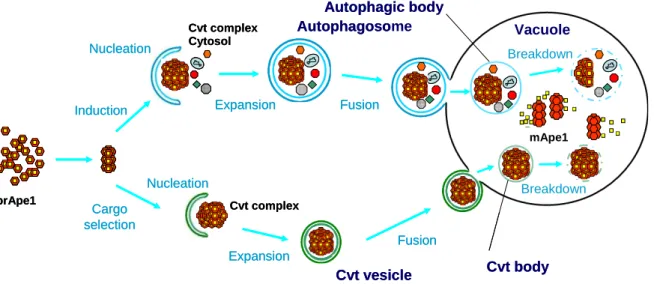
The best-characterized selective type of autophagic process is the Cvt pathway, and its study has provided a possible model for how specific cargoes are enwrapped by autophagosomes (see below). The Cvt pathway is a selective transport for Ape1/Lap4 and Ams1, and occurs constitutively in yeast, Saccharomyces cerevisiae. The classical model for deriver of hydrolases to the vacuole is via a portion of the secretory pathway. Proteins transit from the endoplasmic reticulum (ER) thought the Golgi complex and are sorted away from other proteins in the secretory pathway, diver to the endosome, and then to the vacuole. Analysis of Ape1/Lap4 and Ams1, which are vacuole soluble hydrolases, indicate that they are not transported via secretory pathway (Klionsky et al., 1992; Yoshihisa and Anraku, 1990; Yoshihisa et al., 1988). It is constitutively synthesized as an inactive precursor form (61 kDa) in the cytosol. It is targeted to the vacuole, and then processed by vacuolar proteinase B (Prb1) at its unusual 45-amino-acid-long-N-terminal region to become mature (55 kDa) (Segui-Real et al., 1995).
After synthesis, precursor Ape1 (prApe1) rapidly is self-oligomerized in the cytosol into the dodecamer (Figure 4; Figure 5) (Kim et al., 1997). Then the complex (Cvt complex) is specifically enwrapped by the Cvt vesicles and is delivered directly from cytoplasm to the vacuole; therefore, this pathway utilizes a subset of the autophagy machinery (Harding et al., 1996) (Figure 5). The
complex is enclosed by autophagosome-like membrane and the Cvt bodies inside the vacuole was detected in vacuolar hydrolase deficient cells (Figure 5). The membrane topology between autophagosome and the Cvt vesicles quite similar, although they are made in different sizes. The size of autophagosome is 300-900 nm, whereas that of the Cvt vesicle is 140-160 nm (Baba et al., 1997; Takeshige et al., 1992). Under starvation conditions, since the Cvt complex is enriched in the autophagosome, autophagy carried out the transport of precursor Ape1.
5-2 Mitophagy
Mitochondria are the power stations of the cell, and they generate most of the ATP required to keep the majority of the cellular functions operational. During this process, harmful reactive oxygen species (ROS) are generated. Over time, ROS production increases because aged and/or damaged mitochondria produce and release more ROS, thereby increasing the amount of cellular alterations. In order to maintain healthy cells, defective mitochondria must, therefore, be eliminated. A link between ROS and the induction of autophagy has recently been revealed (Kim et al., 2007). Nutrient starvation stimulates the production of ROS that is essential for the induction of autophagy. Indeed, treatment with antioxidants impairs autophagosome formation. This discovery suggests the possible scenario that damaged mitochondria activate mitophagy by uncontrolled release of ROS. How mitochondria are selectively sequestered into autophagosome has ever been unknown. Recently, studies in S. cerevisiae have shown that ATG11, in which it appears to be a common adaptor protein for specific types of autophagy, is essential for the process (Figure 3; Figure 6) (Kanki and Klionsky, 2008), suggesting that mitophagy is a specific process.
5-3 Pexophagy
The abundance of peroxisomes can rapidly change in response to changing environmental and/or physiological conditions. For example, the number of peroxisomes rapidly increases upon induction of peroxisome proliferation. In rodents, this is observed upon administration of peroxisomes proliferators, whereas in yeast species, peroxisome proliferation is induced during growth of cells on specific carbon sources (e.g., oleic acid or methanol). Generally, these responses are the result of metabolic adaptations to new physiological conditions that require peroxisomal metabolism. The opposite process, a rapid decrease in peroxisome abundance, can also be induced. Thus, when the peroxisome proliferation stimulus is removed, and/or peroxisomal metabolism is not required anymore, peroxisomes are degraded by
generally induced by nutrient starvation.
The abundance of peroxisomes within a cell can rapidly decrease by selective autophagic degradation (also designated pexophagy). Studies in yeast species have shown that at least two modes of peroxisome degradation are employed, namely macropexophagy and micropexophagy (Sakai et al., 2006). During macropexophagy, peroxisomes are individually sequestered by membranes, thus forming a pexophagosome. This structure fuses with the vacuolar membrane, resulting in exposure of the incorporated peroxisome to vacuolar hydrolases. During micropexophagy, a cluster of peroxisomes is enclosed by vacuolar membrane protrusions and/or segmented vacuoles as well as a newly formed membrane structure, the micropexophagy-specific membrane apparatus (MIPA), which mediates the enclosement of the vacuolar membrane. Subsequently, the engulfed peroxisome cluster is degraded.
5-4 ER-phagy
One of the main functions of the ER is the folding and processing of newly synthesized proteins destined for the plasma membrane, the extracellular space and almost all subcellular organelles including the ER itself. These events are co-ordinated by a highly regulated process of quality control performed by a plethora of chaperones, enzymes and other factors present in the ER lumen. In certain stress conditions, the balance between the folding capacity of the ER and the number of nascent proteins is shifted in such a way that there is an excess of unfolded proteins. An ER-to-nucleus signaling pathway, the unfolded protein response (UPR), senses this accumulation (Bernales et al., 2007). The UPR also modulates the expansion of the ER volume by increasing the size of its limiting membrane and that accommodates the folding of a large number of proteins(Bernales et al., 2007). When the UPR system is no longer necessary and becomes inactivated, the ER re-establishes its homeostatic volume. Recently, autophagy participates in the elimination of the superfluous ER (Bernales et al., 2007). atg1Δ, atg8Δ, atg9Δ, and atg16Δ cells lead to a growth defect upon UPR induction (Bernales et al., 2007). ER-Phagy also required additional genes when compared with the non-selective autophagy, including ATG19 and ATG20, two genes also involved in two other selective types of yeast autophagy (Figure 3; Figure 6), suggesting that ER-phagy is a specific process.
5-5 Ribophagy
Starved cells induce autophagy, by which ribosomes are non-selectively delivered to and are degraded in the vacuole, together with other cytoplasmic constituents. In addition, ribosomes are
ribosomal proteins of both 60S and 40S subunits, which were functional and correctly assembled into ribosomes, is delivered to and degraded in the vacuole of nitrogen-starved cells in an autophagy-dependent manner. This is not surprising, given that autophagy randomly delivers cytoplasmic constituents to the vacuole. However, when autophagic degradation of ribosomal proteins is compared with that of other cytoplasmic proteins, ribosomal degradation has occurred more extensively. This suggests that ribosomes are preferentially targeted to the autophagic pathway under conditions of nitrogen starvation. Ubp3/Bre5 is also specifically involved in the uptake of ribosomal 60S subunits into autophagosomes; however, it is not clear how Ubp3/Bre5 mediate selective autophagic degradation of ribosomal 60S subunits.
5-6 Targeting of protein aggregation
Several lines of evidence have indicated that the elimination of the toxic aggregates by autophagy is a selective process (i.e., aggrephagy) (Figure 6). For instance, the study of p62/sequestosome 1 (SQSTM1) has provided a possible model for how protein aggregates are recognized and selectively eliminated by autophagosomes. The p62/SQSTM1 interacts directly with Atg8 (Pankiv et al., 2007). In the absence of p62/SQSTM1, protein aggregates are not enwrapped by autophagosomes, strongly suggesting that p62/SQSTM1 mediates the recognition of polyubiquitinated proteins by double-membrane vesicles (Pankiv et al., 2007). It also became clear that autophagy not only has a role in eliminating aberrant protein complexes in pathological conditions but also its cellular basal activity prevents the formation of aggregates that could cause cell death in neurons (Rubinsztein, 2006). The specific knockout of either ATG5 or ATG7 in murine neurons leads to a time-dependent formation of ubiquitinated protein aggregates that resemble those observed in neurodegenerative disorders. These mice do not express any disease-associated mutant proteins and thus are not particularly prone to form aggregates. This crucial finding demonstrates that autophagy is also of vital importance in sustaining healthy aggregate-free neuronal cells.
6. The Aim of This Study
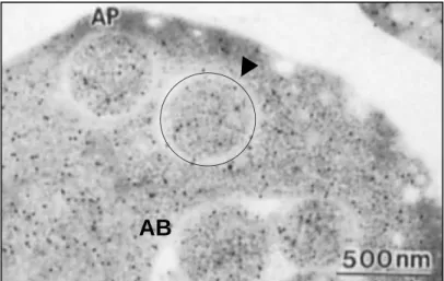
Much progress has been made in the functional analysis of Atg proteins. Atg proteins also function in the steps of autophagosome formation. One of the interesting subjects remained is an issue what is the substrate selectively in autophagy. Ohsumi and co-workers previously revealed that autophagy transports cytoplasmic constituents to the vacuole non-selectively in morphological and biochemical experiments. Immuno-electoron microscopy has shown that
and phosphoglycerate kinase (Pgk1), are present in the autophagosomes and autophagic bodies at the same densities as in the cytosol (Figure 7) (Baba et al., 1994). The measurement of the enzymatic activities of Adh1, Pgk1, glcose-6-phosphate dehydrogenase (G6pdh), and glutamate dehydrogenase also supports this conclusion (T. Noda and Y. Ohsumi., unpublished data) (Takeshige et al., 1992).
If degradation of long-lived proteins is exclusively mediated by autophagy, all proteins might be expected to have similar lifetimes. Long-lived proteins, however, have a variety of lifetimes; thus, the autophagic process might have some selectivity. It is known that Ape1 and Ams1 (via the Cvt pathway) (Figure 5; Figure 6), fructose-1,6-bisphosphatase (Fbp1) via the vacuolar import and degradation pathway; i.e., the Vid pathway (Brown et al., 2008; Chiang and Chiang, 1998; Hoffman and Chiang, 1996) are selectively transported from the cytoplasm to the vacuole. Previously, Onodera and Ohsumi showed that Ald6, alcohol dehydrogenase, is preferentially sequestered in autophagosome (Onodera and Ohsumi, 2005). Chiba et al. observed that autophagosome-like structure selectively sequestered to the part of chloroplasts to the exclusion of thylakoid membranes termed Rubisco containing bodies in wheat leaf cells during natural senescence (Chiba et al., 2003) . Microautophagy may have the substrate selectivity potentially, in fact, peroxisomes and cell nucleus are imported to the vacuole via the process via microautophagy (Sakai et al., 1998; Tuttle and Dunn, 1995), and via piecemeal macroautophagy (Roberts et al., 2003). To investigate the possibility of selective autophagic degradation, I focused on Lap3 in this study. GFP-fused Lap3 forms an aggregate associating with the Cvt complex and then selectively transported to the vacuole together with Ape1. Lap3 is degraded in the vacuole, whereas Ape1 is relatively stable. I propose that Lap3 is a novel target of selective degradation mediated by the machinery of the Cvt pathway and autophagy.
7. References
Baba, M., M. Osumi, S.V. Scott, D.J. Klionsky, and Y. Ohsumi. 1997. Two distinct pathways for targeting proteins from the cytoplasm to the vacuole/lysosome. J. Cell Biol. 139:1687-95.
Baba, M., K. Takeshige, N. Baba, and Y. Ohsumi. 1994. Ultrastructural analysis of the autophagic process in yeast: detection of autophagosomes and their characterization. J. Cell Biol. 124:903-13.
Bernales, S., S. Schuck, and P. Walter. 2007. ER-phagy: selective autophagy of the endoplasmic reticulum. Autophagy. 3:285-7.
Brown, C.R., A.B. Wolfe, D. Cui, and H.L. Chiang. 2008. The vacuolar import and degradation pathway merges with the endocytic pathway to deliver fructose-1,6-bisphosphatase to the vacuole for
Chiang, M.C., and H.L. Chiang. 1998. Vid24p, a novel protein localized to the fructose-1, 6-bisphosphatase-containing vesicles, regulates targeting of fructose-1,6-bisphosphatase from the vesicles to the vacuole for degradation. J. Cell Biol. 140:1347-56.
Chiba, A., H. Ishida, N.K. Nishizawa, A. Makino, and T. Mae. 2003. Exclusion of ribulose-1,5-bisphosphate carboxylase/oxygenase from chloroplasts by specific bodies in naturally senescing leaves of wheat. Plant Cell Physiol. 44:914-21.
Cowles, C.R., G. Odorizzi, G.S. Payne, and S.D. Emr. 1997. The AP-3 adaptor complex is essential for cargo-selective transport to the yeast vacuole. Cell. 91:109-18.
De Duve, C., and C. Hooft. 1968. [Quinquennial prizes of the medical sciences, period 1961-1965. Address by Prof. Chr. De Duve]. Verh K Vlaam Acad Geneeskd Belg. 30:381-8.
Doelling, J.H., J.M. Walker, E.M. Friedman, A.R. Thompson, and R.D. Vierstra. 2002. The APG8/12-activating enzyme APG7 is required for proper nutrient recycling and senescence in Arabidopsis thaliana. J. Biol. Chem. 277:33105-14.
Goldberg, A.L., and J.F. Dice. 1974. Intracellular protein degradation in mammalian and bacterial cells. Annu. Rev. Biochem. 43:835-69.
Harding, T.M., A. Hefner-Gravink, M. Thumm, and D.J. Klionsky. 1996. Genetic and phenotypic overlap between autophagy and the cytoplasm to vacuole protein targeting pathway. J. Biol. Chem. 271:17621-4.
Hershko, A., and A. Ciechanover. 1998. The ubiquitin system. Annu. Rev. Biochem. 67:425-79.
Hochstrasser, M. 1991. Functions of intracellular protein degradation in yeast. Genet. Eng. (N Y). 13:307-29.
Hoffman, M., and H.L. Chiang. 1996. Isolation of degradation-deficient mutants defective in the targeting of fructose-1,6-bisphosphatase into the vacuole for degradation in Saccharomyces cerevisiae. Genetics. 143:1555-66.
Ichimura, Y., T. Kirisako, T. Takao, Y. Satomi, Y. Shimonishi, N. Ishihara, N. Mizushima, I. Tanida, E. Kominami, M. Ohsumi, T. Noda, and Y. Ohsumi. 2000. A ubiquitin-like system mediates protein lipidation. Nature. 408:488-92.
Kanki, T., and D.J. Klionsky. 2008. Mitophagy in yeast occurs through a selective mechanism. J. Biol. Chem. 283:32386-93.
Kim, I., S. Rodriguez-Enriquez, and J.J. Lemasters. 2007. Selective degradation of mitochondria by mitophagy. Arch. Biochem. Biophys. 462:245-53.
Kim, J., S.V. Scott, M.N. Oda, and D.J. Klionsky. 1997. Transport of a large oligomeric protein by the cytoplasm to vacuole protein targeting pathway. J. Cell Biol. 137:609-18.
Kraft, C., A. Deplazes, M. Sohrmann, and M. Peter. 2008. Mature ribosomes are selectively degraded upon starvation by an autophagy pathway requiring the Ubp3p/Bre5p ubiquitin protease. Nat. Cell Biol. 10:602-10.
Levine, B., and D.J. Klionsky. 2004. Development by self-digestion: molecular mechanisms and biological functions of autophagy. Dev. Cell. 6:463-77.
Mizushima, N. 2005. The pleiotropic role of autophagy: from protein metabolism to bactericide. Cell Death Differ. 12 Suppl 2:1535-41.
Mizushima, N., T. Noda, and Y. Ohsumi. 1999. Apg16p is required for the function of the Apg12p-Apg5p conjugate in the yeast autophagy pathway. EMBO J. 18:3888-96.
Mizushima, N., A. Yamamoto, M. Hatano, Y. Kobayashi, Y. Kabeya, K. Suzuki, T. Tokuhisa, Y. Ohsumi, and T. Yoshimori. 2001. Dissection of autophagosome formation using Apg5-deficient mouse embryonic stem cells. J. Cell Biol. 152:657-68.
Mortimore, G.E., and A.R. Poso. 1987. Intracellular protein catabolism and its control during nutrient deprivation and supply. Annu. Rev. Nutr. 7:539-64.
Nair, U., and D.J. Klionsky. 2005. Molecular mechanisms and regulation of specific and nonspecific autophagy pathways in yeast. J. Biol. Chem. 280:41785-8.
Nakatogawa, H., K. Oh-oka, and Y. Ohsumi. 2008. Lipidation of Atg8: how is substrate specificity determined without a canonical E3 enzyme? Autophagy. 4:911-3.
Ohsumi, Y. 2001. Molecular dissection of autophagy: two ubiquitin-like systems. Nat. Rev. Mol. Cell Biol. 2:211-6.
Onodera, J., and Y. Ohsumi. 2005. Autophagy is required for maintenance of amino acid levels and protein synthesis under nitrogen starvation. J. Biol. Chem. 280:31582-6.
Pankiv, S., T.H. Clausen, T. Lamark, A. Brech, J.A. Bruun, H. Outzen, A. Overvatn, G. Bjorkoy, and T. Johansen. 2007. p62/SQSTM1 binds directly to Atg8/LC3 to facilitate degradation of ubiquitinated protein aggregates by autophagy. J. Biol. Chem. 282:24131-45.
Piper, R.C., N.J. Bryant, and T.H. Stevens. 1997. The membrane protein alkaline phosphatase is delivered to the vacuole by a route that is distinct from the VPS-dependent pathway. J. Cell Biol. 138:531-45. Piper, R.C., A.A. Cooper, H. Yang, and T.H. Stevens. 1995. VPS27 controls vacuolar and endocytic traffic
through a prevacuolar compartment in Saccharomyces cerevisiae. J. Cell Biol. 131:603-17. Reggiori, F. 2006. 1. Membrane origin for autophagy. Curr. Top Dev. Biol. 74:1-30.
Reggiori, F., I. Monastyrska, T. Shintani, and D.J. Klionsky. 2005. The actin cytoskeleton is required for selective types of autophagy, but not nonspecific autophagy, in the yeast Saccharomyces cerevisiae. Mol. Biol. Cell. 16:5843-56.
Rubinsztein, D.C. 2006. The roles of intracellular protein-degradation pathways in neurodegeneration. Nature. 443:780-6.
Sakai, Y., A. Koller, L.K. Rangell, G.A. Keller, and S. Subramani. 1998. Peroxisome degradation by microautophagy in Pichia pastoris: identification of specific steps and morphological intermediates. J. Cell Biol. 141:625-36.
Sakai, Y., M. Oku, I.J. van der Klei, and J.A. Kiel. 2006. Pexophagy: autophagic degradation of peroxisomes. Biochim. Biophys. Acta. 1763:1767-75.
Schimke, R.T., and D. Doyle. 1970. Control of enzyme levels in animal tissues. Annu. Rev. Biochem. 39:929-76.
Scott, R.C., G. Juhasz, and T.P. Neufeld. 2007. Direct induction of autophagy by Atg1 inhibits cell growth and induces apoptotic cell death. Curr. Biol. 17:1-11.
Seglen, P.O., and P. Bohley. 1992. Autophagy and other vacuolar protein degradation mechanisms. Experientia. 48:158-72.
Segui-Real, B., M. Martinez, and I.V. Sandoval. 1995. Yeast aminopeptidase I is post-translationally sorted from the cytosol to the vacuole by a mechanism mediated by its bipartite N-terminal extension. EMBO J. 14:5476-84.
Stepp, J.D., K. Huang, and S.K. Lemmon. 1997. The yeast adaptor protein complex, AP-3, is essential for the efficient delivery of alkaline phosphatase by the alternate pathway to the vacuole. J. Cell Biol. 139:1761-74.
Suzuki, K., Y. Kubota, T. Sekito, and Y. Ohsumi. 2007. Hierarchy of Atg proteins in pre-autophagosomal structure organization. Genes Cells. 12:209-18.
Suzuki, K., and Y. Ohsumi. 2007. Molecular machinery of autophagosome formation in yeast, Saccharomyces cerevisiae. FEBS Lett. 581:2156-61.
Takeshige, K., M. Baba, S. Tsuboi, T. Noda, and Y. Ohsumi. 1992. Autophagy in yeast demonstrated with proteinase-deficient mutants and conditions for its induction. J. Cell Biol. 119:301-11.
Tsukada, M., and Y. Ohsumi. 1993. Isolation and characterization of autophagy-defective mutants of Saccharomyces cerevisiae. FEBS Lett. 333:169-74.
Tuttle, D.L., and W.A. Dunn, Jr. 1995. Divergent modes of autophagy in the methylotrophic yeast Pichia pastoris. J. Cell Sci. 108 ( Pt 1):25-35.
Yoshihisa, T., and Y. Anraku. 1990. A novel pathway of import of alpha-mannosidase, a marker enzyme of vacuolar membrane, in Saccharomyces cerevisiae. J. Biol. Chem. 265:22418-25.
Yoshihisa, T., Y. Ohsumi, and Y. Anraku. 1988. Solubilization and purification of alpha-mannosidase, a marker enzyme of vacuolar membranes in Saccharomyces cerevisiae. J. Biol. Chem. 263:5158-63.
Figure 1. Trafficking pathways to and from the vacuole.
Multiple vesicular pathways deliver proteins to and from the vacuole. Resident vacuolar proteins are sent to the vacuole by several biosynthetic pathways, while proteins targeted for degradation may be sent via endocytosis or autophagy. Biosynthetic pathways: The ALP pathway delivers cargo from the late Golgi to the vacuole. The CPY pathway also starts from the late Golgi, but traverses multivesicular bodies (MVBs) before reaching the vacuole. The Cvt pathway delivers biosynthetic cargo from the cytosol to the vacuole in a process that has common components with autophagy. Trafficking pathways away from the vacuole may recycle membrane and proteins to the endocytic or CPY pathways. Degradative pathways: Endocytosis transports both soluble and membrane-bound cargo from the plasma membrane and extracellular space to the vacuole. Cargo progresses from early endosomes to late endosomes (multivesicular bodies) before reaching the vacuole. In macroautophagy, vesicles called autophagosomes engulf cytosolic material for degradation at the vacuole.
Endocytic pathway Autophagy
Cvt pathway
ALP pathway CPY pathway MVB pathway
MVB -late endosome-
-early endosome-
-Golgi- -Plasma membrane-
-Vacuole-
Endocytic pathway Autophagy
Cvt pathway
ALP pathway CPY pathway MVB pathway
MVB -late endosome-
-early endosome-
-Golgi- -Plasma membrane-
-Vacuole-
Figure 2. Conceptual model for autophagy.
The basic mechanism of autophagy is the sequestration of the cargo material (bulk cytoplasm, protein aggregates, and, organelles) by a cytosolic double-membrane vesicle (i.e., autophagosome). Various types of stimuli trigger the expansion of the isolation membrane (step 1). Upon vesicle completion (step 2), the autophagosome docks with the lysosomes/vacuoles and successively fuses with it (step 3). In this way, the inner vesicle (i.e., autophagic body), is liberated inside the vacuole, where it is finally consumed together with the cargo by vacuolar hydrolases (step 4). Pre- autophagosomal structure (PAS) has been used to describe all the autophagosomal intermediates.
Figure 3. The classification of Atg proteins.
The 31 known Atg proteins are sub-grouped based on their essential function in either nonselective or selective types of autophagy (see Figure 6). Eighteen of them are required for both types of autophagy. Conserved Atg proteins are in blue. Atg22 has not been included in this graph because its role in autophagy is controversial. Atg15 is the only Atg protein not involved in the biogenesis of autophagosomes; it participates in the breakdown of autophagic bodies. Atg17, Atg29, and Atg31 are specific factors for (macro)autophagy in yeast. In addition, Atg11 and Atg19 are essential for the Cvt pathway (see Figure 5).
Atg22
31 Atg proteins
Selective autophagy Non-selective
autophagy
Atg17 Atg29 Atg31
Atg1 Atg3 Atg5 Atg7 Atg9 Atg12 Atg14 Atg18
Atg2 Atg4 Atg6 Atg8 Atg10 Atg13 Atg16
Atg15
Atg11 Atg20 Atg23 Atg25 Atg27 Atg30
Atg19 Atg21 Atg24 Atg26 Atg28
Atg22
31 Atg proteins
Selective autophagy Non-selective
autophagy
Atg17 Atg29 Atg31
Atg1 Atg3 Atg5 Atg7 Atg9 Atg12 Atg14 Atg18
Atg2 Atg4 Atg6 Atg8 Atg10 Atg13 Atg16
Atg15
Atg11 Atg20 Atg23 Atg25 Atg27 Atg30
Atg19 Atg21 Atg24 Atg26 Atg28
Figure 4. Temporal order for packing cargo components in the Cvt pathway.
Precursor Ape1 monomers assemble into dodecamers and then into a large Ape1 complex. The receptor protein Atg19 binds to the propeptide of prApe1 to generate the Cvt complex. The binding of Atg11 is necessary for this complex to be targeted to the PAS where the disassembly of Atg11 oligomers occurs. Consequently, Atg8 conjugated to phosphatidylethanolamine binds to Atg19 and also to the growing membrane. In the final step of vesicle formation, Atg8–PE that is not enclosed within the vesicle is cleaved by the action of Atg4 (not shown) and released from the outer membrane.
Figure 5. Schematic representation of autophagy and the Cvt pathway in yeast.
In both autophagy (upper model) and the Cvt pathway (lower model), cargoes are engulfed by double membrane vesicles. During autophagy, various cytoplasmic components such as organelles and cytoplasm are sequestered into a large, double membrane autophagosome that is 300-900 nm in diameter. The Cvt pathway occurs during vegetative conditions; it is a biosynthetic pathway used to deliver the inactive form of the resident hydrolase aminopeptidase 1 (Ape1) to the vacuole. The Cvt vesicle ranges from 140-160 nm in diameter and, in contrast to the autophagosome, appears to exclude bulk cytoplasm. Following completion, the outer membrane of the sequestering vesicle fuses with the vacuolar membrane. The inner vesicle along with its contents is released into the vacuolar lumen where the vesicle membrane is degraded, allowing processing and activation of prApe1. Under starvation conditions, prApe1 is packaged into autophagosomes and is transported to the vacuole along with other cargo.
prApe1
mApe1 Cytosol
Autophagosome
Cvt vesicle
Cvt complex Cvt complex
Induction
Cargo selection
Nucleation
Nucleation
Expansion
Expansion
Fusion
Fusion
Breakdown Breakdown Autophagic body
Cvt body Vacuole
prApe1
mApe1 Cytosol
Autophagosome
Cvt vesicle
Cvt complex Cvt complex
Induction
Cargo selection
Nucleation
Nucleation
Expansion
Expansion
Fusion
Fusion
Breakdown Breakdown Autophagic body
Cvt body Vacuole
Figure 6. Selective types of autophagy.
Cytoplasmic constituents are selectively transported to the vacuole under the various conditions. Red-asterisk groups are transported to the vacuole for degradation. In contrast to reds, blue-asterisk group is selectively transported to the vacuole as a biosynthetic process; Ape1 and Ams1 are vacuole-resident enzymes.
ERs (ER-phagy )
Ape1 and Ams1 (Cvt pathway) Protein aggregates
(i.e., Aggrephagy )
Mitochondria (Mitophagy )
Peroxisomes (Pexophagy )
Yeast Fly Nematode
Mammal
Yeast Mammal
Yeast Mammal
Yeast Mammal
Yeast
p62/SQSTM1
Atg19 Atg20 actin
Atg11 Uth1 Aup1
Atg11 Atg19 Atg20 Atg21
Atg23 Atg24 Atg27
Tig1
Tig2 Vps45
actin Atg11
Atg20 Atg23
Atg24 Atg25 Atg26
Atg28 Atg30 Pex14
actin
*Biosynthetic process Vacuole
*
*
*
*
*
*Degradation
ERs (ER-phagy )
Ape1 and Ams1 (Cvt pathway) Protein aggregates
(i.e., Aggrephagy )
Mitochondria (Mitophagy )
Peroxisomes (Pexophagy )
Yeast Fly Nematode
Mammal
Yeast Mammal
Yeast Mammal
Yeast Mammal
Yeast
p62/SQSTM1
Atg19 Atg20 actin
Atg11 Uth1 Aup1
Atg11 Atg19 Atg20 Atg21
Atg23 Atg24 Atg27
Tig1
Tig2 Vps45
actin Atg11
Atg19 Atg20 Atg21
Atg23 Atg24 Atg27
Tig1
Tig2 Vps45
actin Atg11
Atg20 Atg23
Atg24 Atg25 Atg26
Atg28 Atg30 Pex14
actin Atg11
Atg20 Atg23
Atg24 Atg25 Atg26
Atg28 Atg30 Pex14
actin
*Biosynthetic process Vacuole
*
*
*
*
*
*Degradation
Figure 7. Immunostaining image of alcohol dehydrogenase in yeast cells.
Vacuolar hydrolase deficient cell (pep4Δ) contains autophagic bodies in the vacuole and autophagosomes in cytosol (Baba et al., 1994). AP, autophagosome (arrow head and circle); AB, autophagic bodies. Bar, 500 nm.
AB
MATERIALS AND METHODS
1. Cultivation and Handling of Yeast Strains
Standard techniques were used for yeast manipulation (Suzuki et al., 2007). The yeast strains used in this study are listed in Table I. Prior to experiments, all strains were grown in YPD medium (1% yeast extract, 2% polypeptone, and 2% glucose) and in YPGly medium (1% yeast extract, 2% polypeptone, and 3% glycerol) (Sherman et al., 1991) supplemented with 0.004% adenine sulfate. Since cells were grown in YPGly medium, single colonies were cultured in YPD medium until logarithmic phase (A600 = 1.0) and inoculated in YPGly medium at 0.08 unit of A600 followed by incubation at 30°C in a temperature-controlled dry shaker orbiting at 210 rpm. After cultured in YPGly medium until early logarithmic phase (A600 = 0.5) of growth, cells were inoculated in YPGly medium again at 0.08 unit of A600 and this operation was repeated for 3 times. For nitrogen starvation, SD-N medium (0.17% yeast nitrogen base without ammonium sulfate and amino acids and with 2% glucose) was used.
2. Manipulation of Yeast Strains
The W303-1A strain was transformed with a DNA fragment containing the wild-type ADE2 open reading frame to produce a parental strain (TKY51). A PCR-based gene modification method (Longtine et al., 1998) was used to disrupt ATG genes. Genome DNA extracts from the lab stock were used as the PCR templates. The yeast leucine aminopeptidase III (Lap3) were endogenously tagged with green fluorescent protein (GFP) at the N-terminus by the PCR-based epitope-tagging strategy (Janke et al., 2004). The sequence encoding the GPD promoter and the GFP was amplified with the S1 and S4 primer pair from the pYM-N17 plasmid (Janke et al., 2004). In the resulting strain, the endogenous promoter is replaced with a GPD promoter, and the GFP tag is fused to the N-terminus of the Lap3. The atg1ts allele (Suzuki et al., 2001) was integrated in TKY71 strains. The TKY109 and TKY110 strains were generated by transforming the TKY71 and TKY72 cells with pPS129 digested with AvrII (Stromhaug et al., 2004) (plasmid was a gift from Dr. Daniel J. Klionsky, University of Michigan, USA).
3. Antibodies
Lap3 specific antibodies were prepared as follow. To raise an antiserum against Lap3, I used synthetic peptides corresponding to amino acids 6-20 and 204-217 as antigens (the NIBB
antiserum was described previously (Suzuki and Ohsumi, 2007). An anti-PGK and anti-FLAG antibodies, and an anti-GFP antiserum were purchased from Invitrogen.
4. Immunoblotting Analysis
Whole cell lysates were prepared by NaOH/2-mercaptoethanol extraction with a slight modification (Horvath et al., 1994) and by disrupting cells with glass beads in lysis buffer (50 mM Tris-HCl, pH 8.0, 4 mM pephablochTM and protease inhibitor cocktail, RocheApplied Science). Cells lysates were boiled with SDS-sample buffer for 5 min, and these proteins content was determined by BCA assay kit (Pierce). Total protein (10 μg) was subjected to SDS-PAGE (Suzuki et al., 2007), and transferred to polyvinylidene fluoride membrane (ImmobilionTM-P, Millipore) and detected with a combination of each antibody and peroxidase-conyugated goat anti-rabbit IgG antibody (Jackson) or peroxidase-conyugated goat anti-mouse IgG antibody (Jackson) by the ECL system (Parkinelmer). To examine GFP–Lap3 transport, cells were grown in YPD medium to mid-log phase at 30°C, washed in water two times and then incubated in nitrogen starvation medium (SD-N) at 30°C. To monitor stability of Lap3 in the vacuole, cells harboring the atg1ts allele were cultured in YPD medium to mid-log phase at 23°C. For starvation, cells were washed in water two times, cultured in SD-N medium at 23°C for 5 h, and then incubated at 37°C to block autophagy. At the time points indicated, 1 ml of culture was harvested and used to prepare cell extract. Protein extracts were subjected to SDS-PAGE as described (Suzuki et al., 2007) and were probed with anti-Lap3, anti-Ape1, anti-GFP and anti-Pgk1 antisera or antibodies.
5. Microscopy
Fluorescence microscopy was performed using a TIR-FM system (OLYMPUS) as described (Suzuki et al., 2007). This system was equipped with a 100× objective lens (Apo100 x OHR, NA: 1.65) and a CoolSNAP HQ CCD camera (Nippon Roper). GFP was excited with a mercury lamp and a U-MGFPHQ filter set. For cells labeled with both GFP and RFP, a blue laser (Sapphire 488-20, Coherent) and a yellow laser (85-YCA-010, Melles Griot) were used. A U-MNIBA2, from which the excitation filter was removed, was used for GFP visualization, and an FF593-Di02 dichroic mirror and an FF593-Em02 excitation filter (Semrock) were used to analyze RFP. A UPlanSApo100 x Oil (NA: 1.40) objective lens was used for microscopic observation. Images were acquired using MetaMorph software (Molecular Devices).