Description of the intertidal aleocharine
Halorhadinus sawadai sp.n. from Japan,
with notes on the genus Halorhadinus S AWADA , 1971
(Coleoptera: Staphylinidae)
M. MARUYAMA & M. HAYASHI
Abstract
Halorhadinus sawadai sp.n. (Coleoptera: Staphylinidae: Aleocharinae) is described. It was collected from gravel beaches of the Sea of Japan (Honshû). A revised key of Halorhadinus SAWADA, 1971 is presented based on its general appearance. Halorhadinus species are confined to gravel beaches in western Japan and southern Korea. Currently, Halorhadinus (SAWADA, 1971) is classified as Liparocephalini, but the monophyly of the tribe is doubtful. Halorhadinus might be closely related to Bryothinusa, and is tentatively regarded as a member of Myllaenini.
Key words: Coleoptera, Staphylinidae, Aleocharinae, Liparocephalini, Myllaenini, Halorhadinus, Bryothinusa, intertidal, new species.
Introduction
The diverse habitats of the Japanese coasts, which extend from the sub-arctic northeast (Etorofu- tô [Iturup], Hokkaidô) to the sub-tropic southwest (Yonaguni-jima, Okinawa), host a rich intertidal insect fauna (KAWAI & TANIDA 2005, SAWADA 1995). Since SAWADA’s (1955, 1956, 1971, 1991) initial systematic frameworks, additional taxa of the intertidal aleocharine fauna in Japan have been discovered (AHN & ASHE 1995, AHN et al. 1999, MARUYAMA & AHN 2000a, 2000b, ASSING & MARUYAMA 2002, MARUYAMA 2002, MARUYAMA et al. 2008) and it is suspected that there are further discoveries to be made.
Recently, M. Hayashi collected a particularly large (>5.0 mm) specimen of the genus Halorhadinus (SAWADA, 1971); it is one of the largest intertidal aleocharine species in the world. It was found underground, on gravel beaches of the Sea of Japan, a habitat unexplored by researchers of intertidal insects. Until now, two intertidal species of Halorhadinus, H. aequalis (SAWADA,1971) and H. inaequalis (SAWADA,1971), were known from Japan and Korea. This paper describes the new species and presents a revised key and the bionomics of Halorhadinus. While it has been classified as either Myllaenini (MOORE & LEGNER 1976) or Diglottini (PACE 1999), Halorhadinus was most recently placed in Liparocephalini based on morphological data (AHN 2001). However, the monophyly of Liparocephalini is supported by several characteristics (AHN 2001, 2004, AHN & ASHE 1996, LESCHEN et al. 2002) that are considered to be highly homoplastic and observed in many other aleocharines. In addition, we discuss the systematic position of Halorhadinus in the light of phylogenetic problems regarding Liparocephalini.
Material, methods, and abbreviations
The specimens discussed in this paper were obtained during faunistic research conducted in Shimane-ken, Japan (KAWAKAMI & HAYASHI 2007) and from material submitted by our colleagues. The terminology of the microstructures largely follows SAWADA (1972) and MARUYAMA (2006). The technical procedures are adapted from those used by MARUYAMA
(2006). The number of setae and pores in the descriptions refer to one side of the body. The specimens examined have been deposited in the following collections.
cSaw Private collection of Kohei Sawada, Ôsaka HOWP Hoshizaki Institute for Wildlife Protection, Izumo KUM The Kyûshû University Museum, Fukuoka NHM The Natural History Museum, London NMW Naturhistorisches Museum Wien
Halorhadinus SAWADA, 1971
Halorhadinus SAWADA 1971: 92 [original description; type species: H. aequalis SAWADA, 1971]. AHN 2001: 123 [redescription, key to species].
SUPPLEMENTARY DESCRIPTION: Body length 2.9–5.5 mm. Lateral setae of epipharynx long, almost as long as lateral setae on labrum [illustrated on dorsal side of labrum in AHN
(2001)]. Labial apodeme long, as long as palpus, and its lateral lobes connected at base. Median lobe of aedeagus with “athetine bridge” (SEEVERS 1978), internal sac with complicated sclerites that are partly exposed from apex of median lobe.
REMARKS: AHN (2001) redescribed the genus in detail based on the two known species of the genus. He regarded the contiguous mesocoxal cavities as an important character state of the genus, but in H. sawadai the mesocoxal cavities are well separated by the process of the mesoventrite. Therefore, the contiguous mesocoxal cavities should be excluded from the generic diagnosis.
AHN (2001) provided a key to the species of the genus based on mouthparts and genital characteristics. However, the members of the genus, including the present new species, are easily distinguished by states of general appearances. A revised key of the genus is presented, and each species is diagnosed with more facile distinguishing characters.
Key to species of Halorhadinus based on general appearance
1 Fore body brown. Antennae shorter than fore body length; antennal segments VIII–X as long as wide or slightly longer than wide. Elytra much wider than pronotum. ... inaequalis (Figs. 5–6) – Fore body pale brown. Antennae longer than fore body length; antennal segments VIII–X
twice as long as wide. Elytra almost as wide as pronotum... 2 2 Body ca. 3.5–3.8 mm. Head semicircular, with frons gently rounded ... aequalis (Figs. 3–4) – Body ca. 4.6–5.5 mm. Head elongate, with frons strongly protruding... sawadai (Figs. 1–2)
Halorhadinus sawadai sp.n. (Figs. 1–2, 7–20) Type locality: Japan, Honshû, Shimane-ken.
TYPE SERIES: Holotype ♂: “NIPPON: Shimane-ken, Izumo-shi, Inome-chô, Inomekaigan, 25 VII 2007, leg. Hayashi-M.” (KUM). Paratypes: 3 ♂♂, 2 ♀♀, same data as holotype (HOWP, KUM, NHM, NMW); 1 ♀, “NIPPON: Shimane-ken, Izumo-shi, Taisha-chô, Hinomisaki, 28 VI 2006, leg. Hayashi-M.” (KUM).
DIAGNOSIS: Body ca. 4.6–5.5 mm; fore body and apex of abdomen pale brown; head flattened above, elongate, with frons protruding; antennae long, longer than fore body length; antennal segments VIII–X twice as long as wide; labrum long, almost circular, its anterior margin rounded; left and right mandibles with two teeth and no tooth respectively; lacinial spines irregular in shape and size; elytra almost as wide as pronotum.
DESCRIPTION: Body (Figs. 1–2) elongate, flattened. Fore body and apex of abdomen pale brown; antennae, mouthparts and legs yellowish brown. Head flattened above, elongate, with frons protruding; antennae long, longer than fore body length; antenna with all segments elongate; segments II–V almost equal in length; segment X twice as long as wide. Labrum (Fig. 7) hypertrichous, long, almost circular, its anterior margin rounded, moderately covered with minute pseudopores; epipharynx with six long lateral setae. Left mandible (Fig. 8) with two teeth, crenulate between teeth and apex; right mandible (Fig. 9) without tooth, crenulate. Lacinia (Fig. 10) with spines irregular in shape and size. Mentum (Fig. 11) trapezoidal and its anterior margin deeply emarginate, hypertrichous, densely covered with pseudopores except for lateral margins. Labium (Figs. 12–14): prementum with one setal pore and two real pores, no pseudopores; segment I of palpus (combined actual I and II, Fig. 13) with setae situated basally; segment II of palpus (actual III) with three pores; ligula (Fig. 14) as long as 2/3 of segment I, with three pairs of sensilla. Pronotum (Fig. 1) as long as wide, widest near anterior margin, finely and densely punctate, densely covered with long decumbent setae, and with 8–10 macrosetae on anterior and lateral margins. Elytra parallel-sided, widest at middle, narrowed apically, almost as wide as pronotum, punctation and setation as on pronotum, but setae longer, and with five or six macrosetae laterally. Abdomen widest at segment IV; tergites III–VI densely and somewhat roughly punctate except for basal depressions.
Male: Tergite VIII (Fig. 15) slightly narrowed posteriorly, with 10–13 macrosetae; sternite VIII with posterior margin rounded, with 30–35 macrosetae. Median lobe of aedeagus (Figs. 17–18) with apical lobe sinuate in lateral view and apex protruding. Apical lobe of paramere (Fig. 19) with two long setae near base and two short setae at apex of inner side.
Female: Tergite VIII (Fig. 16) narrowed posteriorly, with 8–10 macrosetae; sternite VIII with posterior margin roundly emarginate medially, with 20–25 macrosetae. Spermatheca (Fig. 20) coiled at base.
MEASUREMENTS: Head width, 0.66–0.71 mm; pronotal length, 0.77–0.83 mm; pronotal width, 0.77–0.83 mm; elytral width, 0.81–0.88 mm; hind tibial length, 0.96–1.02 mm.
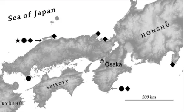
DISTRIBUTION: Shimane-ken, western Japan (Fig. 21).
ETYMOLOGY: Dedicated to Dr. Kohei Sawada for his invaluable contributions to the systematics and morphology of Aleocharinae including Asian intertidal species.
Halorhadinus aequalis SAWADA, 1971 (Figs. 3–4)
TYPE MATERIAL: Paratype, 1 ♀, “Paratype Shingu Wakayama [Shingû-shi, Wakayama-ken] 14. IV. 1970 R. Yosii” (cSaw).
SPECIMENS EXAMINED: JAPAN: 6 exs., Nagashima, Kaminoseki-chô, Yamaguchi-ken, 5.V.2005, leg. Y. Nakase (KUM); 5 exs., Nakayama, Hinomisaki, Taisha-chô, Izumo-shi, Shimane-ken, 9.VII.2006, leg. M. Hayashi (HOWP, KUM, NHM, NMW); 1 ex., same locality but near Hinomisaki-tôdai, Hinomisaki, 7.VII.2006, leg. M. Hayashi (KUM).
DIAGNOSIS: Body ca. 3.5–3.8 mm; fore body and apex of abdomen pale brown; head flattened above, semicircular, with frons gently rounded; antennae long, longer than fore body length; antennal segments VIII–X twice as long as wide; labrum transverse, its anterior margin truncate;
left and right mandibles with one tooth and two teeth respectively; lacinial spines almost same in shape and length; elytra almost as wide as pronotum.
DISTRIBUTION: Western Japan (Fig. 21), southern Korea.
Halorhadinus inaequalis SAWADA, 1971 (Figs. 5–6)
SPECIMENS EXAMINED: JAPAN: 1 ex., Tsumeki-zaki, Suzaki, Shimoda-shi, Shizuoka-ken, 31.X.1999, leg. Y. Tahira (KUM); 1 ex., Shimakage (Kaisuiyokujô), Kurita, Miyazu-shi, Kyôto-fu, Japan, 31.III.2000, leg. K. Yasukawa (KUM); 4 exs., Fukuura, Mihonoseki-chô, Matsue-shi, Shimane-ken, 23.IV.2006, leg. M. Hayashi (KUM, NHM, NMW); 2 exs., near Hinomisaki-tôdai, Hinomisaki, Taisha-chô, Izumo-shi, Shimane-ken, 7.VII.2006, leg. M. Hayashi (KUM).
DIAGNOSIS: Body ca. 2.9–3.5 mm; body brown; head slightly convex above, semicircular, with frons generalized; antennae short, shorter than fore body length; antennal segments VIII–X as long as wide or slightly longer than wide; labrum semicircular, its anterior margin rounded; left and right mandibles with one tooth and no tooth respectively; lacinial spines irregular in shape and size; elytra much wider than pronotum.
DISTRIBUTION: Western Japan (Fig. 21), southern Korea.
Bionomics of Halorhadinus
Three species of Halorhadinus were collected from the intertidal zones of gravel beaches (Figs. 22–25). This zone of the shoreline is characterized by unsorted gravel, cobblestone, and pebbles that are supported by a matrix composed of very coarse sand and granules. At a gravel beach in Fukuura, where only H. inaequalis was found, the ground is rich in shells.
Halorhadinus beetles live in spaces between the gravel and matrix. Most intertidal staphylinids, such as Bryothinusa CASEY,1904, Diaulota CASEY,1904, and Thinobius KIESENWETTER,1844 species, are minute, measuring 1.5–2.5 mm in body length. They live under deeply settled stones on sandy or muddy tidal flats, or in small, dead barnacles on rocks where the spaces are very small. This could be a constraint on their body size. By contrast, the members of Halorhadinus are generally large, particularly H. sawadai. In their habitat, the open spaces of the matrix are much larger than those under stones on tidal flats or dead barnacles, which allows for the larger body size of Halorhadinus species.
In Shimane, Halorhadinus beetles are often found together with centipedes (Geophilomorpha), the earwig Anisolabis maritima, the bug Speovelia maritima, the cricket Caconemobius sazanami, and Luciogobius fishes, although the microhabitat of each species varies slightly. In Nagashima, Yamaguchi, H. aequalis was found together with many Bryothinusa tsutsuii (Aleocharinae), a few Thinobius sp. (Oxytelinae), and Luciogobius gobies (Nakase, personal communication). These intertidal animals are considered indicators of natural shores, and some Luciogobius species are found only on clean gravel beaches.
Current records show that Halorhadinus species are scattered throughout western Honshu (Fig. 21). Although Halorhadinus beetles may be confined to well-preserved environments, research on intertidal insects has not yet focused on gravel beaches as collecting sites; a focus on such sites will likely result in the discovery of further localities in western Japan, as well as in Shikoku and Kyûshû. AHN (2001) reported Halorhadinus species that were collected from a sandy beach together with Cafius (Staphylininae), but this may be a misinterpretation or accidental record. In Japan, no specimen of Halorhadinus has been found in sandy beaches, which are considered an unsuitable habitat for them.
Figs. 1–6: Habitus, dorsal (left) and ventral (right) views, 1–2) Halorhadinus sawadai sp.n., holotype, 3– 4) H. aequalis, 5–6) H. inaequalis. Scale bar: 1.0 mm.
Figs. 7–10: Halorhadinus sawadai sp.n., mouthparts, 7) labrum, setae and pseudopores in left and right sides respectively, 8) left mandible, apical half, dorsal view, 9) right mandible, apical half, dorsal view, 10) lacinia, apical part, ventral view. Scale bars: 0.1 mm.
Figs. 11–14: Halorhadinus sawadai sp.n., mouthparts, 11) mentum, dorsal view, 12) labium, dorsal view, 13) part of prementum and base of labial palpus, 14) ligula. Scale bars: 0.1 mm.
Figs. 15–20: Halorhadinus sawadai sp.n., 15) male tergite VIII, dorsal view, 16) female tergite VIII, dorsal view, 17) median lobe of aedeagus, holotype, lateral view, 18) same, parameral view, 19) apical lobe of paramere, 20) spermatheca. Scale bars: 0.1 mm.
Fig. 21: Distribution of Halorhadinus species in Japan, H. sawadai (black star), H. aequalis (black circle), H. inaequalis (black diamond).
Figs. 22–25: Collecting sites of Halorhadinus species in Shimane-ken, Japan, 22) Inome (H. sawadai), 23) Hinomisaki (H. sawadai, H. aequalis, H. inaequalis), 24) Nakayama (H. aequalis), 25) Fukuura (H. inaequalis).
Systematic position of Halorhadinus
Based on the resemblance of their mouthparts, SAWADA (1971) postulated that Halorhadinus is closely related to the genus Bryothinusa (CASEY, 1904) of the tribe Myllaenini. MOORE & LEGNER (1976) followed SAWADA (1971) and placed Halorhadinus in Myllaenini. PACE (1999) reclassified the genus as a Diglottini, along with Bryothinusa and others, indicating that he also regarded Halorhadinus as closely related to Bryothinusa. By contrast, AHN (2001) transferred Halorhadinus to the tribe Liparocephalini.
In their first attempt to describe the phylogeny of Liparocephalini, AHN & ASHE (1996) listed four character states as autapomorphies of the tribe: 1) the absence of seta V on the mentum; 2) the uniform distribution of setae on the lacinia; 3) the presence of one medial seta on the labium; and 4) contiguous mesocoxal cavities. However, these characteristics are not unique to Liparocephalini, and even tend to vary within tribes of Aleocharinae, including Homalotini, a possible relative of Liparocephalini (AHN & ASHE 1996). Subsequent analyses by AHN (2001, 2004) and LESCHEN et al. (2002) changed the autapomorphies of the tribe on using different outgroups. Consequently, their phylogenetic evidence appears to be unreliable and the monophyly of Liparocephalini is doubtful.
AHN (2001) placed Halorhadinus in Liparocephalini using the following similarities: contiguous mesocoxal cavities, galea with setae only on the mesal surface, and apex with setae. However, these characteristics are homoplastic. The new species, H. sawadai, is characterized by clearly separated mesocoxal cavities, while the other states agree well with the genus concept. The separation of the mesocoxal cavities is probably related to the length or thickness of the legs. In Aleocharinae, long- and stout-legged species tend to have large coxae and their mesocoxal cavities tend to be contiguous or very narrow (Maruyama, unpublished data). Intertidal aleocharines generally have long, stout legs (and long tarsal claws) that are well-adapted to intertidal habitats with tide changes and wave turbulence, and their mesocoxal cavities tend to be contiguous.
AHN (2001) also mentioned that the distribution of the lacinial setae, the ligular shape, the mentum shape, and the gland opening on tergite VII in Halorhadinus are not observed in either Myllaenini or Diglottini. However, we believe that the distribution of the lacinial setae, ligular shape, and gland opening on tergite VII are unreliable diagnostics and even vary within some tribes of Aleocharinae; the mentum shape of Halorhadinus is more similar to that of Bryothinusa than to the core members of Liparocephalini, namely the species of the type genus Liparo- cephalus and its obvious relative Diaulota.
Like SAWADA (1971), we consider Halorhadinus to be more closely related to Bryothinusa than to the core members of Liparocephalini. This is supported by the following characteristics: 1) fused labial palpomeres I and II elongate and 2) narrowing apically; 3) setae are distributed in the basal part of the labial palpus; and 4) the lacinial spines are distributed separately. However, many convergent characteristics are observed in intertidal and coastal aleocharines. This is especially evident in the mouthparts, which are considered the most definitive characteristics for the systematics of Aleocharinae. Therefore, phylogenetic analyses based on morphological data have a number of limitations. Ultimately, phylogenetic analyses based on molecular data will help to give a better idea of the systematic position of Halorhadinus. We tentatively regard Halorhadinus as a member of Myllaenini, as first suggested by SAWADA (1971).
Acknowledgements
We thank Mr. Tateo Ito (Kyoto), Mr. Yuta Nakase (Kyoto University) and Mr. Yoshiaki Tahira (Teiso Kasei co.) for material, and Mrs. Yasuko Kawakami for support in various ways. Special thanks are also due to Dr. Kohei Sawada for his suggestions and encouragement. This paper is supported by Grants-in-Aid for Scientific Research of JSPS (Start-up 20870031) funded to the senior author.
References
AHN, K.-J. 2001: Phylogenetic relationships of the intertidal genus Halorhadinus Sawada and key to the genera of the Liparocephalini (Coleoptera: Staphylinidae: Aleocharinae). – Insect Systematics and Evolution 32: 123–132.
AHN, K.-J. 2004: Moorea zealandica, new genus and species from New Zealand with a discussion of its phylogenetic relationships (Coleoptera: Staphylinidae: Aleocharinae). – New Zealand Journal of Zoology 31: 255–261.
AHN, K.-J. & ASHE, J.S. 1995: Revision of the intertidal aleocharine genus Amblopusa Casey and description of the new genus Paramblopusa (Coleoptera: Staphylinidae). – Journal of New York Entomological Society 103 (2): 138–154.
AHN, K.-J. & ASHE. J. S. 1996. Phylogeny of the intertidal aleocharine tribe Liparocephalini (Coleoptera: Staphylinidae). – Systematic Entomology 21: 99–114.
AHN, K.-J., MARUYAMA, M. & ÔHARA, M. 1999: The intertidal beetle Amblopusa magna Zerche (Coleoptera, Staphylinidae, Aleocharinae), new to Hokkaido, Japan, and Kuril Archipelago, Russia. – Elytra 27 (2): 641–642.
KAWAI, T. & TANIDA, K. (eds.) 2005: Aquatic Insects of Japan: Manual with Keys and Illustrations. – Kanagawa: Tokai University Press, 1360 pp.
KAWAKAMI, Y. & HAYASHI, M. 2007: Faunal studies on the marine Coleoptera of Japan Sea: Part 2. Shimane Peninsula in the San’in District. – Bulletin of the Hoshizaki Green Foundation 10: 37– 76.
LESCHEN, R.A.B., BULLIANS, M.S., MICHAUX, B. & AHN, K.-J. 2002: Systematics of Baeostethus chiltoni, a subantarctic liparocephaline (Coleoptera: Staphylinidae: Aleocharinae): a Pangean relic or a more recent immigrant? – Journal of the Royal Society of New Zealand 32 (2): 189– 201.
MARUYAMA, M. 2002: Rediscovery of Brachypronomaea esakii (Coleoptera, Staphylinidae, Aleochar- inae) and its new record from the island of Okinawa-honto, the Ryukyus. – Elytra 30: 99–100. MARUYAMA, M. 2006: Revision of the Palearctic species of the myrmecophilous genus Pella
(Coleoptera, Staphylinidae, Aleocharinae). – National Science Museum Monographs 32: 1–207. MARUYAMA, M. & AHN, K.-J. 2000a: The intertidal beetle Paramblopusa borealis (Casey) (Coleoptera,
Staphylinidae, Aleocharinae), new to Japan. – Japanese Journal of Systematic Entomology 6 (1): 83.
MARUYAMA, M. & AHN, K.-J. 2000b: Redescription of Liparocephalus litoralis Kirschenblatt, and a key to the species of the genus Liparocephalus Mäklin. – Canadian Entomologist 132: 567–571. MARUYAMA, M., KLIMASZEWSKI, J. & GUSAROV, V. 2008: Osakatheta yasukoae, a new intertidal genus
and species of athetine rove beetles (Coleoptera, Staphylinidae, Aleocharinae) from Japan. – Zootaxa 1683: 39–50.
MOORE, I. & LEGNER, E.F. 1976: Intertidal rove beetles (Coleoptera: Staphylinidae), pp. 521–551. – In: Cheng, L. (ed.): Marine Insects. – Amsterdam: North Holland Publishers.
PACE, R. 1999: Aleocharinae della Namibia raccolte dalla spedizione entomologica “Namibia 1992” del Museo di Storia Naturale di Berlino (Coleoptera: Staphylinidae). – Memorie della Società Entomologica Italiana 77: 161–212.
SAWADA, K. 1955: Marine insects of the Tokara Islands VIII. Family Staphylinidae (Coleoptera). – Publications of the Seto Marine Biological Laboratory 5: 81–87.
SAWADA, K. 1956: A new intertidal species of Staphylinidae from Ishigakijima, Ryukyu Islands (Coleoptera). – Kontyû 24: 197–199.
SAWADA, K. 1971: Aleocharinae (Coleoptera: Staphylinidae) from the intertidal zones of Japan. – Publications of the Seto Marine Biological Laboratory 19: 81–110.
SAWADA, K. 1972: Methodological research in the taxonomy of Aleocharinae. – Contributions from the Biological Laboratory Kyoto University 24: 31–59.
SAWADA, K. 1991: On a new genus and species of intertidal Aleocharinae (Coleoptera: Staphylinidae) and Goniacerinae (Pselaphidae) from Singapore and Japan. – Raffles Bulletin of Zoology 39: 141–152.
SAWADA, K. 1995: Collembola, Thysanura, Dermaptera, Hemiptera, Coleoptera, Diptera. – In: Nishi- mura, S. (ed.): Guide to seashore animals of Japan with color pictures and keys Vol. II: 436–490. – Ôsaka: Hoikusha.
SEEVERS, C. 1978: A generic and tribal revision of the North American Aleocharinae (Coleoptera: Staphylinidae). – Fieldiana: Zoology 71: i–vi+1–275.
Dr. Munetoshi MARUYAMA
The Kyûshû University Museum, Hakozaki 6-10-1, Fukuoka, 812-8581 Japan ([email protected]) Dr. Masakazu HAYASHI
Hoshizaki Green Foundation, Sono-chô 1659-5, Izumo-shi, Shimane, 691-0076 Japan ([email protected])