1
Study on Neural Mechanisms for Reaction Time Facilitation
by Audio-Visual Integration
Junya Hirokawa
Department of Basic Biology, School of Life Science, The Graduate University for
Advanced Studies
Division of Brain biology, National Institute for Basic Biology
March 2008
Contents
Summary ...4
Introduction ...5
Chapter I:
Identification of neural substrates for multisensory facilitation by c-Fos mapping
...8Results 1) Facilitation of reaction time in cAV task but not in iAV task ...8
2) Standardization of rat neocortex and identification of cortical layers and areas...9
3) c-Fos mapping in sensory cortex ...10
4) c-Fos mapping in the superior colliculus (SC)...11
Discussion 1) Use of standardization method of cortical sections for functional mapping...12
2) Neural substrates of multisensory behavioral facilitation ...13
3) Consideration of the retinotopic maps in visual areas and SC...15
Figures...17
3
Chapter II:
Roles of early cortical areas and superior colliculus in multisensory behavior
...24Results 1) Effect of dSC inactivation on the reaction time facilitation...24
2) Effect of V2L inactivation on the reaction time facilitation ...26
3) Effect of inactivation of V2L on intensity-related unimodal facilitation...27
Discussion 1) Functional role of lateral secondary visual area (V2L) ...29
2) Behavioral facilitation mediated by lateral secondary visual area (V2L)...30
3) Anatomical substrates of multisensory facilitation of reaction time ...31
4) Role of superior colliculus (SC) for audiovisual behavioral facilitation of reaction time...31
5) Role of lower sensory area in multisensory facilitation ...33
Figures...35
Materials and Methods ...45
Acknowledgements ...54
References ...55
Abbreviations ...61
Summary
Recent studies reveal that multisensory convergence can occur in early sensory cortical and subcortical
areas. However, the behavioral importance of the multisensory integration in such areas is unknown.
Here, I used c-Fos immunohistochemistry to explore neuronal populations specifically activated
during the facilitation of reaction time induced by the temporally congruent audiovisual stimuli in rats.
My newly developed analytical method for c-Fos mapping revealed a pronounced up-regulation of
c-Fos expression particularly in layer 4 of the lateral secondary visual area (V2L) and the deep layer of
superior colliculus (dSC). A local injection of a GABA A receptor agonist, muscimol, into dSC
deteriorated the reaction time to all kinds of sensory stimuli but the audiovisual facilitation was not
affected, suggesting that the dSC receives the integrated audiovisual information for the facilitated
behavior. On the other hand, injection of muscimol into V2L completely suppressed the audiovisual
facilitation of reaction time without affecting responses to unimodal stimuli. Such a selective
suppression was not found following the injection of muscimol into the primary auditory and visual
cortical areas.
5
Introduction
Animals have the ability to utilize information from different senses to gain behavioral advantages such as rapid responses (e.g. Hughes et al., 1994; Molholm et al., 2002; Corneil et al., 2002; Sakata et al., 2004) and increased accuracy (e.g. Stein et al., 1989; Frens and Van Opstal, 1995; Bolognini et al., 2005). Integration of different sensory information in the brain is considered to be a basic neural mechanism to produce the behavioral facilitation (Stein and Meredith, 1993). Conventionally, it had been considered that multisensory integration only occurs in higher-order association areas after extensive unimodal informational processing (e.g. Felleman and Van Essen, 1991). In agreement with this view, transient inactivation of higher-order cortical areas directly demonstrated their critical roles in the multisensory facilitation of spatial orientation accuracy in cats (Wilkinson et al., 1996; Jiang et al., 2002). However, if only higher cortical areas mediate enhanced responses to multisensory stimuli, such an extensive informational processing would inevitably take a long time. Thus, it may be speculated that there are different neural substrates for facilitating the speed of the behavioral response to multisensory stimuli.
The superior colliculus (SC) is well studied structure for multisensory integration because it contains many multisensory and sensorimotor . Stein and Meredith, 1993; Zangenehpour and Chaudhuri, 2001; Bell et al., 2005; Holmes and Spence, 2005). Previous studies in anesthetized animals noted that the deep layers of the superior colliculus (dSC) showed enhanced or depressed activities to combination of auditory and visual stimuli compared with the sum of those to single auditory or visual stimulus (King and Palmer, 1985; Meredith and Stein, 1986; Meredith et al., 1987; Stein et al., 1988; Peck, 1996). Such firing properties of neurons correlated well with the behavior of animals (Stein et al., 1988; Stein et al., 1989), suggesting that the multisensory integration in the SC is fundamental to produce the behavioral facilitation (Stein and Meredith, 1993; Bell et al., 2005; but see Populin and Yin, 2002; Populin, 2005).
SC in the improvement of the accuracy of spatial orientation behaviors
7
(Sakata et al., 2004), suggesting that the behavioral system is useful to detect the behavioral facilitation by audiovisual integration in rats. In the present study, to identify the neural substrates of audiovisual behavioral facilitation, I conducted two types of experiment. First, I utilized the activity-dependent expression of c-Fos to map the neuronal population activated by the audiovisual facilitation of reaction time in the sensory cortex and subcortical regions (Chapter 1). Then, to determine the functional significance of neuronal activities in the brain regions identified by the c-Fos mapping, the effect of transient inactivation of the brain regions with muscimol on behavioral responses was examined (Chapter 2). I
Chapter I:
Identification of neural substrates for multisensory facilitation by
c-Fos mapping
Results
1) Facilitation of reaction time in a congruent audiovisual stimulus (cAV) task but not in temporally incongruent audiovisual stimulus (iAV) task
In this study, I used some two-alternative-choice tasks based on different audiovisual cue stimuli, slightly modifying the tasks reported in the previous paper (Fig. I-1A, Sakata et al., 2004). In these tasks, rats were trained to wait a stimulus presentation by holding their noses into a central hole (Fig. 1A). After the fore period, auditory and/or visual stimuli were presented from the left or right side. Rats discriminated the direction of the stimulus by poking their noses into a hole ipsilateral to the stimulus. The reaction time and accuracy of the discrimination were measured for each modality stimulus.
To identify neural substrates of the audiovisual facilitation of reaction time by c-Fos mapping, I designed two behavioral tasks (cAV and iAV tasks). In the cAV task, temporally congruent audiovisual stimuli were used as cue stimuli, whereas in the iAV task, the presentation was randomly delayed for 200 ms between auditory and visual stimuli. The delay period was chosen to be long enough to eliminate the audiovisual facilitation of reaction time (Fig. I-1B) but short enough not to cause double
9
reactions to both preceding and delayed stimuli. Two rat groups were trained extensively in either the cAV (n=10) or iAV (n=8) task
to right to form a standardized cortical section. These standardized cortical sections were aligned from the posterior to the anterior cortex to construct a "standardized cortical box" (Fig. I-2D). To analyze the spatial distribution of c-Fos expression at a particular layer, the specific layer fractions were extracted from the standardized cortical box (standardized layer map, Fig. I-2E).
To identify the cytoarchitectonic cortical layers, I analyzed layer profiles of gray level index (GLI) distribution on the standardized cortical sections processed by Nissl staining. GLI indicates the pixel intensity (gray values) of each image of Nissl staining, which reflects the density of neurons (Zilles et al., 1980; Schleicher and Zilles, 1990; Zilles et al., 1984). Because the relative positions of local peaks of GLI layer distribution was very similar across analyzed regions, especially at A1, V1 and V2L, I defined cytoarchitectonic layers in the standardized cortical sections as layer 1 (0-10% cortical depth), layer 2 (10-25% cortical depth), layer 3 (25-35% cortical depth), layer 4 (35-50% cortical depth), layer 5 (50-70% cortical depth) and layer 6 (70-100% cortical depth) (Fig. I-2F). Because the primary sensory areas have higher cell densities in layer 4 (Palomero-Gallagher and Zilles, 2004), the standardized layer-4 map (35-50% cortical depth) was constructed from the sections derived from both groups of rats (n=18) and the borders of cortical areas were delineated on this map by maximum local differences in mean GLI (white line in Fig. I-2G). The primary visual area (V1) and primary auditory areas (A1) were identified as the regions that had the highest cell densities, and these areas were separated by the low-GLI region, V2L. These delineated borders of cortical areas were highly consistent with those of the standard rat brain atlas (Paxinos and Watson, 1997).
3) c-Fos mapping in sensory cortex
I compared the density of c-Fos expression between the rats performed the cAV and iAV tasks by the standardizing method described above. The positive difference in normalized c-Fos-positive cell density between the two groups was evident at 35-50% of the mediodorsal-to-lateroventral distance and at Bregma distances of -6.80 to -6.04 mm, particularly in layer 4 and to a lesser extent in the infragranular layer
11
my statistical criteria (see Materials and Methods), I further analyzed the layer 4 signals. To determine the relationship of c-Fos distribution in layer 4 with cytoarchitectonic areas, I constructed the standardized layer-4 map from both c-Fos-immunostained and Nissl-stained sections for each task group. According to the cytoarchitectonic areal borders, the group-averaged c-Fos densities were particularly high in the primary visual and auditory sensory areas in both groups (cAV and iAV in Fig. I-3B). A prominent positive difference in the region between A1 and V1 was shown by subtracting the average c-Fos density in the iAV task from that in
anterior SC for the cAV and the iAV tasks show that c-Fos in the cAV task is significantly up-regulated in the dSC (Fig. I-4C).
13
statistical significance could be overcome using a larger dataset. Second, my method does not take into account individual variabilities in the size and location of functional areas, which may be caused by different behavioral experiences (Rutkowski and Weinberger, 2005). However, in my experiments the localizations of the averaged cytoarchitectonic areal borders on the standardized layer-4 map of two different groups were very similar (Fig. I-2G), suggesting that averaging cancelled out the individual variability of cytoarchitectonical areal distributions. In conclusion, my cortical standardization method is reliable enough to detect the differences in the expression levels of c-Fos and possibly other staining methods in the cortical areas at high resolution.
2) Neural substrates of multisensory behavioral facilitation 2-1) Lateral secondary visual area (V2L)
To identify the cortical region responsible for multisensory behavioral facilitation, I examined the regions that exhibit enhanced responses to congruent audiovisual stimuli (cAV task) compared with temporally incongruent stimuli (iAV task). In these tasks, experimental conditions such as total amounts of stimuli were set to be constant in two task groups, except for the difference of the stimulus presentation timing. Thus, the result of the subtraction should reflect a population of neurons specifically related to multisensory facilitation of reaction time induced by temporally congruent audiovisual stimuli.
I observed prominent up-regulations of c-Fos expression in the intermediate region between V1 and A1 (Fig. I-3B). Consistent with this result, electrophysiological mapping in anesthetized rats has revealed that the region between the visual and auditory sensory cortices is where superadditive interaction between auditory and visual stimuli occurs (Toldi et al., 1986; Barth et al., 1995; Wallace et al., 2004). In these studies, several distinct integration sites for auditory and visual stimuli are described. In my study, the distribution of c-Fos significantly deviated towards V1 (rightmost panel in Fig. I-3B), which is quite similar to the “POLYvis” area reported by Barth et al. (1995). The extrastriate cortex of rats has at least 7 distinct visual areas (Montero, 1993; Coogan and Burkhalter,
1993; Palomero-Gallagher and Zilles, 2004). Judging from the topographical locations as determined by the Nissl-GLI analysis, the region where the up-regulation of c-Fos expression was observed seems to overlap with the area lateromedial (LM) that occupies the second hierarchical level according to the laminar patterns of its connections with the striate and extrastriate visual areas (Coogan and Burkhalter, 1993).
15
reported in previous studies (Toldi et al., 1986; Barth et al., 1995; Wallace et al., 2004) or to the specific activities derived from known visual areas such as the areas laterointermediate (LI) and anterolateral (AL) (Espinoza and Thomas, 1983; Coogan and Burkhalter, 1993). It is interesting to reveal that the multisensory area corresponds to a specific retinotopic region within known unimodal areas or to a specific region where the multiple sensory modalities spatially overlap beyond the borders of those known areas.
2-3) Superior colliculus (SC)
I also observed a significant and specific c-Fos up-regulation in the dSC in the cAV task compared with that in the iAV task (Fig. I-4). This result is consistent with previous studies, which found that the dSC neurons response to a combination of different sensory stimuli greater than the sum of the responses to each unimodal stimulus (Meredith et al., 1987; Meredith and Stein, 1996). In our behavioral paradigm, the visual and auditory cues are presented in a peripheral space, which is represented in the posterior SC (Stein and Meredith, 1993). However, the major change of c-Fos in the cAV condition is not seen in the posterior dSC but seen in the anterior dSC (Fig. I-4). This result raises a possibility that the c-Fos up-regulation observed in the dSC was related to the maintenance of fixation rather than integration of sensory stimuli. Thus, the activation in anterior dSC may be driven by facilitation of reaction time followed by audiovisual integration in other part of brain. This hypothesis was tested by injecting muscimol into the anterior dSC in Chapter 2.
3) Consideration of the retinotopic maps in visual areas and SC
The differential map of c-Fos expression between the cAV and the iAV tasks also shows up-regulation of c-Fos expression in the layer 4 of the binocular area of V1 (Difference of cAV - iAV in Fig. I-3B), although only a small part of V1 achieved a statistical significance (P-value map in Fig. I-3B). This might indicate that coincident audiovisual stimuli presented from the peripheral visual field (azimuth 90q, elevation 0q) had evoked up-regulation of c-Fos expression in the nasal representation region of
the visual field in V1 and the corresponding region in V2L (the most medial region of V2L) (Espinoza and Thomas, 1983; Olavarria and Montero, 1984; Harvey and Worthington, 1990). This might be because c-Fos induction is highly effective when the dendritic EPSP and the action potential into the cell body are coincident (Mermelstein et al., 2000). Up-regulation of c-Fos in layer-4 neurons of V1 in the cAV task might be achieved by the coincident inputs of feedforward visual information into layer 4 and feedback information from V2L into extra-granular layers in V1 (Johnson and Burkhalter, 1996; Johnson and Burkhalter, 1997). Therefore, it is possible that feedback inputs modulate the visual activations of the nasal representation region in V1 (Coogan and Burkhalter, 1993; Johnson and Burkhalter, 1996; Johnson and Burkhalter, 1997; Angelucci and Bullier, 2003) and in turn, modulate the activity of the medial region of V2L that received feedforward inputs from V1. The medial region of V2L, which represents the nasal visual field, projects to the anterior part of the dSC (Olavarria and Van Sluyters, 1982; Olavarria and Montero, 1984; Harvey and Worthington, 1990), which involves the initiation of the nose-withdrawal response in rats (Sahibzada et al., 1986). Because the reaction time was measured when the animals withdrew their noses from the central hole in our system, the specific neural activation of the medial region of V2L might play an important role in facilitating the initiation of nose-withdrawal responses to audiovisual stimuli through the activation of the dSC (e.g., Meredith and Stein, 1985; King and Palmer, 1985; Bell et al., 2005). In consistent with this hypothesis, I observed up-regulation of c-Fos in the anterior dSC (Fig. I-4A). Further studies are necessary to determine the precise roles of the visual areas and dSC in the multisensory behavioral facilitation using a technique with high temporal resolution.
17
Figures
Figure I-1.
(A) Sequence of events for a trial. This figure is redrawn with slight modification from Fig. 1(A) in Sakata et al. (2004) with the permission of Springer Science and Business Media. The rats received target stimuli (visual and/or auditory) from the left or right side following nose poking into the central hole. They should poke their noses into the hole ipsilateral to the stimuli to receive a reward. (B) The rats (n=6) performed a
a
ntly presented at different times of onset (0, 50, 100, 150, 200, 300 ms). *P<0.05, statistically significant difference from the delay (0) (two-way analysis of variance (ANOVA), Tukey's post hoc analysis). (C) For c-Fos mapping, two task groups were used. In congruent audiovisual task (cAV), auditory and visual stimuli were simultaneously presented from the same place. In incongruent audiovisual task (iAV), auditory or visual stimuli were presented after visual or auditory stimuli, respectively with a delay of 200 ms. In the course of training period both groups of rats were trained by visual task (V) and auditory task (A). (D) The average reaction times in the audiovisual task (cAV or iAV task)
n
d visual task (V)) which were conducted during the course of the training period. *P<0.05.
Figure I-2.
(A) Representative photomicrograph of cortical section stained by antibody against c-Fos protein. (B) The local density of c-Fos-positive cells was computed and pseudocolored. The mediodorsal end (MD), lateroventral end (LV), inner contour (IC) and outer contour (OC) were manually chosen to extract part of the cortex. Scale bars, 1 mm (A, B) and 100 Pm (insets in A, B). (C) The extracted cortex was divided into 100 bins (left; see Materials and Methods), and each bin was converted into a standard rectangle (left to center as shown by thick arrows). The standardized rectangular bins were orderly reassembled into a stripe to form a standardized cortical section (right). (D) Standardized
cortical sections were assembled from the posterior section to the anterior section to form a standardized cortical box. (E) A specific layer fraction (layer 4, for example) from a standardized cortical box was extracted to construct a standardized layer map. (F) The graph shows the average gray level index (GLIs) layer profiles of the Nissl-stained sections (Bregma -5.6) at the lateral secondary visual area (V2L), primary visual area (V1), primary auditory area (A1) and pooled data (all). (G) Standardized layer-4 maps (35-50% in cortical depth) of average Nissl GLI were constructed for the both task groups (all), the congruent audiovisual (cAV) task and temporally incongruent audiovisual (iAV) task groups. Note that areal delineation (white lines and abbreviations) closely matches between the two groups.
Figure I-3.
(A) Each horizontal stripe shows the standardized cortical section of group averaged c-Fos density at the Bregma distance indicated on the left for the congruent audiovisual (cAV) and temporally incongruent audiovisual (iAV) tasks. The right two figures show the differential map (cAV-iAV) and P-value map between the cAV and iAV task groups (P<0.05, t-test). (B) The standardized layer-4 maps of average c-Fos density for each task group (cAV and iAV) and the differential map between them (cAV-iAV) and P-value map (P<0.05, t-test). White lines indicate the boundary of cortical areas, which were identified on the basis of the cytoarchitectonic distribution (Fig. I-2G). (C) Representative photomicrographs of c-Fos staining within the visual cortex of rats from the cAV and iAV task groups. Scale bars, 250 Pm. (D) The layer distribution of the average c-Fos-positive cell density in the medial
region of V2L (Bregma 4.8 to 6.8 mm) in the cAV and iAV tasks were quantified by conventional region of interest (ROI) analysis. Unpaired t-tests were performed for the comparison between the task groups, *P<0.05.
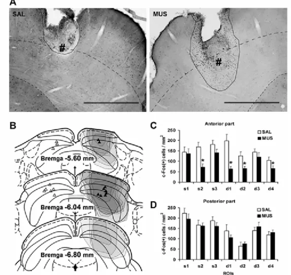
Figure I-4.
(A-B) The left coronal diagrams (adapted from Paxinos and Watson, 2004) show the region of
19
interests (ROIs) for counting c-Fos-positive cells at the anterior (A) and posterior (B) part of superior colliculus (SC). ROIs were selected from the superficial (s1-s3) and deep (d1-d4) layers of SC. The shaded region indicates deep layers of SC (dSC). The right figures show average densities of c-Fos-positive cells in each ROI of SC (s1-s3, d1-d4) after the cAV and iAV tasks at the anterior (A) and posterior (B) part of SC. Unpaired t-tests were performed for the comparison between the task groups, *P<0.05. (C) Representative photomicrographs of c-Fos expression after the cAV (left) and iAV (right) tasks at the anterior part of SC. Dashed lines indicate the borders of the deep layers. Scale bars, 1 mm
Fig. I-1
21
Fig. I-2
Fig. I-3
23
Fig. I-4
24
Chapter II:
Roles of early cortical areas and superior colliculus in
multisensory behavior
Results
1) Effect of dSC inactivation on the reaction time facilitation
To evaluate the contribution of SC to the audiovisual facilitation of reaction time, I injected muscimol into the anterior dSC, where c-Fos expression level specifically increased in the cAV task (Fig. I-4). Since the results in the bilateral inactivation were not consistent and the consequent behavior of the rats was chaotic, probably depending on the balance of muscimol diffusion between the two sides of SC (Kilpatrick et al., 1982), I injected muscimol into one side and saline into the other side to test the performance in the A-V-cAV task
lse alarm", withdrawal from the central hole before the onset of the target stimulus; "false hit", a nose poke into a hole other than the correct hole; and "miss", failure to respond to either hole within 2,500 ms from the onset of the target stimulus. The "false alarm" error did not differ in any conditions and any brain regions between those of the rats injected muscimol and saline (data not shown). The analysis of the error types revealed that the number of "false hits" generally increased in the hemifield contralateral to the muscimol-injection sides in comparison with those in the
25
hemifield ipsilateral to the injection sides (Fig. II-1B). In addition, the numbers of "miss"
essential for the audiovisual facilitation of the reaction time of nose withdrawal.
I examined c-Fos expression after the task performance in subsets of rats treated with muscimol and saline (3 to 4 rats for muscimol and 2 to 3 rats for saline in each injection group), because the use of c-Fos immunohistochemistry to estimate the suppressive effects of muscimol is established (Wang and Redgrave, 1997). The spread of muscimol around the injection sites was estimated by visually assessing the region exhibiting the loss of normal c-Fos staining in the sections. The placements of cannula tips in SC were verified to be within 1 mm of the targeted place of dSC. The inactivated area was primarily restricted to the anterior of the medial SC as verified by c-Fos expression (Fig. II-2A). The comparison of c-Fos expression level between the muscimol- and saline-injected hemispheres revealed that c-Fos expression level decreased statistically significantly in the anterior of the medial dSC as well as in the superficial layer of SC (Fig. II-2B,C). On the other hand, there was no significant decrease in the posterior dSC (Fig. II-2D).
2) Effect of V2L inactivation on the reaction time facilitation
Next, I examined the effect of bilateral injection of muscimol into V2L by the performance of the A-V-cAV task (Fig. II-3A). Because the behavioral consequence of V2L inactivation was unknown, I also injected muscimol into primary auditory (A1) and visual (V1) areas as controls for determining the proper amount of muscimol into cortex (Talwar et al., 2001; Gerstein et al., 2002; Smith et al., 2004). The inactivation of these cortical areas deteriorated the success rate and the reaction time
27
II-3B), but the reaction time for auditory stimuli was not significantly affected (P=0.211, paired t-test, Fig. II-3C).
The injection of the same amount of muscimol into V2L did not significantly affect the success rate for any modality stimuli (Fig. II-3B). In contrast, the reaction time for audiovisual stimuli was specifically increased by muscimol injection into V2L, whereas that for auditory or visual stimuli was not affected (Fig. II-3C). In the case of the saline injection, the cumulative probability of the reaction time in the audiovisual trials was shifted toward shorter-time points than that in the unimodal auditory or visual trials (Fig. II-3D, upper), as previously shown (Sakata et al., 2004), confirming the audiovisual facilitation of the reaction time in the A-V-cAV task. Bilateral muscimol injection into V2L caused a shift of the cumulative probability of the reaction time for audiovisual stimuli to similar probability distribution of faster unimodal responses (Fig. II-3D, lower). In the case of saline injection into V2L, the average FAV was 7.7%, which decreased to 0.4% following the muscimol injection into V2L (Fig. II-3D, P=0.018, Wilcoxon signed rank test). Bilateral muscimol injection into V1 also significantly decreased FAV (7.2 to 2.8%, P=0.028, Wilcoxon signed rank test). In contrast, the decrease of FAV after A1 inactivation was not large and did not reach significance (8.8 to 6.8% P=0.074, Wilcoxon signed rank test).
Injection of muscimol produced loss of c-Fos expression around cannula tips (Fig. II-4A). The center of the inactivation common to all rats was located within the target regions (Fig. II-4B for summary diagrams and Fig.
Patching and Quinlan, 2004). Indeed, the reaction time to unimodal stimuli was delayed when the intensities of the stimuli were lowered (Fig.II-5B). I named this type of response facilitation induced by the increased intensities of unimodal stimuli as FUNI (intensity-related unimodal facilitation), which is in contrast to audiovisual facilitation (FAV) (see Materials and Methods). In the A/V/cAV task, the amount of stimuli in the cAV trial, which received auditory plus visual stimuli, can be larger than those in the A and the V trials, which received only auditory and visual stimuli, respectively. Thus this raised a question as to whether the facilitation of reaction time is due to the convergence of cross modal stimuli or due to the increased intensity of two modal stimuli. I therefore examined the effect of muscimol injection into V2L on the intensity-related unimodal facilitation. In a session in which normal- or low-intensity sensory stimuli were randomly presented (Fig. II-5A), both audiovisual and intensity-related unimodal facilitations were observed (Fig. II-5B and C, left panel). The muscimol injection into V2L suppressed audiovisual facilitation (Fig. II-5C right panel) in comparison with the saline injection. On the other hand, the intensity-related unimodal facilitation was not affected by the muscimol injection into V2L (Fig. II-5B right panel). These results suggest that audiovisual and intensity-related unimodal facilitations are controlled by different neural mechanisms and that V2L specifically mediates the audiovisual facilitation. Histological analysis confirmed the suppressed region of c-Fos expression by muscimol was restricted in the vicinity of V2L (Fig. II-6A) and the c-Fos expression level at V2L in the muscimol-injected animals was significantly lower than that in the saline-injected animals (Fig. II-6B).
29
Discussion
1) Functional role of lateral secondary visual area (V2L)
Bilateral injections of muscimol into primary sensory cortical areas reduced the success rate and delayed the reaction time in a modality specific manner. Because errors made by rats after inactivation of primary sensory areas were mostly false-hit errors, rats randomly poked their noses regardless of the side of holes rather than idled when they could not discriminate the direction of the stimuli. Therefore, inactivation of primary cortical areas did not deteriorate their motor function or motivation. These sensory-specific effects confirmed that the amount of muscimol used was appropriate for examining the selective function of cortical areas.
The same amount of muscimol into the region including the medial region of V2L specifically suppressed the audiovisual facilitation of the reaction time without affecting the reaction time to unimodal stimuli, suggesting a specific role of V2L in multisensory behavioral facilitation. Lesion studies of the region including the medial part of V2L demonstrated the role of the area in visual complex pattern discrimination but not simple ones (McDaniel et al., 1982; Pinto Hamuy et al., 2004). Because pattern discrimination ability was not required for rats in my task, it is not surprising that rats can still respond to visual stimuli after inactivation of V2L in my experiments. The extent of muscimol spread which was determined on the basis of c-Fos expression was affected not only in the medial region of V2L but also in the lateral region: the potential association areas adjacent to A1 (including parietal association cortex (PtA), secondary auditory area, dorsal cortex (AuD) and temporal association cortex (TeA) in Paxinos and Watoson (1997)) (Fig. II-4B). Therefore, although it is possible that the suppression of bimodal-stimulus-specific responses is attributed to the dysfunction of these association areas, the injection of muscimol targeting A1, which significantly affected c-Fos expression in those areas (Fig. II-4B), did not affect the reaction time to bimodal stimuli (Fig. II-3C).
Taken together with the c-Fos mapping data, the medial region of V2L is a critical site for producing facilitated reaction time responding to bimodal stimuli.
2) Behavioral facilitation mediated by lateral secondary visual area (V2L)
It has been argued that the bimodal facilitation that shortens the reaction time for either modality stimuli does not necessarily require bimodal integration in the brain. For example, the facilitation of reaction time to audiovisual stimuli may be simply due to the increased probability of neuronal responses owing to redundant cues (i.e. auditory plus visual stimuli). This type of statistical facilitation can be expressed by the race model (e.g. Miller, 1982; Wenger and Townsend, 2000; Molholm et al., 2002; Patching and Quinlan, 2004), in which two modal sensory signals race along separatepathways toward a common site that generates a response, and the winning modality triggers the response. Using this model, the probabilities of facilitated responses to bimodal stimuli are predicted by a linear summation of unimodal response probabilities. Although the averaged cumulative probability of reaction time to bimodal stimuli was within the prediction of the model (data not shown), this does not necessarily mean that neural interactions between auditory and visual information did not occur. Indeed, we found that V2L inactivation specifically inhibited bimodal responses without affecting unimodal responses. This phenomenon cannot be explained by the race model, because it assumes that a bimodal response is produced by one of two unimodal stimuli. One could also argue that the facilitation was caused by a higher intensity of stimuli regardless of their cross-modality. This was not the case either, because the inactivation of the V2L region specifically suppressed enhanced responses induced by the combination of different modality stimuli but not by that of unimodal stimuli with a high degree of intensity. Taken together, our experiments suggest that the facilitation of reaction time is produced by multisensory interaction of auditory and visual stimuli in the brain and V2L plays a critical role in this process.
31
3) Anatomical substrates of multisensory facilitation of reaction time
As consistent with previous studies (e.g. Corneil et al., 2002; Hairston et al., 2006; Whitchurch and Takahashi, 2006), I confirmed that the spatial localization accuracy (measured as success rate) and response speed (measured as reaction time) did not correlate in the performance of A-V-cAV task, suggesting that these two performances are controlled by different neural mechanisms.
stimuli without affecting the facilitation of reaction time. These results suggest that auditory information and visual information are not integrated in dSC but the audiovisual integration is already conducted in regions upstream of dSC such as V2L and the convergent information in turn drive the activities in dSC to generate facilitated response for head withdrawal from the central hole, resulting in the facilitation of reaction time. To support this idea, c-Fos up-regulation was found in anterior dSC (Fig. I-4) and also in infragranular layers of V2L (Fig. I-3A), which projects to dSC (Thong and Dreher, 1986; Harvey and Worthington, 1990).
33
affected by the activities of different brain regions, it is highly likely that the audiovisual facilitation of these different aspects are also differently controlled by different brain regions. My data strongly suggest that the facilitation of reaction time is critically regulated by V2L, whereas SC is critically important for the facilitation of spatial accuracy to audiovisual stimuli as suggested by others (Burnett et al., 2004). In this study, I did not analyze the facilitation of spatial orientation accuracy, because I used relatively high intensity of stimuli, which were easily detected by rats. It is necessary to reveal the differences of neural substrates for the facilitations of different aspects of behavior.
5) Role of lower sensory area in multisensory facilitation
Accumulating evidence suggests multisensory integration in secondary sensory cortical areas in anesthetized rats (Di et al., 1994; Barth et al., 1995; Brett Green et al., 2003; Brett Green et al., 2004; Wallace et al., 2004). However, the behavioral importance of the integration in such early cortical areas was unclarified. Using my newly developed analytical technique for c-Fos mapping, I found that temporally congruent audiovisual stimuli specifically activated the medial region of V2L. Muscimol injection into the region including the medial region of V2L specifically blocked the facilitation of reaction time related to audiovisual integration. I, therefore, suggest that multisensory integration in the early visual area plays an important role on facilitation of reaction time to temporally congruent audiovisual stimuli. This idea is consistent with recent findings that multisensory interactions in the lower-order sensory cortex are characterized by a high degree of temporal precision (Ghazanfar et al., 2005; Lakatos et al., 2007).
One of the major questions concerning multisensory integration is the difference in multisensory processes between the higher- and lower-order sensory cortices (Ghazanfar and Schroeder, 2006). Our results suggest that certain lower-order cortical areas in rats specifically contribute to rapid responses to temporally coincident multisensory stimuli, whereas higher-order cortical areas such as the posterior parietal cortex (area AM in rats (Sanchez et al., 1997; Nakamura, 1999; Tees, 1999)) may
play roles in integrating spatial information on different modality stimuli. Although the role of higher-order cortical areas in our behavioral system remains to be studied, our findings provide evidence for a functional role of a rapid multisensory integration in the lower-order visual cortex.
35
Figures
Figure II-1. (A) The auditory
e
(SAL) and muscimol (MUS) into the deep layers of the superior colliculus (dSC). (B-D) The average frequencies of errors (false hit and miss, (B)), the average movement time (C) and the average reaction time (D) after unilateral injections of muscimol into one side and that of saline into the other side. Paired t-test was performed for the hemisphere ipsilateral and contralateral to the muscimol injection side. (E) Cumulative probabilities for each type of stimulus are presented for the hemifields ipsilateral (left) and contralateral (right) to the muscimol-injection side. The inset in the right figure indicates the audiovisual facilitation index (FAV) for the hemifields ipsilateral and contralateral to the muscimol-injection side. Wilcoxon signed rank test was performed for the responses in the ipsilateral and contralateral hemifields. N.S. = not significant.
Figure II-2.
(A) Representative photomicrographs of the superior colliculus (SC) of rats that performed the auditory, visual and congruent audiovisual (A-V-cAV) task after injecting saline (left) and muscimol (right). # indicates the location of injection tip. Black lines cover the regions where c-Fos expression in grial cells was observed. Black dotted lines indicate the border of deep layers of the superior colliculus (dSC). Scale bars, 1 mm. (B) Coronal diagrams (adapted from Paxinos and Watson, 1997) showing location of injection sites and spread (shown by shaded areas) of muscimol within SC. The injection sites are indicated for the saline-injected (left) and muscimol-injected (right) hemispheres. The extent of c-Fos expression inactivation was identified only in the hemisphere injected with
muscimol. Different densities of shading indicate the degree of suppression of c-Fos expression in regions of individual animals. (C-D) The average densities of c-Fos-positive cells in each ROI (s1-d4, Fig. I-4A) of the anterior (C) and posterior (D) SC of each hemisphere during the performance of the A-V-cAV task after unilateral injections of muscimol and saline into dSC (n=9). Paired t-tests were conducted for saline- and muscimol-injected hemispheres. *P<0.05.
Figure II-3. (A) The auditory
37
muscimol for each rat in V2L, primary visual area (V1), and primary auditory area (A1). The regions showing decreased c-Fos expression by muscimol injection are indicated by different densities of shading in individual animals. (C) The bars show the average densities of c-Fos-positive cells in layer 4 of V1, V2L and A1 induced by the performance of the A-V-cAV task after bilateral injections of muscimol (black boxes) or saline (white boxes) into the regions indicated above. Unpaired t-tests were performed for saline and muscimol injection conditions in each area. *P<0.05.
Figure II-5.
(A) Six kinds of cue stimuli (the normal- or low-intensity auditory, visual and congruent audiovisual stimuli) were randomly presented in a session as cue stimuli (N/L A-V-cAV task) to test the effect of the bilateral injections of saline (SAL) and muscimol (MUS) into the lateral secondary visual area (V2L). (B) Reaction times of average unimodal responses under normal- and low-intensity stimulus conditions were compared (Hatched areas show intensity-related unimodal facilitation index (FUNI), see Materials and Methods). The inset in the right figure indicates the FUNI for each experimental animal. (B) Reaction times for audiovisual and faster unimodal responses were compared (Shadowed areas show audiovisual facilitation index (FAV)). "Faster (A,V)" indicates maximum unisensory probability at each time point. The insets in the right figure indicate the FAV for each experimental animal. Wilcoxon signed rank test was performed between the saline and muscimol injection sessions.
*P<0.05.
Figure II-6.
(A) Coronal diagrams show the location of the injection sites and the diffusion (shaded areas) of muscimol for each rat in the lateral secondary visual are (V2L). The regions showing decreased c-Fos expression by muscimol injection are indicated by different densities of shading in individual animals. (B) The bars show the average densities of c-Fos-positive cells in layer 4 of V1, V2L and A1 induced
by the performance of the A-V-cAV task after bilateral injections of muscimol (black boxes) or saline (white boxes) into the regions indicated above. Unpaired t-tests were performed for saline and muscimol injection conditions in each area. *P<0.05.
39
Fig. II-1
Fig. II-2
41
Fig. II-3
Fig. II-4
43
Fig. II-5
Fig. II-6
45
Materials and Methods
Animal
a 12-h
n
s. All experiments were conducted in accordance with the Guide for the Care and Use of Laboratory Animals (NIH publication number 86-23, 1985) and the guidelines of Okazaki National Research Institutes in Japan.
3
501 K; Ohara & Co., Ltd., Tokyo, Japan; 320 × 160 × 450 mm), which was enclosed in a soundproof box (Japan Shield Enclosure, Osaka, Japan) and illuminated by an 8-V direct-current bulb as the house light. The apparatus used in behavioral testing was identical to that used in the previous study (Sakata et al., 2004). Briefly, the apparatus consists of three holes for nose
i
rect current bulb placed immediately in front of a loudspeaker so that both visual and auditory stimuli were presented from the same location. The position of each animal's head was controlled such that the animal was able to receive the stimuli at equal distances from the left and right sides when it poked its nose into the central hole. Two intensities of stimuli ("normal" and "low") were used in this study, which were obtained by manipulating applied voltage. Because I focus on the audiovisual facilitation of reaction time rather than two-alternative choice in this study, I used the stimuli that were easily discriminated by rats. The normal-intensity stimuli were set to 11.2 lx for luminescence and 57 dB SPL for sound (broadband noise). The intensities were chosen as high-above-threshold intensity
stimuli with which the rats performed the auditory, visual
47
between auditory and visual stimuli. When the rats reached a success rate higher than 75% after more than three training sessions, final test sessions were performed. On testing, all the animals were removed from their home cages with minimal handling and transferred to the task apparatus. About one hour after the beginning of the final test session, rats were immediately perfused for c-Fos immunohistochemistry. The success rate and average reaction time (excluding those in incorrect trials) in the last session were averaged for each group.
Suppression experiments
The rats were trained to perform the A-V-cAV task (Fig. II-1-4) or the normal- or low-intensity auditory, visual, congruent audiovisual (N/L A-V-cAV) task (Fig. II-5). After the animals had mastered each task, they were chronically implanted with cannulae for muscimol application. Each rat performed two to three test sessions under both saline and muscimol injection conditions. After the final test session, rats were sacrificed for c-Fos immunohistochemistry. Behavioral performance was separately analyzed depending on the injection condition, modality and stimulus intensity. The cumulative probability of reaction time was computed for each condition of each rat, and then averaged across rats under the same condition in the same group. The index of audiovisual facilitation of reaction time
The rats were deeply anesthetized with sodium pentobarbital (50 mg/kg body weight) and transcardially perfused with 0.9% saline followed by 4% paraformaldehyde. Every fifth coronal section
49
the major and minor axes of an equivalent ellipse, which was fitted to each signal by a least-squares fitting technique using a customized software program designed by LabVIEW. Parameters for the identification of c-Fos-positive nuclei were set such that the automated counts became close to the careful manual counts of several representative sections.
Standardization of images of cortex
The sections that contained artifacts such as tearing or bubbles in the cortex were excluded from the analysis. The entire cortical section was automatically reconstructed from four to eight images (Cecchi et al., 1999). To construct a local density map of c-Fos-positive cells, local c-Fos densities were calculated and mapped using a moving window operator (100 × 100 Pm), which scanned pixelwise through an entire image (see Fig. I-2A, B). Image processing was automatically carried out using a customized software program designed by LabVIEW except for identifying the structural landmarks and cutting out image of a large part of the cortex using Adobe Photoshop. Visualizations of standardized maps were carried out using Matlab 7.0 (Mathworks). The standardized cortical section (100 × 1000 pixels, depth × width, respectively) was assigned to one of 16 figures (from Figures 35 to 50; Bregma -4.3 to -8.0 mm) of the standard rat brain atlas (Paxinos and Watson, 1997), as determined from the order of serial sections and the shape of the hippocampus. The averages of 11.5 r1.58 (mean rs.d.) sections stained by the anti-c-Fos antibody in each animal were used for this analysis. When
there were no available sections that corresponded to one of the 16 figures, those data were generated as the standardized cortical sections by linearly interpolating adjacent serial sections. Post hoc smoothing (spatial averaging) of standardized layer map (1000 × 247 pixels) (width × Bregma distance) was achieved using a moving window operator (20 × 20 pixels). To correct intersubject differences in staining intensity in c-Fos immunohistochemistry, the densities of c-Fos-positive cells in each animal were normalized by linearly adjusting mean - 2 s.d. as 0 % and mean + 2 s.d. as 100 (%).
essentially the same results (data not shown).
51
(Bregma 4.8 to 6.8 mm) (Palomero-Gallagher and Zilles, 2004). Because I found that the decrease in c-Fos expression level after muscimol injection was particularly evident in layer 4, c-Fos-positive cell density (cells /mm2) in layer 4 was automatically quantified using the identical quantification criteria described above. Minimum of five sections from both hemispheres of each rat were pooled and group-averaged for a given structure for each injection condition. The implantation of cannulae often caused dense c-Fos labeling only around the injector track (see Fig. II-2,4), which may be due to the activation of glial growth (Wang and Redgrave, 1997). When I took ROIs for c-Fos quantification in a section that contained cannula tracks, I avoided those artifacts that were easily discriminated from neurons on the basis of the intensity, location, and morphology of the signal. Some sections were excluded from the quantitative analysis because the artifacts extended into entire target regions.
Surgery and cannulation
The rats (n=35) were anesthetized with sodium pentobarbital (50 mg/kg body weights). I targeted V2L, where an increase in c-Fos expression level was observed after the cAV task, for the inactivation with muscimol. Stainless steel guide cannulae (27 gauge, 0.45 mm o.d.) were bilaterally implanted into one of the three brain regions using the following coordinates: (i) V2L (n=7 for Exp. 2 and n=7 for Exp. 3, respectively), 5.8 mm caudal to Bregma, 6.0 mm lateral to the midline, and 0.6 mm ventral from the dural surface; (ii) A1 (n=6), 5.6 mm caudal to Bregma, 6.7 mm lateral to the midline, and 1.4 mm ventral from the dural surface; (iii) V1 (n=6), 7.3 mm caudal to Bregma, 3.3-3.7 mm lateral to the midline, and 0.6 mm ventral from the dural surface; (iv) anterior dSC (intermediate/deep layer of superior colliculus, n=9), 5.8 mm caudal to Bregma, 1.4 mm lateral to the midline, and 3.2 mm ventral to the dural surface. Note that I set the target region of V1 away fromV2L
located within targeted structures were included in the study.
Muscimol microinfusion
I analyzed the effect of reversible inactivation of brain sites on the performance of behavioral tasks using muscimol, which transiently suppresses the activity of excitatory neurons by activating GABA A receptors on the surface of neurons
53
54
Acknowledgements
This thesis was supported by many people.
I greatly thank Prof. Yamamori in the National Institute for Basic Biology (NIBB) for providing the chance and environment to investigate exciting research. I thank Dr. Sakata in the Rutgers University for his general and technical advices. I thank Dr. Bosch in brain institute at MIT for his helpful advices and technical contribution for muscimol experiment. I thank Prof. Sakurai in Kyoto University for encouragement and technical suggestions. I thank Toru Nakamura for valuable help with initiating the muscimol experiment and Drs. Yasunori Hayashi and Akiya Watakabe for helpful comments on early
drafts of the manuscript. their
wisdom and technical supports and advices. And I greatly appreciate my parents, grandparents and brothers for their financial and mental supports all through my life.
Finally, I am grateful and pray for the sacrifice of many experimental animals.
55
References
Angelucci A, Bullier J (2003) Reaching beyond the classical receptive field of V1 neurons: horizontal or feedback axons? J Physiol Paris 97:141-154.
Barth DS, Goldberg N, Brett B, Di S (1995) The spatiotemporal organization of auditory, visual, and auditory-visual evoked potentials in rat cortex. Brain Res 678:177-190.
Bell AH, Meredith MA, Van Opstal AJ, Munoz DP (2005) Crossmodal integration in the primate superior colliculus underlying the preparation and initiation of saccadic eye movements. J Neurophysiol 93:3659-3673.
Berninger B, Guillemot F, Gotz M (2007) Directing neurotransmitter identity of neurones derived from expanded adult neural stem cells. Eur J Neurosci 25:2581-2590.
Bolognini N, Frassinetti F, Serino A, Ladavas E (2005) "Acoustical vision" of below threshold stimuli: interaction among spatially converging audiovisual inputs. Exp Brain Res 160:273-282. Brett Green B, Fifkova E, Larue DT, Winer JA, Barth DS (2003) A multisensory zone in rat
parietotemporal cortex: intra- and extracellular physiology and thalamocortical connections. J Comp Neurol 460:223-237.
Brett Green B, Paulsen M, Staba RJ, Fifkova E, Barth DS (2004) Two distinct regions of secondary somatosensory cortex in the rat: topographical organization and multisensory responses. J Neurophysiol 91:1327-1336.
Bronchti G, Heil P, Sadka R, Hess A, Scheich H, Wollberg Z (2002) Auditory activation of "visual" cortical areas in the blind mole rat (Spalax ehrenbergi). Eur J Neurosci 16:311-329.
Brosch M, Selezneva E, Scheich H (2005) Nonauditory events of a behavioral procedure activate auditory cortex of highly trained monkeys. J Neurosci 25:6797-6806.
Burnett LR, Stein BE, Chaponis D, Wallace MT (2004) Superior colliculus lesions preferentially disrupt multisensory orientation. Neuroscience 124:535-547.
Bushnell PJ, Benignus VA, Case MW (2003) Signal detection behavior in humans and rats: a comparison with matched tasks. Behav Processes 64:121-129.
Calvert GA, Bullmore ET, Brammer MJ, Campbell R, Williams SC, McGuire PK, Woodruff PW, Iversen SD, David AS (1997) Activation of auditory cortex during silent lipreading. Science 276:593-596.
Campi KL, Karlen SJ, Bales KL, Krubitzer L (2007) Organization of sensory neocortex in prairie voles (Microtus ochrogaster). J Comp Neurol 502:414-426.
Cecchi GA, Ribeiro S, Mello CV, Magnasco MO (1999) An automated system for the mapping and quantitative analysis of immunocytochemistry of an inducible nuclear protein. J Neurosci Methods 87:147-158.
Coogan TA, Burkhalter A (1993) Hierarchical organization of areas in rat visual cortex. J Neurosci 13:3749-3772.
Corneil BD, Van Wanrooij M, Munoz DP, Van Opstal AJ (2002) Auditory-visual interactions
subserving goal-directed saccades in a complex scene. J Neurophysiol 88:438-454.
Dean P, Redgrave P (1984) The superior colliculus and visual neglect in rat and hamster. II. Possible mechanisms. Brain Res 320:143-153.
Dean P, Mitchell IJ, Redgrave P (1988) Responses resembling defensive behaviour produced by microinjection of glutamate into superior colliculus of rats. Neuroscience 24:501-510.
Dean P, Mitchell IJ, Redgrave P (1988) Contralateral head movements produced by microinjection of glutamate into superior colliculus of rats: evidence for mediation by multiple output pathways. Neuroscience 24:491-500.
Di S, Brett B, Barth DS (1994) Polysensory evoked potentials in rat parietotemporal cortex: combined auditory and somatosensory responses. Brain Res 642:267-280.
Espinoza SG, Thomas HC (1983) Retinotopic organization of striate and extrastriate visual cortex in the hooded rat. Brain Res 272:137-144.
Farivar R, Zangenehpour S, Chaudhuri A (2004) Cellular-resolution activity mapping of the brain using immediate-early gene expression. Front Biosci 9:104-109.
Felleman DJ, Van Essen DC (1991) Distributed hierarchical processing in the primate cerebral cortex. Cereb Cortex 1:1-47.
Frens MA, Van Opstal AJ (1995) A quantitative study of auditory-evoked saccadic eye movements in two dimensions. Exp Brain Res 107:103-117.
Gerstein GL, Kirkland KL, Musial PG, Talwar SK (2002) Recordings, behaviour and models related to corticothalamic feedback. Philos Trans R Soc Lond B Biol Sci 357:1835-1841.
Ghazanfar AA, Maier JX, Hoffman KL, Logothetis NK (2005) Multisensory integration of dynamic faces and voices in rhesus monkey auditory cortex. J Neurosci 25:5004-5012.
Ghazanfar AA, Schroeder CE (2006) Is neocortex essentially multisensory? Trends Cogn Sci. 10:278-285.
Giard MH, Peronnet F (1999) Auditory-visual integration during multimodal object recognition in humans: a behavioral and electrophysiological study. J Cogn Neurosci 11:473-490.
Goodale MA, Murison RC (1975) The effects of lesions of the superior colliculus on locomotor orientation and the orienting reflex in the rat. Brain Res 88:243-261.
Hairston WD, Hodges DA, Burdette JH, Wallace MT (2006) Auditory enhancement of visual temporal order judgment. Neuroreport 17:791-795.
Harvey AR, Worthington DR (1990) The projection from different visual cortical areas to the rat superior colliculus. J Comp Neurol 298:281-292.
Holmes NP, Spence C (2005) Multisensory integration: space, time and superadditivity. Curr Biol 15:R762-764.
Hughes HC, Reuter Lorenz PA, Nozawa G, Fendrich R (1994) Visual-auditory interactions in sensorimotor processing: saccades versus manual responses. J Exp Psychol Hum Percept Perform 20:131-153.
Jiang W, Jiang H, Stein BE (2002) Two corticotectal areas facilitate multisensory orientation behavior. J Cogn Neurosci 14:1240-1255.
57
Johnson RR, Burkhalter A (1996) Microcircuitry of forward and feedback connections within rat visual cortex. J Comp Neurol 368:383-398.
Johnson RR, Burkhalter A (1997) A polysynaptic feedback circuit in rat visual cortex. J Neurosci 17:7129-7140.
Kayser C, Petkov CI, Augath M, Logothetis NK (2005) Integration of touch and sound in auditory cortex. Neuron 48:373-384.
Kayser C, Petkov CI, Augath M, Logothetis NK (2007) Functional imaging reveals visual modulation of specific fields in auditory cortex. J Neurosci 27:1824-1835.
Kilpatrick IC, Collingridge GL, Starr MS (1982) Evidence for the participation of nigrotectal gamma-aminobutyrate-containing neurones in striatal and nigral-derived circling in the rat. Neuroscience 7:207-222.
Kim J, Ragozzino ME (2005) The involvement of the orbitofrontal cortex in learning under changing task contingencies. Neurobiol Learn Mem 83:125-133.
King AJ, Palmer AR (1985) Integration of visual and auditory information in bimodal neurones in the guinea-pig superior colliculus. Exp Brain Res 60:492-500.
Koch M, Schnitzler HU (1997) The acoustic startle response in rats--circuits mediating evocation, inhibition and potentiation. Behav Brain Res 89:35-49.
Komura Y, Tamura R, Uwano T, Nishijo H, Ono T (2005) Auditory thalamus integrates visual inputs into behavioral gains. Nat Neurosci 8:1203-1209.
Lakatos P, Chen CM, O'connell MN, Mills A, Schroeder CE (2007) Neuronal oscillations and multisensory interaction in primary auditory cortex. Neuron 53:279-292.
Lein ES, Hawrylycz MJ, Ao N, Ayres M, Bensinger A, Bernard A, Boe AF, Boguski MS, Brockway KS, Byrnes EJ, Chen L, Chen L, Chen TM, Chi Chin M, Chong J, Crook BE, Czaplinska A, Dang CN, Datta S, Dee NR et al. (2006) Genome-wide atlas of gene expression in the adult mouse brain. Nature 445:168-176.
Leitner DS, Cohen ME (1985) Role of the inferior colliculus in the inhibition of acoustic startle in the rat. Physiol Behav 34:65-70.
Macaluso E, Frith CD, Driver J (2000) Modulation of human visual cortex by crossmodal spatial attention. Science 289:1206-1208.
Majchrzak M, Di Scala G (2000) GABA and muscimol as reversible inactivation tools in learning and memory. Neural Plast 7:19-29.
Martin JH, Ghez C (1999) Pharmacological inactivation in the analysis of the central control of movement. J Neurosci Methods 86:145-159.
McDaniel WF, Coleman J, Lindsay JF Jr (1982) A comparison of lateral peristriate and striate neocortical ablations in the rat. Behav Brain Res 6:249-272.
Meredith MA, Stein BE (1985) Descending efferents from the superior colliculus relay integrated multisensory information. Science 227:657-659.
Meredith MA, Stein BE (1986) Visual, auditory, and somatosensory convergence on cells in superior colliculus results in multisensory integration. J Neurophysiol 56:640-662.
Meredith MA, Nemitz JW, Stein BE (1987) Determinants of multisensory integration in superior colliculus neurons. I. Temporal factors. J Neurosci 7:3215-3229.
Meredith MA, Stein BE (1996) Spatial determinants of multisensory integration in cat superior colliculus neurons. J Neurophysiol 75:1843-1857.
Mermelstein PG, Bito H, Deisseroth K, Tsien RW (2000) Critical dependence of cAMP response element-binding protein phosphorylation on L-type calcium channels supports a selective response to EPSPs in preference to action potentials. J Neurosci 20:266-273.
Miller J (1982) Divided attention: evidence for coactivation with redundant signals. Cognit Psychol 14:247-279.
Molholm S, Ritter W, Murray MM, Javitt DC, Schroeder CE, Foxe JJ (2002) Multisensory
auditory-visual interactions during early sensory processing in humans: a high-density electrical mapping study. Brain Res Cogn Brain Res 14:115-128.
Montero VM (1993) Retinotopy of cortical connections between the striate cortex and extrastriate visual areas in the rat. Exp Brain Res 94:1-15.
Murray MM, Foxe JJ, Higgins BA, Javitt DC, Schroeder CE (2001) Visuo-spatial neural response interactions in early cortical processing during a simple reaction time task: a high-density electrical mapping study. Neuropsychologia. 39:828-844.
Murray MM, Molholm S, Michel CM, Heslenfeld DJ, Ritter W, Javitt DC, Schroeder CE, Foxe JJ (2005) Grabbing your ear: rapid auditory-somatosensory multisensory interactions in low-level sensory cortices are not constrained by stimulus alignment. Cereb Cortex 15:963-974.
Nakamura K (1999) Auditory spatial discriminatory and mnemonic neurons in rat posterior parietal cortex. J Neurophysiol 82:2503-2517.
Nguyen PT, Holschneider DP, Maarek JM, Yang J, Mandelkern MA (2004) Statistical parametric mapping applied to an autoradiographic study of cerebral activation during treadmill walking in rats. Neuroimage 23:252-259.
Olavarria J, Van Sluyters RC (1982) The projection from striate and extrastriate cortical areas to the superior colliculus in the rat. Brain Res. 242:332-336.
Olavarria J, Montero VM (1984) Relation of callosal and striate-extrastriate cortical connections in the rat: morphological definition of extrastriate visual areas. Exp Brain Res 54:240-252.
Palomero-Gallagher, N., Zilles, K. (2004) The rat isocortex. In: Paxinos, G.: The Rat Nervous System. Academic Press, San Diego pp :729-757.
Patching GR, Quinlan PT (2004) Cross-modal integration of simple auditory and visual events. Percept Psychophys 66:131-140.
Paxinos G, Watson C (1997) The Rat Brain in Stereotaxic Coordinates (3rd ed.). Academic Press, :Sydney.
Peck CK (1996) Visual-auditory integration in cat superior colliculus: implications for neuronal control of the orienting response. Prog Brain Res 112:167-177.
Piche M, Robert S, Miceli D, Bronchti G (2004) Environmental enrichment enhances auditory takeover of the occipital cortex in anophthalmic mice. Eur J Neurosci 20:3463-3472.
59
Pinto Hamuy T, Montero VM, Torrealba F (2004) Neurotoxic lesion of anteromedial/posterior parietal cortex disrupts spatial maze memory in blind rats. Behav Brain Res 153:465-470.
Populin LC, Yin TC (2002) Bimodal interactions in the superior colliculus of the behaving cat. J Neurosci 22:2826-2834.
Populin LC (2005) Anesthetics change the excitation/inhibition balance that governs sensory processing in the cat superior colliculus. J Neurosci 25:5903-5914.
Redgrave P, Dean P, Souki W, Lewis G (1981) Gnawing and changes in reactivity produced by microinjections of picrotoxin into the superior colliculus of rats. Psychopharmacology (Berl) 75:198-203.
Rutkowski RG, Weinberger NM (2005) Encoding of learned importance of sound by magnitude of representational area in primary auditory cortex. Proc Natl Acad Sci USA. 102:13664-13669. Sahibzada N, Dean P, Redgrave P (1986) Movements resembling orientation or avoidance elicited by
electrical stimulation of the superior colliculus in rats. J Neurosci 6:723-733. Sakata S, Kitsukawa T, Kaneko T, Yamamori T, Sakurai Y (2002) Task-dependent and
cell-type-specific Fos enhancement in rat sensory cortices during audio-visual discrimination. Eur J Neurosci 15:735-743.
Sakata S, Yamamori T, Sakurai Y (2004) Behavioral studies of auditory-visual spatial recognition and integration in rats. Exp Brain Res 159:409-417.
Sakowitz OW, Quian Quiroga R, Schurmann M, Basar E (2005) Spatio-temporal frequency characteristics of intersensory components in audiovisually evoked potentials. Brain Res Cogn Brain Res 23:316-326.
Sanchez RF, Montero VM, Espinoza SG, Diaz E, Canitrot M, Pinto Hamuy T (1997) Visuospatial discrimination deficit in rats after ibotenate lesions in anteromedial visual cortex. Physiol Behav 62:989-994.
Sanderson KJ, Dreher B, Gayer N (1991) Prosencephalic connections of striate and extrastriate areas of rat visual cortex. Exp Brain Res 85:324-334.
Schleicher A, Zilles K (1990) A quantitative approach to cytoarchitectonics: analysis of structural inhomogeneities in nervous tissue using an image analyser. J Microsc 157:367-381.
Schroeder CE, Foxe JJ (2002) The timing and laminar profile of converging inputs to multisensory areas of the macaque neocortex. Brain Res Cogn Brain Res 14:187-198.
Schroger E, Widmann A (1998) Speeded responses to audiovisual signal changes result from bimodal integration. Psychophysiology 35:755-759.
Smith AL, Parsons CH, Lanyon RG, Bizley JK, Akerman CJ, Baker GE, Dempster AC, Thompson ID, King AJ (2004) An investigation of the role of auditory cortex in sound localization using
muscimol-releasing Elvax. Eur J Neurosci 19:3059-3072.
Stein BE, Huneycutt WS, Meredith MA (1988) Neurons and behavior: the same rules of multisensory integration apply. Brain Res 448:355-358.
Stein BE., Meredith MA., Huneycutt WS. & McDade L. (1989) Behavioral indices of multisensory integration: orientation to visual cues is affected by auditory stimuli. J. Cogn. Neurosci., 1:12-24.