The mechanism of oxygen sensing and signal transduction in the
heme-based oxygen sensor protein HemAT from Bacillus subtilis
Hideaki Yoshimura
2006
Contents
Contents ... 2
Chapter 1: General Introduction ... 5
Introduction ...5
Heme-based CO and NO sensor proteins ...6
CO sensor protein CooA...8
NO sensor protein sGC ...10
Heme-based O2 sensor proteins ... 11
O2 senso p otein FixLr r o s s ...12
The heme-based aerotactic signal transducer HemAT ...13
Chemotaxis signal transduction system in bacteria ...18
The purpose of this work...22
REFERENCES ...22
Chapter 2: Specific hydrogen-bonding networks responsible for selective O2 sensing of HemAT-Bs ... 25
Abstract...25
Introduction ...26
Experimental Procedure ...29
Protein expression and purification ...29
Sample preparation for measurements...30
Spectrosc pic methods ...30
Results...32
Absorption spectra of the CO- and NO-bound HemAT-Bs ...32
EPR spectra of the NO-bound HemAT-B ...33
Resonance Raman spectroscopy for the CO-bound HemAT-Bs...34
Resonance Raman spectroscopy for the NO-bound HemAT-B ...37
Discussion ...43
REFERENCES ...46
Chapter 3: Signal transduction through the proximal heme pocket in HemAT-Bs upon ligand binding revealed by time-resolved resonance Raman spectroscopy ... 49
Abstract...49
Introduction ...50
Experimental Procedure ...53
Protein expression and purification ...53
Time-resolv d res nance Raman spectra measuremente o o s ...54
Results...55
The time-resolved resonance Raman spectra in the low-frequency region ...55
The νFe—His band of WT HemAT-Bs ...56
The νFe—His band of Y133F HemAT-Bs ...58
The νFe—His band of the sensor d main of WT HemAT-B ...58
Discussion ...60
REFERENCES ...66
Chapter 4: Summary and Future Perspectives ... 69
List of Publications... 72
Acknowledgements... 74
Chapter 1: General Introduction
Introduction
All of organisms and cells must sense and respond to various stimuli from their outer environment in order to survive and maintain their homeostasis. There are a wide variety of the stimuli, such as nutrients, harmful matters, pH, light, temperature, pressure, etc. Organisms and cells recognize these stimuli, and display some responses and/or adaptations for respective stimulus. To accept the respective stimulus, organisms and cells have various receptor (sensor) proteins.
In general, receptor proteins consist of several domains including a sensor domain and a functioning domain. The sensor domain senses the corresponding effector molecule or the external signal exclusively. The selectivity in the binding of the effector is achieved by the formation of multiple and specific interactions between the receptor protein and the effector molecules. When a receptor protein binds its cognate effector, a conformational alteration of the protein is induced by the interaction with the effector. As the result of transmission of the conformational alteration from the sensor domain to the functioning domain, the functioning domain expresses a respective role, such as enzymatic activity, transcriptional regulation, and controlling the activity of another enzyme.
In the case that the effector molecule is extremely small, the receptor protein must take a special strategy to recognize and discriminate the small effector molecule. It is difficult to form multiple and specific interactions between the effector and the receptor protein. Thus, the discrimination mechanism of small molecules by the cognate receptor protein is especially interesting subject to investigate.
Another interesting subject in the investigation on receptor proteins is the mechanism of signal transduction from the effector binding site to the functioning domain. Although there are a variety of structures in the sensor domain and the functioning domain depending on the effector molecules and the functions, the intramolecular signal transduction from the sensor domain to the
functioning domains is a crucial process for all receptor proteins to express their functions. Therefore, elucidating this signal transduction mechanism is very important to study the structure and function relationships of the receptor proteins.
In this work, the author chose an O2 sensor protein HemAT from Bacillus subtilis to study the molecular mechanism of O2 sensing and signal transduction. HemAT is a member of heme-based sensor proteins where a heme acts as the active site for sensing the effector molecule. In addition, HemAT functions as the aerotactic signal transducer, that is, HemAT controls the chemotaxis of B. subtilis toward O2.
This thesis consists of four chapters. In Chapter 1, the general introduction is described. This chapter contains the background of this work and some properties of HemAT previously reported. In Chapter 2, the author describes the molecular mechanism of O2 sensing by HemAT-Bs revealed by mutagenesis and spectroscopic studies including resonance Raman, EPR, and UV-Vis spectroscopies. In Chapter 3, the author describes the signal transduction mechanism of HemAT-Bs revealed by time-resolved resonance Raman spectroscopy. Finally, the summary of this work and the future prospects are presented in Chapter 4.
Heme-based CO and NO sensor proteins
While gas molecules are known to be the substrates and/or the products of several enzymatic reactions, a new physiological function of gas molecules have been found recently, in which some diatomic gas molecules function as physiological effector molecules to regulate various biological functions in many organisms. Oxygen (O2), carbon monoxide (CO), and nitric oxide (NO) are the typical gas molecules functioning as signaling molecules.
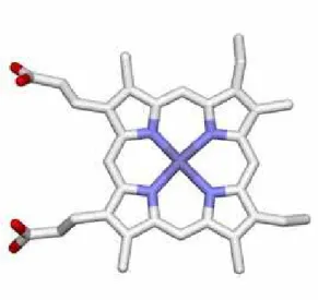
O2, CO, and NO cannot be bound to protein directly. The sensor proteins for these gas molecules must therefore have a cofactor that functions as the binding site of these gas molecules. Many gas sensor proteins utilize a heme as the binding site of the effector gas molecule. Typical
heme-based gas sensor proteins are summarized in Table 1. Heme is a complex of an iron ion and a protoporphyrin (Figure 1). In most of heme proteins, one or two amino acids are coordinated to the heme iron as the axial ligands. The axial ligand tethers the heme to the protein matrix. In addition to this coordination, the high hydrophobicity of the porphyrin plane helps the heme to stay in the protein matrix stably. The cytoplasm in a cell is usually in reducing conditions, which usually keeps the heme iron in heme proteins in the Fe(II) state (ferrous state). The Fe(II) heme (ferrous state) can bind O2, CO and NO reversibly. These properties of a heme are convenient for gas sensor proteins to utilize a heme as the binding site of the gas molecule. On the other hand, these gas molecules are difficult to form multiple and specific interactions with the protein matrix of the corresponding receptor proteins because of their small sizes. Moreover, a ferrous heme binds all of O2, CO, and NO equally. These features of a heme make the selective molecular recognition difficult. As described above, a sensor protein must discriminate its cognate effector molecule with high specificity. If this discrimination is absent, the receptor protein will lose its functions and/or be out of control. To avoid the disruption of selectivity, heme-based gas sensor proteins adopt a unique mechanism for selective gas sensing, which will be discussed in the following sections.
Figure 1. The molecular structure of heme.
CO sensor protein CooA
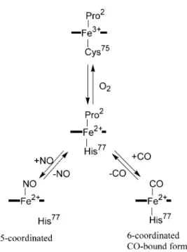
CooA is a typical heme-based CO sensor protein that functions as a transcriptional regulator in CO metabolizing bacteria. CooA activates the expression of the proteins required for the CO metabolism including CO dehydrogenase and hydrogenease in the presence of CO under anaerobic condition.1 Only CO-bound CooA can bind to the target DNA to act as a transcriptional activator, but the ferric and ferrous CooA cannot. A conformational change is induced by the CO binding to the heme in CooA, which is the trigger of the activation of CooA by CO. The mechanism of CO sensing has been extensively studied for CooA from Rhodospirillum rubrum (Rr-CooA) and Carboxydothermus hydrogenoformans (Ch-CooA).
Rr-CooA and Ch-CooA contain a b-type heme (protoheme) as the active site for sensing CO. The heme in Rr-CooA has two axial ligands, (Pro2 and Cys75) and (Pro2 and His77) for the ferric and ferrous hemes, respectively. Pro2 is the N-terminal residue because Met1 is removed by post-translational modification. When ferrous CooA reacts with CO, Pro2 is replaced by CO to form CO-bound CooA.
Ch-CooA also has a 6-coordinated heme in the ferric, ferrous, and CO-bound forms, but its coordination structure is slightly different from that of Rr-CooA. While the ligand exchange takes place between Cys75 and His77 upon the change in the oxidation state of the heme in Rr-CooA, such a ligand exchange does not occur in Ch-CooA. In Ch-CooA, the N-terminal α-amino group is coordinated to the ferric and ferrous hemes as the distal 6th ligand. The α-amino group coordinated to the ferrous heme is replaced by CO upon the CO binding to the heme in Ch-CooA.
Although the N-terminal amino acid residue is not conserved between Rr-CooA and Ch-CooA, the coordination of the N-terminus, the N-terminal Pro and α-amino group in Rr-CooA and Ch-CooA, respectively, is conserved between them. The more important property conserved between Rr-CooA and Ch-CooA is that the N-terminus coordinated to the ferrous heme is replaced by CO upon the CO binding. The dissociation of the N-terminus from the heme takes place upon the CO binding. The dissociation of the N-terminus from the heme upon the CO binding will induce a conformational change around the heme, which is a trigger of the activation of CooA by CO.
When ferrous CooA reacts with O2, a rapid autoxidation proceeds to form the ferric form without the formation of a stable O2-bound form. As described above, ferric CooA is an inactive form disable to binding to the target DNA. Therefore, O2 cannot activate CooA.
NO also cannot activate Rr-CooA, though NO can bind to the heme in Rr-CooA. When Rr-CooA reacts with NO, a 5-coordinated nitrosyl heme is formed, where all of the endogenous axial ligands are dissociated from the heme. The dissociation of the proximal His upon the NO binding will result in the different conformation of NO-bound Rr-CooA compared with CO-bound CooA, because the proximal His is retained in CO-bound CooA. In the case of Ch-CooA, the dissociation of proximal His does not occur upon NO binding. As the result, Ch-CooA exhibits some activity upon the NO binding. Thus, the dissociation of proximal His in Rr-CooA is essential for the discrimination between CO and NO.
Figure 2. The coordination structures of the heme in Rr-CooA. Pro2 is the N-terminal residue because Met1 is removed by post-translational modification.
NO sensor protein sGC
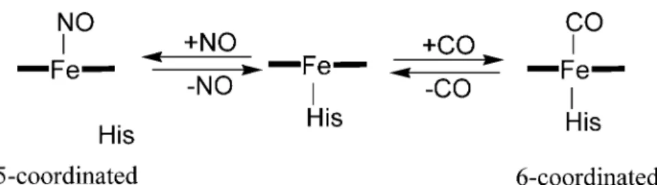
sGC from mammal is a typical NO sensor protein, which catalyze the formation of cGMP from GTP.2 The enzymatic product, cGMP, functions as a second messenger that controls various function of the mammal. The enzymatic activity of sGC is activated by NO binding to the heme in sGC by 200~300 fold. The alteration in the coordination structure of the heme in sGC upon CO and NO binding is similar to that of CooA. In ferrous state without external ligands, the heme in sGC takes a 5-coordinated structure with a histidine as an axial ligand. Upon CO binding to the heme, the heme takes a 6-coordinated structure with His and CO as the axial ligands. However, the activity of sGC is enhanced only by 4~5 fold upon CO binding. As described above, NO activates sGC by 200~300 fold. These differences result from the difference of the coordination structure of the heme in CO- and NO-bound forms. When NO binds to the heme in sGC, the dissociation of the Fe—His bond takes place to form a 5-coordinated nitrosyl heme. This dissociation of the Fe—His bond will
induce a conformational change of sGC to be a trigger of the activation of sGC by NO. Because the dissociation of the Fe—His bond does not occur upon the CO binding, CO cannot activate sGC as does NO. Thus, sGC discriminates CO from NO by the different coordination structure.
O2 is not bound to the heme in sGC even in the environment with O2 because of low affinity of O2 to the heme. Although the detail mechanism of this low O2 affinity is not clear, sGC discriminates O2 from NO by the different binding affinity.
Figure 3. The coordination structure of the heme in sGC.
Heme-based O
2sensor proteins
Aerobes require O2 to obtain the energy for growth by aerobic respiration where O2
functions as the final electron acceptor in the electron transport chain. Moreover, O2 is necessary for oxidation reaction by oxidases and oxygenases such as P450s, tyrosinase, and catechol dioxygenase. Thus O2 is one of the essential substrates for many organisms to maintain their lives. On the other hand, excess O2 sometimes behaves as a harmful matter in biological systems by the formation of reactive oxygen species. Organisms must therefore sense the suitable O2 concentration and make an exact response and adaptation. Some bacteria sense the O2 concentration, and exhibit a chemotactic response toward or against O2, which called aerotaxis. In the case of mammal, hypoxic condition of the cell stabilizes hypoxia inducible factor 1α (HIF1α) that is a transcriptional regulator inducing the expression of genes required for the response to hypoxia3. The stability of HIF1α is controlled whether it is hydroxylated or not. Proline hydroxylase catalyzes the hydroxylation of HIF1α with O2
under aerobic condition, but the hydroxylation is stopped by hypoxia. Thus proline hydroxilase acts as an O2 sensor in this system.
However, for selective O2 sensing, heme-based O2 sensor proteins must have peculiar mechanism in different manner from that in CO and NO sensor proteins. CO and NO sensor proteins utilize the lower affinity of O2 to the heme. This feature of O2 is, however, just a disadvantage to O2
sensing and discrimination. Heme-based O2 sensor proteins cannot take a ligand discrimination strategy by exclusive binding of O2. In fact, the affinity of CO to heme without protein matrix is reported 27,000 times larger than that of O24. Even in myoglobin, in which the affinity for O2 is increased by hydrogen bond formation between the heme-bound O2 and a histidine residue in the heme distal pocket, and that for CO is remarkably decreased by a static hindrance by amino acid, the affinity of CO is still 25 times higher than that of O25. In general the affinity of NO to the heme is still higher than that of CO. These facts suggest that heme-based O2 sensor proteins cannot avoid the binding of CO and NO. Therefore, heme-based O2 sensor proteins, must equip special mechanisms for selective O2 sensing. These mechanisms have not been fully understood yet, but partially elucidated in some O2 sensor proteins.
O2 sensor protein FixL
FixL is a sensor kinase of the FixL/FixJ two-component system that regulates the expression of nif genes responsible for N2 fixation in rhizobia.6 The kinase activity of FixL is regulated by O2, in which FixL senses O2 by means of a heme. When O2 is bound to the heme in FixL, the kinase activity of FixL is inhibited. Once O2 is dissociated from the heme, FixL is activated as the kinase to carry out the autophosphorylation and then the phosphoryl transfer to FixJ.
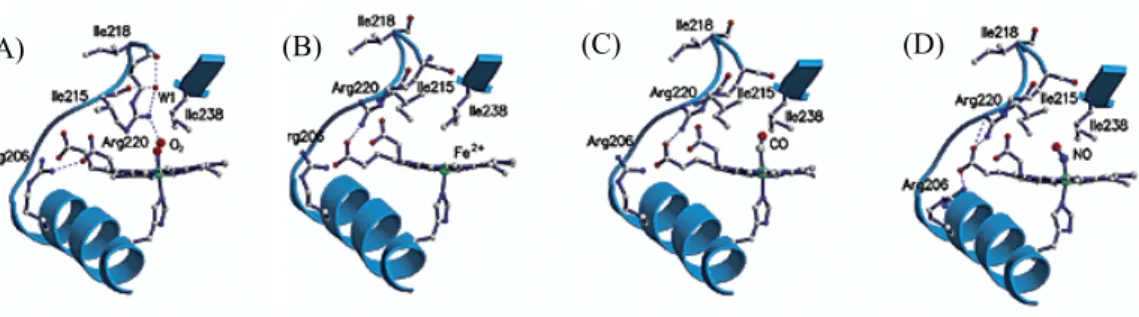
The crystal structures of FixL from different sources have been solved in various forms, providing much information to consider the mechanism of selective O2 recognition. The crystal structures of FixL from Bradyrhizobium japonicuum (BjFixL) are shown in Figure 4 for the deoxy O2-, CO-, and NO-bound forms. In the case of BjFixL, Arg220 forms a hydrogen bond to the heme propionate 7 in the ferrous deoxy state. This hydrogen bond is present in CO- and NO-bound forms. Only in the O2-bound form, the hydrogen bond between Arg220 and the heme propionate 7 is broken,
resulting in the movement of Arg220 into the heme distal pocket where the heme-bound O2 is present. And then Arg220 forms a hydrogen bond to the heme-bound O2. This specific hydrogen-bonding pattern in the O2-bound form is considered to be essential for the O2 discrimination by FixL.
The movement of the FG-loop where Arg220 is located will be induced by the reconstruction of the hydrogen bonding network triggered by O2 binding, which may induce the structural change responsible for kinase inactivation. Though CO and NO bind to the distal position of the heme as does O2, they do not induce the reconstruction of the hydrogen bonding network in the distal heme pocket. These results are consistent with the fact that CO and NO are weak inhibitors for FixL while O2 is a strong one.
Figure 4. The reported crystal structures of FixL from Bradyrhizobium japonicum in the (A) O2-bound, (B)
The heme-based aerotactic signal transducer HemAT
HemAT is a heme-based O2 sensor protein responsible for the aerotaxis control in some bacteria
been completed, and 10 putative genes (D)
(B) (C) (A)
frerrous, (C) CO-bound, and (D) NO bound forms7. Arg220 forms a hydrogen bond to the heme-bound ligand only in the O2-bound form. This specific hydrogen bond formation would essential for the selective O2 sensing and induce the signaling event.
. While the putative gene of HemAT has been reported in tens of bacteria so far,8 just a few HemAT are confirmed to function as the aerotactic signal transducer. B. subtilis is one of such bacteria containing HemAT as an aerotactic signal transducer.
In 1997, the whole genome analysis of B. subtilis has
were id
(residues 1-178) and the C-termin
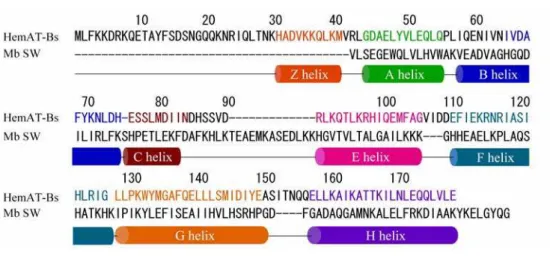
entified as chemotactic signal transducer proteins. 9 Alam and co-workers found that the N-terminal region in the one of these proteins, YhfV, displays a limited homology to myoglobin (Mb), and named it HemAT-Bs.10 The amino acid sequence of Mb and HemAT-Bs are shown in Figure 5. The recombinant HemAT-Bs expressed in Escherichia coli (E. coli) contains a heme and shows similar UV/Vis spectra to Mb. While the aerotactic response was lost in the B. subtilis ∆ten strain where all of the putative 10 chemotactic signal transducer proteins were deleted, the expression of HemAT-Bs in the ∆ten strain rescued the aerophilic response of this bacterium. These results clearly show the function of HemAT-Bs as the aerotactic signal transducer protein.
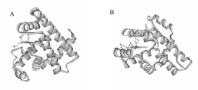
HemAT-Bs consists of two domains: the N-terminal sensor domain
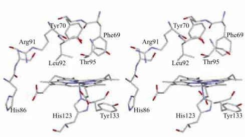
al signaling domain (residues 198-432). Phillips and co-worker reported the crystal structure of the sensor domain (Figure 6B).11 The sensor domain of HemAT-Bs indeed shows a globin fold similar to that of Mb (Figure 6). The crystal structure of the sensor domain of HemAT-Bs reveals that the homology alignment of HemAT-Bs and Mb previously presented by Alam and co-workers is considerably different from the actual structural alignment. A few difference of the structural feature of HemAT-Bs and Mb is observed. HemAT-Bs lacks the D helix, and an extra helix is present in the N-terminal, named the Z helix. Phe69 and Tyr70 are present in the B helix in HemAT-Bs, which are not present in Mb and mammalian Hb, but are highly conserved in Hb from some bacteria, plant, and protozoa. In addition, HemAT-Bs has no distal histidine residue that stabilizes the heme-bound O2 in the case of Mb and Hb (Figure 7,8). These facts indicate that HemAT-Bs will belong to the ancient globin family rather than the globins from higher animals.
Figure 5. The amino acid sequences of the sensor domain in HemAT and sperm whale Mb.
Among the heme-based O2 sensor proteins, HemAT-Bs is the first discovered globin-coupled sensor protein. The investigation about HemAT may, therefore, give a novel mechanism of selective O2 sensing. Moreover, we may be able to reveal the essential factor for selective O2 sensing with comparing the mechanisms of HemAT-Bs and those of other heme-based O2 sensor proteins.
As mentioned above, heme-based O2 sensor proteins must discriminate O2 from other small molecules, such as CO and NO. However, it is hard for heme proteins to discriminate O2 among these molecules, because all of these molecules have an almost similar shape and size. Though O2, CO and NO can bind to a heme, the affinity of these molecules to a heme is different. CO and NO show higher affinity than O2 does. Because of this difference of affinity, some heme-based CO and NO sensor proteins such as CooA and sGC can avoid the binding of O2 to their heme to discriminate their effector from O2. This lower affinity of O2 to the heme, however, is unfavorable for heme-based O2
sensor proteins to sense O2 selectively. In principle, all of O2, CO, and NO are bound to the heme in the heme-based O2 sensor proteins. Thus, heme-based O2 sensor proteins must adopt a special mechanism for selective O2 recognition.
A B
Figure 6. The crystal structure of (A) myoglobin and (B) the A subunit of HemAT sensor domain.
Tyr70 Thr95
← hydrogen bond His64
Figure 7. The crystal structure around the heme in (A) O2-bound myoglobin and (B) the A subunit of deoxy HemAT-Bs
Arg91
His123 Tyr133 His86
Thr95 Leu92
Tyr70
Phe69
Arg91
Tyr133 His123
His86
Thr95 Leu92
Tyr70
Phe69
Figure 8. The stereo view of the crystal structure around the heme in the subunit A of ferrous HemAT-Bs (PDB: 1OR6).
The protein structure of the sensor domain, especially the heme distal structure, would be important to achieve the selective O2 sensing by the heme-based O2 sensor proteins because only the amino acid residues in the distal heme pocket can interact to the heme-bound O2. As described above, investigations on the heme-based O2 sensor protein FixL gives an example of the specific interaction between the heme-bound O2 and an amino acid residue in the distal heme pocket.
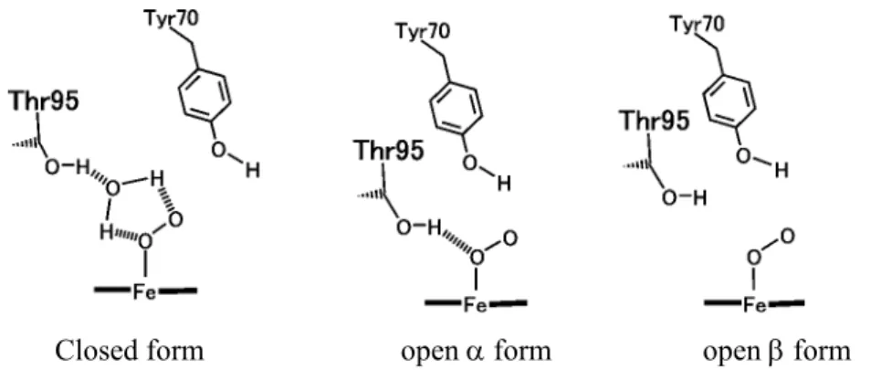
The distal heme pocket in HemAT-Bs includes two polar amino acid residues, Tyr70 in the B helix and Thr95 in the E helix (Figure 8). Resonance Raman spectroscopy has shown that there are three conformers with different hydrogen bonding pattern to the heme-bound O2 (Figure 9):12 a conformer with hydrogen bonds from Thr95 to both of the O atoms of the heme-bound O2 via a water molecule (closed form), a conformer with a direct hydrogen bond from Thr95 to the proximal O atom of the heme-bound O2 (open α form), and a conformer without hydrogen bonds to the heme-bound O2 (open β form). Thus, the hydrogen bond between the heme bound O2 and Thr95 would be responsible for the selective O2 recognition and signal generation. However, the O2 discrimination mechanism of HemAT-Bs is not obvious.
Closed form open α form open β form
Figure 9. The three conformations of the O2-bound HemAT-Bs
Chemotaxis signal transduction system in bacteria
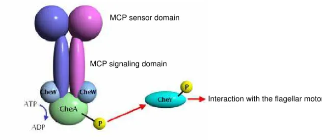
HemAT-Bs is a signal transducer protein in aerotaxis (chemotaxis toward O2) control system in B. subtilis. In this section, a background of the chemotaxis system in bacteria is described. Most of bacteria exhibit chemotactic responses when the bacteria sense attractants or repellents. In general, one bacterium has several chemotactic sensor proteins to sense various effectors for chemotaxis. The most typical proteins for chemotactic receptor are methyl-accepting chemotaxis proteins (MCPs), which are generally transmembrane proteins consisting of the sensor domain in the periplasm and the signaling domain in the cytoplasm.13 The amino acid sequence of the HemAT signaling domain exhibits high homology to other MCP signaling domains despite of that HemAT is a soluble protein. The amino acid sequence of the signaling domain is 30% identical to the signaling domain of Tsr from E. coli, a typical bacterial chemotactic receptor protein called methyl-accepting chemotaxis protein (MCP). Therefore HemAT-Bs can be considered as a member of MCPs.
The chemotaxis control system consists of MCP, CheW, CheA and CheY (Figure 7).13 When an effector molecule is bound to the sensor domain of the MCP, a structural alteration of the sensor domain induced by the effector binding is transmitted to the signaling domain. The structural alteration of MCP regulates the activity of the histidine kinase protein CheA through the adaptor
protein CheW. CheW is necessary for the signal transduction from MCP to CheA, although CheW is not required to form the complex between MCP and CheA. In the most bacteria including E. coli, the CheA activity is down regulated, that is, CheA activity is “on” state without the binding of the effector molecule to the MCP sensor domain, while the binding of the effector to MCP turn off the kinase activity of CheA. On the other hand, some bacteria, for example B. subtilis, exhibit the opposite system of the CheA regulation; the binding of the effector to MCP activates the CheA kinase. The phosphate group on the activated CheA is transmitted to CheY, a response regulator. And then, the phosphorylated CheY interacts with the flagellar motor to control the direction of the flagellar motor rotation.
MCP signaling domain MCP sensor domain
Interaction with the flagellar motor
Figure 9. A schematic model of the aerotactic signaling system with MCP, CheW, CheA, and CheY.
The amino acid sequences of the MCP signaling domains show substantial homology (Figure 10), and the signaling domain can be considered to have a conserved structure regardless of the effectors. The crystal structure of the signaling domain of the serine receptor from E. coli (Tsr) and another MCP from Thermotoga maritima are solved by X-ray crystallography.14,15 As shown in Figure 8B, the MCP signaling domain forms a dimer that constructs a huge four-helix-bundle with
about the length of 200 Å and the diameter of 20 Å. Each subunit contributes two helices with a U-turn tip, where the amino acid sequence is especially conserved. The tip would be the interface to CheW and CheA.
A B
Figure 10. (A) Amino acid sequence alignment in the signaling domain of Tsr from E. coli, McpB from B. subtilis, and HemAT-Bs. (B) the reported crystal structure of the signaling domain of MCP from Thermotoga maritina (PDB: 2CH7).
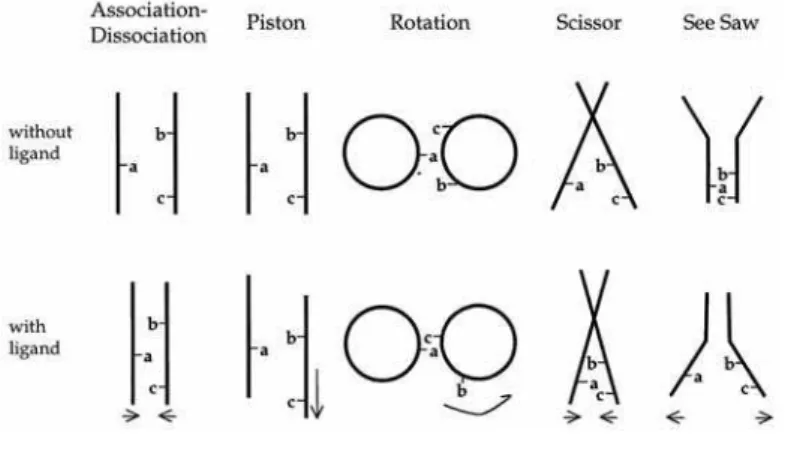
The periplasmic sensor domain and the cytoplasmic signaling domain of MCP are connected by the transmembrane domain. The movement of the transmembrane region should be important for the signal transduction from the sensor domain to the signaling domain. This region consists of helices called TM1 and TM2, which form a helix-bundle motif. In the most cases, TM1 and TM2 are the N and the C terminal helices of the sensor domain, respectively. TM2 is connecting with the extended helix of the signaling domain. Koshland and co-worker estimated the movement of the
transmembrane region of E. coli aspartate receptor, Tsr, using a site-directed spin-labeling method.16 They conclude that the helix bundle of the transmembrane region exhibits an about 1 Å “piston-like movement” (Figure 11). On the other hand, Ordal and co-worker performed a disulfide crosslinking study on the aspartate receptor, McpB, from B. subtilis, and showed a rotational movement on the TM1 (Figure 11).17 The similar helix bundle motif is observed not only in MCPs but also in many other receptor proteins. It is not determined at present which model is correct for signal transduction. There are some reports that chimera proteins consisting of MCP sensor domain and the signaling domain of another receptor protein retain the signaling ability. Therefore, the information from the investigation about MCP would useful to resolve the general mechanism of receptor proteins.
Figure 11. The models for the patterns of conformational alteration in a helix bundle by Koshland et al. 16
The purpose of this work
Since HemAT belongs to the MCP family, the conformational change of HemAT upon O2
binding would be in the same pattern to other MCP proteins. Although the movement of the region between the sensor and signaling domain is proposed for the intramolecular signal transduction for MCP, the detail mechanism is not clear yet. Among MCPs, HemAT is an excellent model for investigation of MCP because HemAT-Bs is the only soluble protein among MCPs so far isolated. Moreover, HemAT-Bs has a heme, which is a good probe for several spectroscopic studies. These features would also help the investigation especially about the mechanism of signal transduction upon effector binding.
There are two purposes in this work on HemAT-Bs. One is the elucidation of the mechanism of the selective O2 sensing, and the other is the elucidation of the intramolecular signal transduction mechanism. To elucidate these mechanisms, the author has done the mutagenesis and spectroscopic studies on HemAT-Bs
REFERENCES
1. Aono, S (2003) Biochemical and biophysical properties of the CO-sensing transcriptional activator CooA, Acc. Chem. Res. 36, 825-831
2. Pal, B. and Kitagawa, T. (2005) Interactions of soluble guanylate cyclase with diatomics as probed by resonance Raman spectroscopy, J. Inorg. Biochem., 99, 267-279
3. Berra, E., Ginouves, A. and Pouyssegui, J. (2006) The hypoxia-inducible-factor hydroxylases bring fresh air into hypoxia signalling, EMBO Rep., 7, 41-45
4. Traylor, T. G., Mitchell, M. J., Tsuchiya, S., Campbell, D. H., Stynes D. V. and Koga, N. (1981) Cyclophane hemes. 4. Steric effects on dioxygen and carbon monoxide binding to hemes and
heme proteins, J. Am. Chem. Soc. 103, 5234-5236
5. Olson, J. S. and Phillips, G. N. Jr. (1997) Myoglobin discriminates between O2, NO, and CO by electrostatic interactions with the bound ligand, J.Biol. Inorg. Chem., 2, 544-552
6. Chan, M. K. (2001) Recent advances in heme-protein sensors, Curr. Opin. Chem. Res. 5, 216-222
7. Hao, B., Isaza, C., Arndt, J., Soltis, M. and Chan, M. K. (2002) Structure-based mechanism of O2 sensing and ligand discrimination by the FixL heme domain of Bradyrhizobium japonicum, J. Am. Chem. Soc. 126, 15000-15001
8. Allen, T., Freitas, K. and Alam, M. (2003) The diversity of globin-coupled sensors, FEBS Lett., 552, 99-104
9. Kunst, F. et al. (1997) The complete genome sequence of the gram-positive bacterium Bacillus subtilis, Nature 309, 249-256
10. Hou, S., Larsen, R. W., Boudko, F., Riley, C. W., Karatan, E., Zimmer, M., Ordal, G. W. and Alam, M. (2000) Myoglobin-like aerotaxis transducers in Archaea and Bacteria, Nature 403, 540-544
11. Zhang, W. and Phillips, G. N. Jr. (2003) Structure of the oxygen sensor in Bacillus subtilis: signal transduction of chemotaxis by control of symmetry, Structure 11, 1097-1110
12. Ohta, T., Yoshimura, H., Yoshioka, S., Aono, S. and Kitagawa, T. (2004) Oxygen-sensing mechanism of HemAT from Bacillus subtilis: A resonance Raman spectroscopic study, J. Am. Chem. Soc. 126, 15000-15001
13. Szurmant, H. and Ordal, G. W. (2004) Diversity in chemotaxis mechanisms among the bacteria and archaea, Microbiol. Mol. Biol. Rev. 68, 301-319
14. Kim, K. K., Yokota, H. and Kim, S. H. (2000) Four-helix bundle structure of the cytoplasmic domain of a serine chemotaxis receptor, Nature 400, 787-792
15. Park, S. Y., Borbad, P. P., Gonzalez-Bonet, G., Bhatnager, J., Pollard, A. M., Freed, J. H., Bilwes, A. M. and Crane, B. R. (2006) Reconstruction of the chemotaxis receptor-kinase assembly, Nat.
Struct. Mol. Biol 13, 400-407
16. Ottemann, K. M., Xiao, W., Shin, Y. and Koshland, D. E. Jr. (1999) A piston model for transmembrane signaling of the aspartate receptor, Science, 285, 1751-1754
17. Szurmant, H., Bunn, M. W., Cho, S. H. and Ordal, G. W. (2004) Ligand-induced conformational changes in the Bacillus subtilis chemoreceptor McpB determined by disulfide crosslinking in vivo, J. Mol. Biol., 344, 919-928
Chapter 2: Specific hydrogen-bonding networks responsible for selective
O
2sensing of HemAT-Bs
Biochemistry, 45, 8301-8307 (2006)
Abstract
HemAT-Bs discriminates its physiological effector, O2, from other gas molecules to generate the aerotactic signal, but the detailed mechanism of the selective O2 sensing is not obvious. In this study, the author measured electronic absorption, electron paramagnetic resonance (EPR), and resonance Raman spectra of HemAT-Bs to elucidate the mechanism of the selective O2 sensing by HemAT-Bs. The resonance Raman spectroscopy revealed that a hydrogen bond is formed between His86 and the heme propionate only in the O2-bound form, in addition to that between Thr95 and the heme-bound O2. Disruption of this hydrogen bond by the mutation of His86 caused disappearance of a conformer with a direct hydrogen bond between Thr95 and the heme-bound O2 that is present in WT HemAT-Bs. On the basis of these results, the author proposes a model for the selective O2
sensing by HemAT-Bs as follows. The formation of the hydrogen bond between His86 and the heme propionate induces a conformational change of the CE-loop and the E-helix, by which Thr95 is located at the proper position to form the hydrogen bond with the heme-bound O2. This stepwise conformational change would be essential to the selective O2 sensing and the signal transduction by HemAT-Bs.
Introduction
The diatomic gas molecules such as dioxygen (O2), carbon monoxide (CO), and nitric oxide (NO) can work as signaling molecules with the cognate receptor proteins in biological systems. The heme-based sensor proteins are the most general receptor proteins for these gas molecules.1-3 The heme in these proteins functions as the binding site for the effector gas molecules. These heme-based gas sensor proteins must discriminate the effector from other molecules so as to take a specific conformation only upon the binding of the effector molecule. However, the discrimination of these gas molecules is difficult for heme proteins because all of O2, CO, and NO have similar size and can be bound to a reduced heme with a similar coordination structure. The ligand discrimination mechanism of the heme-based gas sensor proteins is not fully understood yet.
HemAT is a heme-based O2 sensor protein that functions as a signal transducer for bacterial aerotaxis.4-7 HemAT monomer consists of two domains, a sensor domain and a signaling domain. The sensor domain shows a globin fold containing a heme that acts as the O2 binding site. The signaling domain of HemAT interacts with a histidine kinase protein CheA, a component of the CheA/CheY two-component signal transduction system that regulates the direction of the flagellar motor rotation.8-10
A specific conformational change of HemAT will occur around the heme upon O2 binding, and then intramolecular signal transduction takes place from the sensor domain to the signaling domain. As a result, the self-kinase activity of CheA is regulated by a change in the HemAT-CheA interaction via the conformational change of HemAT. This signaling event takes place only with O2, but not with other gas molecules. However, the detailed mechanisms of the selective O2 sensing and the signal transduction of HemAT remain to be elucidated.
To elucidate these mechanisms of HemAT, it is necessary to characterize the heme environmental structure including the coordination structure of the heme and hydrogen-bonding pattern around the heme and the ligand. The crystal structure of the sensor domain of HemAT from Bacillus subtilis (HemAT-Bs) reveals that Tyr70 and Thr95 in the distal heme pocket are possible candidates of amino acid residues to form hydrogen bonds with the heme-bound ligand (Figure 1).11
Resonance Raman spectroscopy has revealed that Thr95 forms hydrogen bonds to the heme-bound O2, but Tyr70 does not.12 Oxygen-bound HemAT-Bs has three different conformers with different hydrogen bonding pattern around the heme-bound O2. The hydrogen-bonding interaction between Thr95 and the heme-bound O2 would be responsible for the selective O2 sensing and the intramolecular signal transduction upon O2 binding.
Figure 1. Stereo views of the heme environmental structures of deoxy HemAT-Bs (A) subunit A and (B) subunit B (PDB: 1OR6).9 Depicted residues are Tyr70, Leu92, Thr95, His123, and the amino acids with a polar side chain that are located within 5 Å from the heme propionates.
In this study, the author characterized the coordination structure of the heme, and the hydrogen-bonding pattern on the heme-bound ligands and on the heme propionates in O2-, CO-, and NO-bound forms of wild type (WT) and several mutants of full-length HemAT-Bs to investigate the mechanism of the selective O2 sensing by HemAT-Bs. On the basis of these results, the author shows that the formation of a hydrogen bond between His86 and the heme propionate 6 upon O2
binding causes a protein conformational change that allows Thr95 to form a specific hydrogen bond to the heme-bound O2.
Experimental Procedure
Protein expression and purification
In this study, the author used full-length HemAT-Bs with a C-terminal His6-tag, which was expressed in E. coli BL21(DE3) under the control of T7 promoter in pET-24(+) vector (Novagen). Site-directed mutagenesis was carried out using QuikChange Site-directed Mutagenesis Kit (Stratagene). For the expression of HemAT-Bs, the E. coli cells were grown aerobically at 37°C for 4 hours in Terrific Broth containing 30 µg/mL kanamycin. The expression was induced by addition of isopropyl-β-D-thiogalactopyranoside to a final concentration of 1 mM, and then the cultivation was continued at 22°C for 18 hours. The cells were harvested by centrifugation at 4,000 × g and were stored at –78°C until use.
The cells were thawed and resuspended in the buffer A (50 mM Tris-HCl buffer (pH 8.0) containing 15 mM glycine and 1 M NaCl), and then were broken by sonication. The resulting suspension was centrifuged at 100,000 × g for 20 minutes, and the supernatant was loaded on a Ni2+-charged HiTrap Chelating column (GE Healthcare). After washing the column with the buffer A, and then with 50 mM Tris-HCl buffer (pH 8.0), the adsorbed proteins were eluted by 50 mM Tris-HCl buffer (pH 8.0) containing 100 mM imidazole. The fractions containing HemAT-Bs were combined and loaded on a HiTrap Q HP column (GE Healthcare). The column was washed with 50 mM Tris-HCl buffer (pH 8.0) containing 100 mM NaCl, and then HemAT-Bs was eluted by increasing the concentration of NaCl in the buffer. The purity and yield of the sample were checked by SDS-PAGE. A typical result is shown in Figure 2.
198,000 120,000 91,000 63,000
← HemAT-Bs 47,000
37,000
28,000
19,000
Figure 2. The SDS-PAGE of the purified HemAT-Bs with a Ni2+-charged HiTrap Chelating column and a HiTrap Q HP column.
Sample preparation for measurements
HemAT-Bs was oxidized by adding a small amount of potassium ferricyanide. Excess potassium ferricyanide was removed by using a PD10 column (Amersham Biosciences Corp.) with 50 mM Tris-HCl buffer (pH 8.0). Five times excess amount of sodium dithionite was added into degassed ferric HemAT-Bs solution to make ferrous HemAT-Bs. To prepare CO-, NO-, and O2-bound HemAT-Bs, the ferrous HemAT-Bs solution was exposed to CO, NO, and O2 gas, respectively.
Spectroscopic methods
EPR spectra of NO-bound HemAT-Bs were measured at 20 K on a Bruker ESP300E spectrometer with the microwave frequency of 9.5 GHz and the modulation amplitude of 5.0 G.
Resonance Raman spectra of HemAT-Bs were measured as reported previously (12,13). Excitation source were a Kr+ laser (Spectra Physics 2060) at 413.1 nm and 406.7 nm for the O2- and
CO-bound forms, and for the NO-bound form, respectively. The scattered light was dispersed with a single polychromator (Ritsu DG-1000) equipped with a liquid nitrogen-cooled charge-coupled device camera. Obtained Raman shifts were calibrated using indene and aqueous solution of potassium ferrocyanide. The laser power was 0.2 mW for the CO-bound form and 1 mW for the O2- and NO-bound forms at the sample point.
In the resonance Raman spectra of O2-bound HemAT-Bs, some isotope (16O2 and
18O
2)-insensitive bands overlap with the Fe—O2 stretching bands. Gaussian-bands fitting analyses of the resonance Raman spectra of the O2-bound forms were carried out with Igor Pro 5.03 for distinguishing the Fe—O2 modes from other isotope-insensitive modes.12 The width, peak position, and intensity ratio for the isotope-insensitive modes were fixed for the fitting of 16O2 and 18O2
spectra.
Results
Absorption spectra of the CO- and NO-bound HemAT-Bs
The electronic absorption spectra of the CO-bound form of WT, T95A, and Y70F HemAT-Bs showed the Soret, β and α peaks at 422, 542, and 566 nm, respectively (Figure 3A). These spectra are typical of CO-bound heme proteins with a 6-coordinated, low-spin heme. All of the NO-bound form of WT, T95A, and Y70F HemAT-Bs gave the same spectra with the Soret, β, and α peaks at 419, 548, and 575 nm, respectively (Figure 3B). These spectra are typical of a 6-coordinate, low-spin Fe(II) nitrosyl heme with a proximal histidine ligand, as seen in NO-bound myoglobin (Mb).14
Figure 3. Electronic absorption spectra of (A) CO-bound and (B) NO-bound HemAT-Bs. (a) WT, (b) T95A, and (c) Y70F HemAT-Bs
EPR spectra of the NO-bound HemAT-Bs
Figure 3 shows the EPR spectra of NO-bound HemAT-Bs. The first derivative spectrum with g = 2.08, 2.01, and 1.97 was typical of a 6-coordinated Fe(II) nitrosyl heme (Figure 4A).15 In the second derivative spectrum, nine hyperfine splitting signals (a triplet of triplets) were observed as the result of the hyperfine interaction with both of 14N nuclei of NO (A1) and the trans axial His (A2). The hyperfine coupling constants derived from the nitrogen atoms of the heme-bound NO and the proximal histidine were determined to be A1 = 2.2 mT and A2 = 0.6 mT, respectively (Figure 4B). The EPR spectra of T95A and Y70F HemAT-Bs were the same as that of WT (Figure 4C). These results indicate the formation of a 6-coordinated nitrosyl heme with a proximal histidine in the NO-bound form, which is consistent with the results of the electronic absorption spectroscopy.
Figure 4. (A) The first derivative EPR spectrum and (B) the second derivative EPR spectrum of NO-bound WT HemAT-Bs in 50 mM Tris-HCl buffer (pH 8.0). These spectra were recorded at 20K with microwave frequency of 9.5 GHz, and modulation amplitude of 5.0 G.
Resonance Raman spectroscopy for the CO-bound HemAT-Bs
Fe—C and C—O stretching frequencies of CO-bound heme proteins, νFe—CO and νC—O, are sensitive and reliable markers for the electrostatic environment around the heme-bound CO.16 The νFe—CO and νC—O bands of CO-bound WT HemAT-Bs were observed at 495 cm-1 and 1966 cm-1, respectively (Figure 5). These values were similar to those of H64I (νFe—CO = 490 cm-1, νC—O = 1968 cm-1) and H64L (νFe—CO = 490 cm-1, νC—O = 1965 cm-1) Mbs,17 but different from those of WT Mb (νFe—CO = 512 cm-1, νC—O = 1944 cm-1).18 These results indicate that the heme-bound CO is in a hydrophobic environment without any electrostatic interaction in WT HemAT- Bs.
T95A HemAT-Bs gave the similar νFe—CO and νC—O values to those of WT HemAT-Bs, indicating that the environment around the CO in T95A HemAT-Bs is also hydrophobic without electrostatic interaction on the CO and that the mutation of Thr95 makes little effect on the environment around the heme-bound CO. On the other hand, Y70F HemAT-Bs showed different resonance Raman spectra from those of WT and T95A HemAT-Bs. Two νC—O bands were observed at 1961 and 1945 cm-1 in Y70F HemAT-Bs (Figure 5). Furthermore, a weak shoulder around 510 cm-1 was observed in the νFe—CO region of Y70F HemAT-Bs, which may correlate with the new νFe—CO band observed at 1945 cm-1. These results suggest the existence of two conformers of Fe-CO unit with different environments around the heme-bound CO in this mutant.
In the crystal structure of the subunit A in the deoxy form of the HemAT-Bs sensor domain,11 the phenyl oxygen atom of Tyr70 side chain is just above the heme iron atom with the distance of 5.8 Å. This fact implies that the non-bonding electron pair on the oxygen atom of the phenyl group of Tyr70 would interact with the oxygen atom of the heme-bound CO. The 5 cm-1 low-frequency shift seen in the νC—O band of Y70F HemAT-Bs (the νC—O at 1961 cm-1) is consistent with the disappearance of this interaction by the mutation. In addition, the oxygen atom of Tyr70 side chain is close to the carbonyl oxygen atom of Leu92 with the distance of 2.9 Å, which is consistent with the presence of a hydrogen bond between Tyr70 and Leu92.11 Disappearance of this hydrogen bond in Y70F mutant would cause a considerable conformational perturbation in the distal heme pocket. The new νC—O band at 1945 cm-1 and a weak shoulder at ca 510 cm-1 seen in this mutant might be caused by this structural perturbation.
Zhang et al. reported that Tyr70 mutants of the HemAT-Bs sensor domain showed the large dissociation constants for the heme-bound O2, and proposed that Tyr70 would form a hydrogen bond to the heme-bound O2 on the basis of these results.6 Our previous results, however, have shown that Tyr70 does not form such a hydrogen bond in the full-length of HemAT-Bs.12 Their results will imply that Tyr70 is responsible for the fixation of the position of Thr95 that forms the hydrogen bond with the heme-bound O2. Leu92 and Thr95 are located in the same helix, the E-helix. If the hydrogen bond between Leu92 and Tyr70 disappear, the position of the E-helix would be perturbed, resulting in low stability of the O2-bound form of Tyr70 mutants. In addition, the author have also shown that hydrogen-bonding pattern around the heme-bound O2 is different between the full-length and the truncated sensor domain of HemAT-Bs.12 These results suggest that the affinity of O2 and the hydrogen-bonding interaction around the heme-bound O2 are different between the truncated sensor domain and the full-length of HemAT-Bs.
Figure 5. Resonance Raman spectra of CO-bound (a) WT, (b) T95A, and (c) Y70F HemAT-Bs. (A) Fe—CO stretching bands in the low frequency region. (B) C-O stretching bands in the high frequency region.
Resonance Raman spectroscopy for the NO-bound HemAT-Bs
The high frequency region in the resonance Raman spectra of heme proteins contain some marker bands sensitive to the oxidation state (ν4), and the spin and coordination state (ν2 and ν3) of the heme iron. In the case of NO-bound HemAT-Bs, the ν4, ν3, and ν2 band were observed at 1373, 1498, and 1577 cm-1, respectively (Figure 6). These results are typical of the 6-coordinated low-spin heme, and consist with the results from UV-Vis and EPR spectroscopies.
Figure 6. The resonance Raman spectrum of NO-bound HemAT WT in the high frequency region.
N—O stretching frequency, νN—O, of NO-bound heme proteins is also affected by the heme distal environments.19 The νN—O band of WT HemAT-Bs was observed at 1636 cm-1 (Figure 7). This value was similar to that of the νN—O of H64L Mb (νN—O = 1635 cm-1), but not to that of WT Mb (νN—O = 1613 cm-1).19 These results suggest that the heme-bound NO in WT HemAT-Bs is in a hydrophobic environment without any electrostatic interaction with the heme-bound NO.
The Fe—NO stretching mode, νFe—NO, of NO-bound WT HemAT-Bs was observed at 545 cm-1 (Figure 7). The νFe—NO band is not sensitive to the electrostatic environment of the distal heme pocket.19 Rather, the νFe—NO frequency is reported to be dependent on the Fe-N-O angle.20 This
frequency is lowered by decreasing of the Fe-N-O angle. 20 The νFe—NO frequency of NO-bound HemAT-Bs (545 cm-1) was lower than that of NO-bound Mb (558 cm-1),19 suggesting that Fe-N-O angle in NO-bound HemAT-Bs is lower than that in NO-bound Mb.
Figure 7. Resonance Raman spectra of NO-bound HemAT-Bs. (a) 14NO-bound WT HemAT-Bs, (b)
15NO-bound WT HemAT-Bs, (c) (a)-(b) difference spectrum.
The heme propionate bending mode, δ(CβCcCd), in the resonance Raman spectra of HemAT-Bs
The bending mode of the heme propionate, δ(CβCcCd), in the resonance Raman spectra of O2-bound HemAT-Bs showed a different feature from those of the ferrous, CO-, and NO-bound forms. The resonance Raman spectra of ferrous, CO-, and NO-bound HemAT-Bs showed one δ(CβCcCd) band at 365, 373, and 375 cm-1, respectively (Figure 8). On the other hand, in the O2-bound form, two δ(CβCcCd) bands were observed at 370 and 383 cm-1, suggesting that there are two species with different conformations around the heme propionates in O2-bound HemAT-Bs.
The δ(CβCcCd) band is sensitive to electrostatic interaction on the heme propionate(s), e.g. hydrogen bonds and salt bridges.21 The stronger electrostatic interaction between the heme propionate(s) and surrounding residue(s), the higher frequency of the δ(CβCcCd) band.21 The δ(CβCcCd) band at 365, 373, 375, and 370 cm-1 in ferrous, CO-, NO-, and O2-bound HemAT-Bs, respectively, are lower than those of the corresponding forms of Mb (371, 377, 378, and 378 cm-1 for the ferrous, CO-, NO-, and O2-bound forms, respectively) in which moderate hydrogen bonds exist on the heme propionate 7.19,22-24 These results indicate that the conformers showing these δ(CβCcCd) bands have no or a weak hydrogen bond on the heme propionate.
The additional δ(CβCcCd) band at 383 cm-1 in O2-bound HemAT-Bs indicates the existence of a stronger hydrogen bond or a salt bridge on the heme propionate. This conformer was observed only in the O2-bound form. To determine the amino acid residue(s) responsible for the 383 cm-1 band, the author prepared the following five mutants: H86A, S87A, R91A, R118A, and R125A, in which every polar residue within 5 Å from the heme propionates is mutated to alanine (Figure 1). The δ(CβCcCd) band at 383 cm-1 disappeared only in O2-bound H86A HemAT-Bs, while other mutants (S87A, R91A, R118A and R125A HemAT-Bs) showed almost the same δ(CβCcCd) band at 383 cm-1 as that of WT HemAT-Bs (Figure 9). The 370 cm-1 band seen in the spectrum of WT exhibited 5 cm-1 upshift by the mutation of His86 and Ser87, both of which are located in the CE-loop near the heme propionate. This upshift of the 370 cm-1 band might be caused by a structural perturbation around the heme propionate. These results indicate that His86 is the residue that forms the hydrogen bond with the one of the heme propionate, probably the one at the position 6,
and that the residues in the CE-loop are responsible to maintain the proper structure around the heme propionate.
Figure 8. Resonance Raman spectra of WT HemAT-Bs in the (a) ferrous, (b) O2-bound, (c) NO-bound, and (d) CO-bound forms.
Figure 9. Resonance Raman spectra of O2-bound form of (a) R125A, (b) R118A, (c) R91A, (d) S87A, (e) H86A, and (f) WT HemAT-Bs.
The hydrogen bonding pattern to the heme-bound O2 in the O2 bound form of H86A HemAT-Bs
The Fe—O2 stretching frequency region of the resonance Raman spectrum of H86A HemAT-Bs was also different from that of WT HemAT-Bs. Three νFe—O2 bands were observed at 554, 566, and 572 cm-1 in O2-bound WT HemAT-Bs, as shown in the Introduction. In H86A HemAT-Bs, the band at 566 cm-1 disappeared, and only the bands at 557 and 572 cm-1 were observed as shown in Figure 10. These bands were shifted to 538 and 554 cm-1, respectively, upon 18O2
substitution. These isotope shifts of 19 and 18 cm-1 were consistent with the previously reported values for WT HemAT-Bs and in agreement with a calculated shift value of 21 cm-1.12 The 16O2 -
18O
2 difference spectrum was also consistent with the result of the Gaussian fitting analysis (Figure 10(C)). The difference in the Fe—O—O bond angle affects the shift of the Fe—O2 stretching mode upon the isotope substitution of O2, which will cause a slight difference between the experimental and calculated values of the isotope shift. These results indicate that the loss of the hydrogen bond between His86 and the heme propionate 6 affects the hydrogen-bonding interaction between the heme-bound O2 and Thr95.
Figure 10. Resonance Raman spectra of (A) 16O2-bound H86A HemAT-Bs, (B) 18O2-bound H86A HemAT-Bs. The bold solid lines are the raw spectra. All traces except for the bold solid lines were obtained by Gaussian band fitting analysis. The thin solid lines denote contribution from νFe—O2, which were sensitive to the isotope substitution of O2. The thin dotted lines are due to the isotope-insensitive modes. The bold dotted lines are the sum of the component bands. (C) The 16O2 – 18O2 difference spectra. The broken line in the spectrum (C) is the difference spectrum obtained the Gaussian band fitting analysis. The bold solid line is the experimental difference spectrum.
Discussion
It is a critical requirement for the heme-based gas sensor proteins to discriminate between the effector gas molecule and other gas molecules. Some heme-based gas sensor proteins, such as CooA and sGC, alter the coordination number (5-coordinate vs. 6-coordinate) of the heme depending on the species of heme-bound gas molecules.25-27 The proper coordination structure with the effector molecule causes a protein conformational change that is essential to a specific response to the cognate effectors. The heme of HemAT-Bs, however, has a 6-coordinate structure in all of the O2-, CO-, and NO-bound forms, indicating that HemAT-Bs does not adopt this strategy for the selective O2 sensing.
Another possible strategy for the heme-based gas sensor proteins to discriminate the effector gas molecule is to form a specific interaction between the heme-bound effector molecule and neighboring amino acid residues. The author have reported that Thr95 forms the hydrogen bonds to the heme-bound O2 in the two conformers among the three ones in the O2-bound form.12 On the other hand, the author has revealed here that no hydrogen bond is formed on the heme-bound ligand in the case of CO- and NO-bound HemAT-Bs. These results show that the hydrogen bonding between a heme-bound ligand and Thr95 is specific in the O2-bound form, which supports the idea that this hydrogen bonding interaction is essential for the selective O2 sensing by HemAT-Bs.
The crystal structure of the HemAT-Bs sensor domain in the ferrous form shows that the distances between the hydroxyl oxygen atom of Thr95 and the heme iron atom are 6.8 and 7.6 Å in the subunits A and B, respectively.11 To form the direct hydrogen bond to the heme-bound O2, Thr95 has to be located within 3 Å from the heme iron. Although the above distances in the ferrous form are too long to form a hydrogen bond, a direct hydrogen bond is certainly formed between Thr95 and the “proximal” oxygen atom of the heme-bound O2.12 These results indicate that the formation of the hydrogen bond between Thr95 and the proximal oxygen atom requires a preceding conformational change of the distal heme pocket that moves Thr95 to the suitable position to form the hydrogen bond.
Because the O2-bound H86A HemAT-Bs cannot form the conformer with the direct hydrogen bond between Thr95 and the proximal oxygen atom, the preceding conformational alteration would be induced by the formation of the hydrogen bond between His86 and the heme propionate 6, as discussed below. His86 is located in the CE-loop, which is adjacent to the E-helix that contains Thr95. The formation of the hydrogen bond between His86 and the heme propionate 6, which takes place only upon O2 binding, will induce a conformational change of the CE-loop. And then, this conformational alteration of the CE-loop will propagate to the E-helix, finally shifting Thr95 to the proper position to form the direct hydrogen bond to the proximal oxygen atom. Thus, HemAT-Bs would cause a stepwise conformational change upon O2 binding.
If Thr95 were located in advance at the suitable position to form the hydrogen bond to the heme-bound O2 in ferrous form, Thr95 would also form a similar hydrogen bond to the heme-bound CO and NO. In this situation, the hydrogen bond between Thr95 and the heme-bound O2 could not be used for the selective O2 sensing. The stepwise conformational change upon O2 binding allows the hydrogen bond between Thr95 and the heme-bound ligand to be specific for the O2-bound form.
On the basis of these results, the author proposes the heme environmental structure of O2-bound HemAT-Bs as shown in Figure 11. Three conformers exist in O2-bound HemAT-Bs with different hydrogen bonding interactions on the heme-bound O2, corresponding to the closed, the open α, and the open β forms.12 Hydrogen bonding interaction on the heme propionate is also different among these three conformers. His86 forms the hydrogen bond to the heme propionate in the open α form in which the direct hydrogen bond is formed between Thr95 and the heme-bound O2. His86 does not form the hydrogen bond with the heme propionate in the closed and the open β forms in which Thr95 does not come close to the heme-bound O2.
Figure 11. The conformations around the heme in the O2-bound form of HemAT-Bs
HemAT-Bs should undergo a sufficient conformational change upon O2 binding for generati
for the selective O2 sensing and the sign
ng the aerotactic signal. The formation of the open α form involves a considerable conformational change of the CE-loop and the E-helix, suggesting that the open α form is the
“on-state” for the signaling. In the closed and open β forms, the conformational change of the CE-loop and the E-helix will not be so large because the hydrogen bond is not formed between His86 and the heme propionate. Therefore, the closed and the open β forms will be the “off-state”. It remains to be elucidated why the on- and off-states exist together in O2-bound HemAT-Bs, and whether the closed and open β forms have any physiological role.
In summary, the author proposes the following mechanism
al transduction in HemAT-Bs. When O2 is bound to the heme in HemAT-Bs, the hydrogen bond forms between His86 and the heme propionate 6. The formation of this hydrogen bond makes a conformational alteration in the distal heme pocket, especially on the position of the E-helix. The change in the position of the E-helix makes Thr95 to be located at the suitable position to form the hydrogen bond with the proximal oxygen atom of the heme-bound O2. As the hydrogen bond is not formed between His86 and the heme propionate 6 upon CO and NO binding, the proper conformational change does not occur in these cases. The proper conformational change around the heme upon O2 binding will propagate to the signaling domain of HemAT-Bs, and then activate CheA.
REFERENCES
1. Rodgers, K.R. (1999) Heme-based sensors in biological systems, Curr. Opin. Chem. Biol. 3, 158-167
2. Chan, M. K. (2001) Recent advances in heme-protein sensors, Curr. Opin. Chem. Biol. 5, 216-222
3. Jain, R., and Chan, M. K. (2003) Mechanisms of ligand discrimination by heme proteins, J Biol. Inorg. Chem. 8, 1-11
4. Hou, S., Larsen, R. W., Boudko, F., Riley, C. W., Karatan, E., Zimmer, M., Ordal, G. W. and Alam, M. (2000) Myoglobin-like aerotaxis transducers in Archaea and Bacteria, Nature 403, 540-544
5. Aono, S., Kato, T., Matsuki, M., Nakajima, H., Ohta, T., Uchida, T., and Kitagawa, T., (2002) Resonance Raman and ligand binding studies of the oxygen-sensing signal transducer protein HemAT from Bacillus subtilis, J. Biol. Chem. 277, 13528-13538
6. Zhang, W., Olson, J. S. and Phillips, G. N. Jr. (2005) Biophysical and kinetic characterization of HemAT, an aerotaxis receptor from Bacillus subtilis, Biophys. J. 88, 2801-2814
7. Ohta, T. and Kitagawa, T. (2005) Resonance Raman investigation of the specific sensing mechanism of a target molecule by gas sensory proteins, Inorg. Chem. 44, 758-769
8. Szurmant, H. and Ordal, G. W. (2004) Diversity in chemotaxis mechanisms among the bacteria and archaea, Microbiol. Mol. Biol. Rev. 68, 301-319
9. Bischoff, D. S. and Ordal, G. W (1992) Bacillus subtilis chemotaxis: a deviation from the Escherichia coli paradigm, Mol. Microbiol. 6, 23-28
10. Garrity, L. F. and Ordal G, W. (1995) Chemotaxis in Bacillus subtilis: how bacteria monitor environmental signals, Pharmac. Ther. 68, 87-104
11. Zhang, W. and Phillips, G. N. Jr. (2003) Structure of the oxygen sensor in Bacillus subtilis: signal