CBF-1 Controls the Retinotectal Topographic
Map along the Anteroposterior Axis through
Multiple Mechanisms
Hiroo Takahashi
DOCTOR OF PHILOSOPHY
Department of Molecular Biomechanics
School of Life Science
The Graduate University for Advanced Studies
2003
- 1 -
Abstract
Topographic maps with a defined spatial ordering of neuronal connections are a key feature of the brain’s organization. The most widely used model for studies of the formation of topographic maps is the retinotectal projection. Chick brain factor-1 (CBF-1), a nasal retina-specific winged- helix transcription factor, is known to prescribe the nasal specificity which leads to the formation of the precise retinotectal map especially along the antero-posterior (A-P) axis. However, the molecular mechanisms by which CBF-1 controls the expression of topographic molecules have not been elucidated. In the present study, to gain insight into the downstream target genes of CBF-1, I employed electroporation of a retroviral vector carrying the CBF-1 gene into the optic vesicle, and examined effects of the misexpression of CBF-1 on the expression of topographic molecules and other asymmetrically distributed molecules. The in ovo electroporation of retrovirus allows the immediate and sustained expression of a transgene. Since endogenous CBF-1 begins to be topographically expressed prior to Hamburger-Hamilton (HH) stage 11 in native embryos, at which stage the polarity along the nasotemporal (N-T) axis appears to be determined (Dütting and Thanos, 1995; Thanos et al., 1996), this in ovo electroporation system is suitable for the functional study of CBF-1. Here, I show that ectopic expression of CBF-1 in the temporal retina represses expression of EphA3 and CBF-2, and induces that of SOHo-1, GH6, ephrin-A2, and ephrin-A5. A chimeric protein that consists of an even-skipped repression domain and CBF-1 DNA binding domain exerted the same effects as the wild-type CBF-1 on the expression of SOHo-1, GH6, EphA3, CBF-2, and ephrin-A5, but not on that of ephrin-A2. On the other hand, a CBF-1
- 2 -
mutant deficient in DNA-binding activity exerted the same effects as the wild-type CBF-1 on the expression of SOHo-1, GH6, EphA3, CBF-2, and ephrin-A2, but not ephrin-A5. These results suggest that CBF-1 controls ephrin-A5 by a DNA binding-dependent mechanism, ephrin-A2 by a DNA binding-independent mechanism, and CBF-2, SOHo-1, GH6, and EphA3 by dual mechanisms.
Our lab recently identified a novel secreted molecule, Ventroptin, which is an antagonist of bone morphogenetic protein 4 (BMP-4) in the retina, and demonstrated that Ventroptin is implicated in the retinotectal topographic projection along both the dorsoventral (D-V) and A-P axes (Sakuta et al., 2001). Asymmetrical expression of Ventroptin along the N- T axis regulates the graded expression of ephrin-A2 along this axis but not of ephrin-A5 or EphA3 in the retina, which is associated with the retinotectal mapping along the A-P axis (Sakuta et al., 2001). Vneroptin expression is ventral-specific, complementary to the dorsal-specific BMP-4 expression at the early developmental stage of the retina. However, along with a decline of BMP-4 expression from the dorsal retina, the expression of Ventroptin forms a double-gradient pattern along the two axes from E6. This suggests that a member of the TGF-β family other than BMP-4 should appear in a temporal high-nasal low gradient along the N-T axis with a complementary pattern to the Ventroptin expression (Sakuta et al., 2001). In this study, I found that a TGF-β family member, BMP-2, is expressed in a pattern complementary to that of Ventroptin, with a double-gradient along the two axes from E6 onward. Ventroptin antagonizes BMP-2 as well as BMP-4. Misexpression of BMP-2 in the developing retina repressed expression of Ventroptin and ephrin-A2. Moreover, I demonstrate that CBF-1 perturbs BMP-2 signaling through a DNA binding-independent mechanism, which resultantly causes the induction of ephrin-A2 expression. These results thus suggest that CBF-1 is located at the top of the gene
- 3 -
cascade for the regional specification along the N-T axis in the retina and distinct BMP signals play pivotal roles in the topographic projection along both axes.
- 4 -
Acknowledgements
I wish to thank Prof. M. Noda for his guidance and support in my thesis work as well as for passing onto me a wealth of scientific knowledge and experience. I thank all the current and past members of the same lab. In particular, I would like to thank Drs, T Shintani and H Sakuta for their assistance and encouragement, and Mr. A. Kato, Dr. R. Suzuki and Mr. T. Ohkawara for their suggestions about my experiments. I also thank Ms. M. Matsui, M. Gotoh, Y. Ayabe, S. Usami and Mr. H. Kuribayashi for technical assistance, and Ms. A. Kodama for secretarial assistance. I thank Dr. K.W.Y. Choi for the plasmid pCS2eve. On a more personal basis I must thank my parents for their educational and economical support.
- 5 -
Table of contents
Abstract ...1
Acknowledgements ...4
Table of contents ...5
Abbreviations ...6
List of Figures ...8
Chapter I Introduction ...9
Chapter II CBF-1 regulates the expression of asymmetrically distributed molecules through multiple mechanisms Introduction ...20
Materials and Methods ...21
Results ...24
Discussion ...31
Chapter III BMP signaling plays a pivotal role in the topographic mapping along both axes Introduction ...47
Materials and Methods ...48
Results ...50
Discussion ...53
Chapter IV References ...61
- 6 -
Abbreviations
A-P axis anteroposterior axis
ALK-3 activin-like kinase-3
BF-1 brain factor-1
BMP bone morphogenetic protein
CBF-1 Chick brain factor-1
CBF-2 Chick brain factor-2
CR cysteine-rich repeat
cVax chick ventral anterior homeobox-containing gene
DiI 1,1’-dioctadecyl-3,3,3’,3’-tetramethylindocarbocyamine perchlorate D-V axis dorsoventral axis
E embryonic day
EDTA ethylendiamine tetraacetic acid
GAPDH glyceraldehyde phosphate dehydrogenase
GCL ganglion cell layer
GDF growth differentiation factor
GH6 Gallus gallus homeobox protein 6
GPI glycosyl-phoshpatidyl-inositol
HEK293 human embryonic kidney fibroblast 293
HH Hamburger-Hamilton
HNF hepatocyte nuclear factor
INL innear nuclear layer
N-T axis nasotemporal axis
PCR polymerase chain reaction
- 7 -
RGM repulsive guidance molecule
Tbx5 T-box gene 5
TGF-β transforming growth factor-β
SC superior colliculus
SOHo-1 sensory organ homeobox-1
WH domain winged helix domain
- 8 -
List of Figures
Chapter I
Fig. 1. Schematic representation of topographic retinotectal projection.
Fig. 2. Schematic representation of domain structure of Eph receptor and ephrin ligands.
Fig. 3. Summary of the expression pattern of EphA family members in the retina and the tectum.
Chapter II
Fig. 4. Gene transfer by electroporation into the developing chick retina. Fig. 5. Expression of ephrin-A5 and ephrin-A2 in the developing chick retina. Fig. 6. Misexpressed CBF-1 induces expression of SOHo-1 and GH6, and
represses expression of EphA3 and CBF-2.
Fig. 7. Misexpressed CBF-1 induces expression of ephrin-A5 and ephrin-A2, and represses that of EphA3 and CBF-2.
Fig. 8-I, II. Misexpression of CBF-1 repressing form (CBF-1-eve) alters the expression of SOHo-1, GH6, EphA3, CBF-2, and ephrin-A5, but not ephrin-A2.
Fig. 9-I, II. Misexpression of CBF-1AA mutant proteins alters expression of SOHo-1, GH6, EphA3, CBF-2, and ephrin-A2, but not ephrin-A5.
Chapter III
Fig. 10. CBF-1 inhibits BMP signaling.
Fig. 11. Expression of BMP-2 and Ventroptin in the E8 chick retina. Fig. 12. BMP-2 controls eprhin-A2 expression along the N-T axis.
Fig. 13. The molecular mechanisms by which CBF-1 controls the expression of topographic molecules
- 9 -
Chapter I
Introduction
- 10 -
Introduction
One of the most common characteristics of axonal connection patterns in the nervous system is their organization into topographic maps, in which the spatial order of neurons at the origin is reflected in the order of their axon terminals in the target. The projection from the retina to the tectum, or its mammalian homologue, the superior colliculus (SC), has been a good model system for studies of topographic map formation. In this system, retinal ganglion cell axons from the anterior (nasal) retina project to the posterior region of the tectum (or SC), while axons from the posterior (temporal) retina project to the anterior tectum (Fig. 1). Along the dorsoventral (D-V) axis of the retina, orthogonal to the anterposterior (A-P) axis, the dorsal and ventral retinal axons are connected to the ventral (lateral) and dorsal (medial) tectum (SC), respectively (Fig. 1). Sperry proposed in his chemoaffinity hypothesis that graded distributions of topographic molecules along the A-P and D-V axes in the retina and tectum confer the positional information and control the topographical projection of retinal axons (Sperry, 1963). Recent studies have implicated the Eph family of receptor tyrosine kinases and their ligands, ephrins, as the topographic molecules in the retinotectal system.
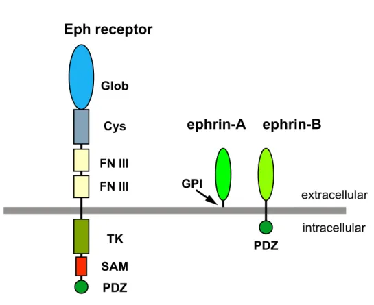
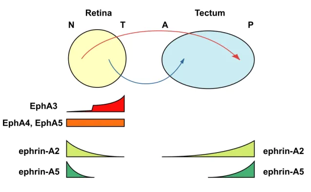
The Eph family of receptors, the largest subgroup of receptor tyrosine kinases, are classified into two subfamilies, EphA and EphB, according to their preference for either glycosyl-phoshpatidyl-inositol (GPI)-anchored ephrin-A ligands or transmembrane ephrin-B ligands (Eph Nomenclature Committee, 1997; Fig. 2). In chick, the EphA3 receptor is expressed in a temporal high-nasal low gradient in the retinal ganglion cells (Cheng and Flanagan, 1994; Fig. 3), and ephrin-A2 and ephrin-A5 are expressed in posterior high-anterior low gradients in the tectum (Cheng and Flanagan,
- 11 -
1994; Drescher et al., 1995; Feldheim et al., 1998; Frisén et al., 1998; Fig. 3). Several lines of evidence suggest that ephrin-A ligands are repulsive to EphA-expressing axons (Cheng and Flanagan, 1994; Drescher et al., 1995; Feldheim et al., 1998; Frisén et al., 1998): According to the expression level of EphA3 in growth cones, growing retinal axons stop at different A-P positions due to the repellent signals from ephrin-A2 and ephrin-A5 graded on the tectum. Consistent with this concept, analyses of ephrin-A2–/– and ephrin-A5–/– mutant mice show that ephrin-As play an important role in the formation of the topographic map along the A-P axis in the retino-collicular system (Frisén et al., 1998; Feldheim et al., 2000). Further, by ectopic expression of the EphA3 receptors in a subset of retinal ganglion cells, it has been shown that EphA3 mediates the repellent action of ephrin-As (Brown et al., 2000).
In addition to EphA3, four EphA receptors (EphA4, EphA5, EphA6 and EphA7) are uniformly expressed in the chick retina (Monschau et al., 1997; Connor et al., 1998; Fig. 3). On the other hand, ephrin-A2 and ephrin-A5 are expressed in nasal high-temporal low gradients in the retina (Marcus et al., 1996; Connor et al., 1998; Hornberger et al., 1999; Fig. 3). Overexpression of ephrin-As in temporal axons leads to errors in the topographic targeting of temporal axons (Dütting et al., 1999; Hornberger et al., 1999), suggesting a role for retinal ephrin-As in the formation of topographic projections. Endogeneous coexpression of EphAs and ephrin-As on the nasal axons causes Eph receptors to autophosphorylate at tyrosine residues (Connor et al., 1998; Hornberger et al., 1999). This phosphorylation is considered to attenuate sensitivity to the repellent cues, and accommodate nasal axons invading into tectal regions expressing substantial amounts of ephrin-A2 and ephrin-A5. Thus, EphAs uniformly expressed in the retina are also thought to be involved in the topographic projection along the A-P axis.
- 12 -
On the other hand, EphB receptors and ephrin-B ligands are known to exhibit a graded distribution along the D-V axis in the retina. EphB2 and EphB3 receptors are expressed in a ventral high-dorsal low gradient (Holash and Pasquale, 1995; Braisted et al., 1997; Connor et al., 1998), and ephrin-B1 is expressed in a dorsal high-ventral low gradient (Braisted et al., 1997). In EphB2-/-/EphB3-/- double mutant mice, the topographic projection along D-V axis is aberrant, suggesting that these EphB receptors are required for the formation of the topographic map (Hindges et al., 2002). Thus, Eph receptors and ephrins play pivotal roles in the axon guidance for the topographic map formation along both axes. However, the mechanisms regulating the expression patterns of Eph receptors and ephrins have not been fully revealed in the retina.
The achievement of graded distributions of topographic molecules along the A-P and D-V axes in the retina and tectum during development is a crucial step in the formation of the topographic map. From the early developmental stages of the retina and tectum, a number of morphogens and transcription factors show asymmetrical expression patterns along the two axes. Studies have demonstrated that these asymmetrically expressed molecules regulate the graded distributions of topographic molecules along the two axes in the retina and optic tectum, which finally leads to the topographic projection. Our lab previously found that two winged-helix transcription factors, chick brain factor 1 and 2 (CBF-1 and CBF-2), are expressed specifically in the nasal and temporal regions of the developing chick retina, respectively (Yuasa et al., 1996). Our lab further demonstrated that ectopic misexpression of CBF-1 and CBF-2 reversed the topographic map in the retinotectal system along the A-P axis (Yuasa et al., 1996). In the embryo infected with CBF-1 recombinant virus, temporal axons frequently passed their appropriate terminal zones of the rostral tectum, and extended caudally (Yuasa et al., 1996). On the other hand, in
- 13 -
embryos infected with the CBF-2 recombinant virus, anterograde-labelled nasal axons frequently arborized in inappropriate regions of the rostral tectum, in addition to the caudal area (Yuasa et al., 1996). Although CBF- 1 and CBF-2 are supposed to determine the regional specificity along the N-T axis in the retina through the regulation of expression of their downstream target genes, their modes of action remain largely unknown. In addition to CBF-1, two homeobox transcription factors, SOHo-1 and GH6, are also expressed specifically in the nasal region of the developing retina (Schulte and Cepko, 2000). Misexpression of these molecules in the retina results in projection errors of retinal axons along the A-P axis, due to the repression of EphA3 expression (Schulte and Cepko, 2000). Since CBF-1 is expressed in the retina before the expression of SOHo-1 and GH6 starts, there is the possibility that these homeobox transcription factors are downstream target genes of CBF-1. However, the relationship between CBF-1 and these homeobox proteins has not been fully investigated.
On the other hand, Along the D-V axis, Tbx5, a dorsal-specific T-box transcription factor, or cVax, a ventral-specific homeobox transcription factor, are known to show asymmetrical expression patterns in the retina. Misexpression of these molecules alters positive regions of EphBs and ephrin-Bs along the D-V axis, which is associated with errors in the targeting by retinal axons of the tectum (Schulte et al., 1999; Koshiba- Takeuchi et al., 2000). Our lab recently identified a novel secreted molecule, Ventroptin, which is an antagonist of bone morphogenetic protein 4 (BMP-4) in the retina, and demonstrated that Ventroptin is implicated in the retinotectal topographic projection along both the dorsoventral (D-V) and A-P axes (Sakuta et al., 2001).
Bone morphogenetic proteins (BMPs) are secreted growth factors, which, based on the amino acid homology of a highly conserved seven-
- 14 -
cysteine domain in the carboxy-terminal region of the proteins, form a subgroup of the transforming growth factor-β (TGF-β) superfamily (Kingsley, 1994). They are dimeric proteins with a single interchain disulfide bond, and this dimeric conformation is an absolute requirement for the biological action of BMPs (Eimon and Harland, 1999). BMPs were originally identified as molecules that can induce ectopic bone and cartilage formation in rodents (Wozney et al., 1988; reviewed in Hogan, 1996). But it became rapidly evident that BMPs also act as multifunctional regulators in morphogenesis during development in vertebrates and invertebrates (reviewed in Hogan, 1996a,b; Graff, 1997; Ebendal et al., 1998; Wozney, 1998). At the early developmental stages of the eye, the dorsal retina-specific expression of BMP-4 has been implicated in the dorsalization in the retina (Koshiba-Takeuchi et al., 2000).
Research in recent years has shown that the BMP signaling pathway is often regulated at both extracellular and intracellular levels. Extracellulary, the activity of BMPs can be regulated by secreted proteins such as Noggin, Chordin, Follistatin, Cerberus, and Gremlin (Smith., 1999). These BMP antagonists specifically bind to BMPs and prevent their binding to specific receptors or their signaling. Ventroptin has a characteristic motif called the cysteine-rich repeat (CR), which is conserved in some proteins, including von Willebrand factor and Chordin. Three CRs of Ventroptin are significantly homologous with those of Chordin, although the remaining region has no homology. Ventroptin binds with high affinity to BMP-4 and with lower affinity to a BMP-4/7 heterodimer, but not at all to BMP-7, TGF-β, or activin (Sakuta et al., 2001). In the retina, Ventroptin can inhibit the function of BMP-4, and regulate the retinotectal topographic projection along both the D-V and A-P axes (Sakuta et al., 2001). At the early developmental stages of the eye, Ventroptin is specifically expressed in the ventral retina, with a
- 15 -
complementary pattern to the dorsal-specific expression of BMP-4 (Sakuta et al., 2001). The counteraction between Ventroptin and BMP-4 governs the regional specification along the D-V axis by regulating the distributions of downstream target genes, such as Tbx5 and cVax, along the D-V axis (Sakuta et al., 2001). At the later stages (E6 onward), Ventroptin begins to be expressed in a nasal high-temporal low gradient, in addition to a ventral high-dorsal low gradient. This is the first demonstration of a molecule with a double-gradient pattern of expression in the retina. Since expression of BMP-4 declines from E5, a member of the TGF-β family other than BMP-4 is supposed to appear in a temporal high-nasal low gradient along the N-T axis with a complementary pattern to the Ventroptin expression (Sakuta et al., 2001). Asymmetrical expression of Ventroptin along the N-T axis regulates the graded expression of ephrin-A2 along this axis but not of ephrin-A5 or EphA3 in the retina, which is associated with the retinotectal mapping along the A-P axis (Sakuta et al., 2001).
extracellular intracellular
Eph receptor
ephrin-A
Glob
GPI
TK FN III
Cys
FN III
ephrin-B
SAM PDZ
PDZ
Fig. 2. Schematic representation of the domain structure of the Eph receptor and ephrin ligands.
The domain structure of Eph receptors is conserved from sponges up to humans. The two ephrin classes differ in their type of membrane attachment, that is, GPI- anchored (ephrin-A) versus transmembrane region (ephrin-B). The cytoplasmic domain of ephrin-Bs contains conserved tyrosine phosphorylation motifs and a PDZ motif. Glo, globular; Cys, cysteine-rich; FNIII, fibronectin type III; TK, tyrosine kinase motif.
Retina Tectum
A P
EphA3 EphA4, EphA5
ephrin-A2 ephrin-A5
ephrin-A2 ephrin-A5
N T
Fig. 3. Summary of the expression pattern of EphA family members in the retina and the tectum.
The EphA receptors EphA4 and EphA5 are uniformly expressed in the retina, whereas EphA3 is expressed in the temporal retina in a gradient, and there is little or no expression in the nasal retina. Ephrin-A2 and ephrin-A5 are expressed in the retina in a high-nasal-to-low-temporal gradient, whereby the expression domain of ephrin-A5 is restricted more to the nasal retina than that of ephrin-A2. Both ligands are expressed in the tectum in a high-caudal-to-low-rostral gradient. The expression domain of ephrin-A5 is restricted more to the caudal half of the tectum than that of ephrin-A2. The projection of temporal axons onto the rostral tectum and the projection of nasal axons onto the caudal tectum are indicated. N, nasal; T, temporal; A, anterior; P, posterior.
- 19 -
Chapter II
CBF-1 regulates the expression of asymmetrically distributed
molecules through multiple mechanisms
- 20 -
Introduction
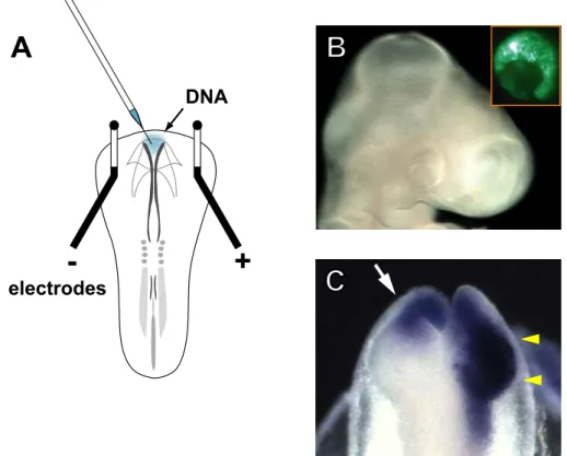
Chick brain factor-1 (CBF-1), a nasal retina-specific winged-helix transcription factor, is known to prescribe the nasal specificity which leads to the formation of the precise retinotectal map especially along the antero- posterior (A-P) axis (Yuasa et al., 1996). However, the molecular mechanisms by which CBF-1 controls the expression of topographic molecules have not been elucidated. In the present study, to gain insight into the downstream target genes of CBF-1, I employed, for the first time, electroporation of a retroviral vector carrying the CBF-1 gene into the optic vesicle, and examined effects of the misexpression of CBF-1 on the expression of topographic molecules and other asymmetrically distributed molecules. The in ovo electroporation of retrovirus vector at Hamburger- Hamilton (HH) stage 8 (Hamburger and Hamilton, 1951) allows the immediate (at stage 10) and sustained expression of a transgene, as shown in Fig. 4C. Here, I show that ectopic expression of CBF-1 in the temporal retina represses expression of EphA3 and CBF-2, and induces that of SOHo-1, GH6, ephrin-A2, and ephrin-A5. The mode of action of CBF-1 on these molecules has been revealed to be classified into three distinct categories. Based on these results I suggest that CBF-1 plays pivotal roles in the determination and maintenance of the N-T specificity in the retina by integratively regulating the expression of asymmetrically distributed molecules through multiple mechanisms.
- 21 -
Materials and Methods
Plasmids
To prepare the CBF-1/RCAS construct, the coding region of CBF-1 was amplified from a retroviral construct expressing CBF-1 (CBF-1/RCAS (B): Yuasa et al., 1996), subcloned once into a shuttle vector SLAX-NS containing the 5’-noncoding region of the Src gene (Suzuki et al., 2000), and then transferred into the RCAS-NS retrovirus vector (Suzuki et al., 2000).
To make CBF-1-eve/RCAS, the repression domain of the Drosophila even-skipped protein (Han et al., 1993) from pCS2eve was first fused with the CBF-1 DNA binding domain (amino acid residues 139 to 253). It was inserted into SLAX-NS myc, which was prepared with a shuttle vector SLAX-NS by inserting double myc-epitope tags at the NcoI-EcoRI site, and then subcloned into the RCAS-NS retroviral vector using NotI and SpeI sites. The CBF-1AA mutant was generated by substitutions of asparagine 189 and histidine 193 with alanines by PCR, and cloned into RCAS-NS or pcDNA3.1(+) (Invitrogen).
In ovo electroporation
In ovo electroporation was performed as described previously (Sakuta et al., 2001; Fig. 4). Retrovirus constructs for the electroporation were suspended at a concentration of 0.1 – 1.0 µg/µl in 10 mM Tris-HCl, 0.25 mM ethylendiamine tetraacetic acid (EDTA), pH 8.0 containing 0.05%(w/v) Fast Green (Sigma). Embryos were electroporated at HH stage 8 and incubated in a humidified incubator. The embryos of normal size and morphology were used for assays.
In situ hybridization and riboprobes
- 22 -
Section in situ hybridization and whole-mount in situ hybridization were carried out as described previously (Suzuki et al., 2000). Templates used for digoxigenine-labeled RNA probes were as follows: The 516bp fragment of chick CBF-1 (nucleotide residues 1252-1761; GenBank accession number U47275), the 656bp fragment of chick CBF-2 (1191- 1846; U47276), the 724bp fragment of chick EphA3 (2267-2990; M68514), the 541bp fragment of chick ephrin-A2 (94-634; L40932), the 681bp fragment of chick ephrin-A5 (25-705; X90377), the 782bp fragment of chick SOHo-1 (34-815; S69380), the 567bp fragment of chick GH6 (4-570; AF227921) and the 1179bp fragment of chick BMP-2 (1-1179; AY237249). The template for the probe of Ventroptin was described previously (Sakuta et al., 2001). Image acquisition and figure processing of sections and whole-mount samples were performed as reported (Suzuki et al., 2000).
Northern blot analysis
Total RNA was prepared from one-third of the nasal or temporal E8 chick retina electroporated with CBF-1/RCAS, with Trizol Reagent (Invitrogen) following the manufacturer’s protocol. Control retinae were prepared from the left eye of the same embryos. Northern blot analysis with 20 µg of total RNA was performed as described previously (Suzuki et al., 2000). Templates used for preparing the probe were as follows: the same fragments of chick EphA3, ephrin-A2, and ephrin-A5 used for in situ hybridization, the 300bp fragment of chick CBF-2 (nucleotide residues 1418-1717; GenBank accession number U47276), and the 477bp fragment of chick glyceraldehyde-3-phosphate dehydrogenase (GAPDH) (218-794; K01458).
DNA pull-down assay
Chick embryonic fibroblasts were transfected with retroviral vectors for
- 23 -
myc-tagged CBF-1 or myc-tagged CBF-1AA mutant using Lipofectamine plus (Invitrogen), and cultured for a week. The preparation of nuclear extract and DNA pull-down assay were performed as described previously (Mukai et al., 2002). For DNA pull-down assays, biotininylated double- stranded oligonucleotides derived from the sequence named B2 in the HNF1 promoter were synthesized by Qiagen: B2 is known as the BF-1 binding sequence (Li et al., 1995). The eluates of precipitates were subjected to SDS-PAGE, followed by immunoblot analyses with the anti- myc primary antibody 9E10 (Santa Cruz Biotechnology) and horseradish peroxidase-conjugated secondary antibody (Amersham Biosciences). CBF-1 or its was visualized by chemiluminiscence using ECL plus (Amersham Biosciences) and the lumino-image analyzer LAS-1000plus (Fujifilm).
- 24 -
Results
Expression patterns of asymmetrically distributed molecules along the N-T axis in the retina
During retinal development, two winged-helix transcription factors, CBF-1 and CBF-2, begin to be expressed in the nasal and temporal regions, respectively, prior to HH stage 10 (Yuasa et al., 1996; Yamagata et al., 1999). After that, two homeobox transcription factors, SOHo-1 and GH6 (from HH stage 12-14), and then EphA3 (from HH stage 15) begin to be asymmetrically expressed along the N-T axis in the retina (Yamagata et al., 1999; Schulte and Cepko, 2000).
Although ephrin-A5 and ephrin-A2 are also known to be expressed in nasal high-temporal low gradients (Cheng and Flanagan, 1994), their spatial and temporal expression patterns in the retina had not been fully investigated. Thus, we examined their expression during development of the chick retina by whole-mount and section in situ hybridization. Expression of ephrin-A5 was first detected in the optic vesicle at HH stage 12, while asymmetrical distributions were not detected (Fig. 5A). At stage 18, expression of ephrin-A5 was absent in the dorsal and ventral regions, but observed in the nasal and temporal retina (Fig. 5Ba). At this stage, the expression was significantly stronger on the nasal than temporal side (Fig. 5Ba). It was evident also by section in situ hybridization. In a section along the N-T axis, ephrin-A5 expression was observed in the nasal and temporal third of the retina, although the expression was stronger on the nasal than temporal side (Fig. 5Bb). Thus, ephrin-A5 was not expressed in a nasal-specific fashion at stage 18 (E3). At E6, a nasal high- temporal low gradient was clearly visible for the first time, although the expression was also observed in the periphery of the temporal retina (data not shown). At E8, a similar nasal high-temporal low expression gradient
- 25 -
and expression in the temporal periphery was observed (Fig. 5Ca-5Cc). At this stage, ephrin-A5 transcripts in the central retina were mainly distributed in the ganglion cell layer (GCL), while those in the peripheral regions were observed in all cell layers like the retina at early stages (Fig. 5Ca-5Cc).
In contrast, the onset of ephrin-A2 expression in the retina was quite late: Expression of ephrin-A2 was not detected until E4 (Fig. 5D) and began to be observed from E5 in the nasal retina (data not shown). From E6 onward, a nasal high-temporal low gradient of ephrin-A2 expression was observed predominantly in the GCL (Fig. 5Ea-5Ec): The gradient of ephrin-A2 expression in the GCL appeared to be slighter than that of ephrin-A5 expression at E6 and E8 (Fig. 5Eb and data not shown). Although ephrin-A2 expression was also detected in cell layers other than the GCL, the expression appeared to be rather uniform along the N-T axis, as compared with that of ephrin-A5 (compare Fig. 5Cb, 5Cc with Fig. 5Eb, 5Ec).
Misexpression of CBF-1 affects expression patterns of asymmetrically distributed molecules along the N-T axis in the retina
I investigated the expression of the topographic molecules mentioned above when CBF-1 was misexpressed into the area of the eye. As shown in Fig. 4B, in ovo electroporation gives rise to ectopic expression of a transgene (EGFP) all over the retina. In the present study, to sustain the transgene expression, I attempted to transfect a replication-competent avian retrovirus RCAS-BP vector carrying the CBF-1 gene (CBF-1/RCAS) into the right optic vesicle of HH stage 8 embryos by electroporation. In embryos transfected with CBF-1/RCAS, the expression of CBF-1 was observed in the whole of the right optic vesicle by in situ hybridization at 6 h post-electroporation as early as HH stage 10, prior to the stage when the
- 26 -
polarity along the N-T axis appears to be determined (Fig. 4C and data not shown). Signals in the anterior optic vesicle represents endogenous expression of CBF-1, as it is evident from the untransfected left side in which only the endogenous expression in the anterior optic vesicle is detected (Fig. 4C).
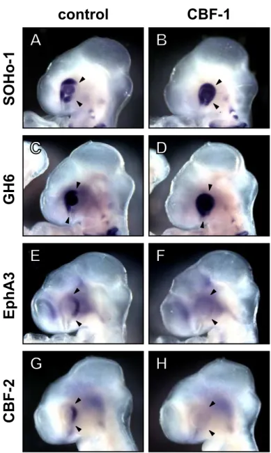
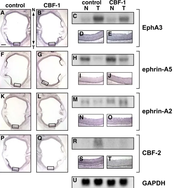
At first, I examined the expression of SOHo-1, GH6, EphA3, and CBF-2 at HH stage 18-20 (E3) in the electroporated embryos, because these molecules clearly show graded distributions along the N-T axis in the native embryos at this stage. At E3, SOHo-1 and GH6 were expressed mainly in the nasal retina and the lens ectoderm but not in the temporal retina (Fig. 6A and 6C), while EphA3 expression was observed in the temporal retina (Fig. 6E). When CBF-1 was misexpressed in the retina, the expression of SOHo-1 (Fig. 6B; 5 out of 8 embryos) and GH6 (Fig. 6D; 4 out of 6 embryos) was significantly induced in the temporal retina. In contrast, misexpression of CBF-1 resulted in loss of EphA3 expression in the temporal retina (Fig. 6F; 8 out of 9 embryos). Our lab previously reported that misexpressed CBF-1 repressed the expression of the CBF-2 gene (Yamagata et al., 1999). Consistent with this observation, misexpression of CBF-1 completely repressed the temporal-specific expression of CBF-2 at E3 (Fig. 6H; 5 out of 5 embryos).
I next examined the expression of EphA3, ephrin-A5, ephrin-A2, and CBF-2 at E8. At this stage, these molecules distribute asymmetrically along the N-T axis in the retina. Misexpression of CBF-1 continuously repressed the expression of EphA3 in the GCL in the temporal retina (Fig. 7B and 7E; 4 out of 5 embryos). In some embryos, a complete loss of EphA3 expression was observed (2 out of 5 embryos). Conversely, CBF-1 misexpression induced ectopic expression of ephrin-A5 (Fig. 7G and 7J; 5 out of 5 embryos) and ephrin-A2 (Fig. 7L and 7O; 5 out of 7 embryos) in the GCL in the temporal retina: In the control retina, the expression of these
- 27 -
ephrins is weak in the GCL in the temporal retina (Fig. 7I and 7N). The expression of CBF-2 in the temporal retina was completely repressed by the misexpression of CBF-1 (Fig. 7Q and 7T: 5 out of 6 embryos). I further examined the effects of misexpressed CBF-1 on expression of the EphA4 receptor, which is uniformly expressed in the developing retina (Holash and Pasquale, 1995; Dütting et al., 1999; Hornberger et al., 1999). When CBF-1 was misexpressed in the developing retina, the expression of EphA4 was not affected (data not shown).
The effects of misexpressed CBF-1 on the expression of EphA3, ephrin-A5, ephrin-A2, and CBF-2 were verified by Northern blot analysis. Expression of the EphA3 transcript was significantly repressed on the temporal side of the retina by misexpression of CBF-1 (Fig. 7C). In contrast, CBF-1 misexpression induced expression of ephrin-A5 and ephrin-A2 transcripts on the temporal sides (Fig. 7H and 7M). Interestingly, the expression of ephrin-A2 and ephrin-A5 was also enhanced in the nasal retina, where these ephrins were endogenously expressed. From the in situ hybridization, however, this effect was not clearly detectable due to homogeneous enhancement. Finally, consistent with the results of in situ hybridization, the expression of CBF-2 in the temporal retina was completely abolished by the misexpression of CBF-1 (Fig. 7R).
Repressing construct of CBF-1 regulates expression of SOHo-1, GH-6, EphA3, CBF-2, and ephrin-A5 similar to the wild-type, but not
expression of ephrin-A2
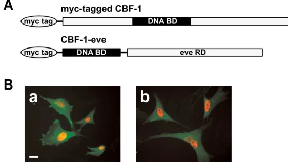
I next analyzed the mode of action of CBF-1. Since BF-1 is suggested to act as a transcriptional repressor (Li et al., 1995; Bourguignon et al., 1998), we examined whether this function is essential to regulate the expression of SOHo-1, GH-6, EphA3, CBF-2, ephrin-A5 and ephrin-A2 in the developing retina. I designed a RCAS vector (CBF-1-eve/RCAS) expressing a
- 28 -
chimeric protein that consists of an even-skipped repression domain (Han et al., 1993) and a winged helix DNA binding domain (WH domain) of CBF-1 as shown in Fig. 8-I A. It has been demonstrated that nuclear localization of CBF-1 is determined by a sequence within the WH domain (Chang et al., 1996). Consistent with this report, the chimeric protein was found to be concentrated in the nucleus when expressed in the chick embryonic fibroblasts (Fig. 8-I B), and in the chick retinal cells (data not shown).
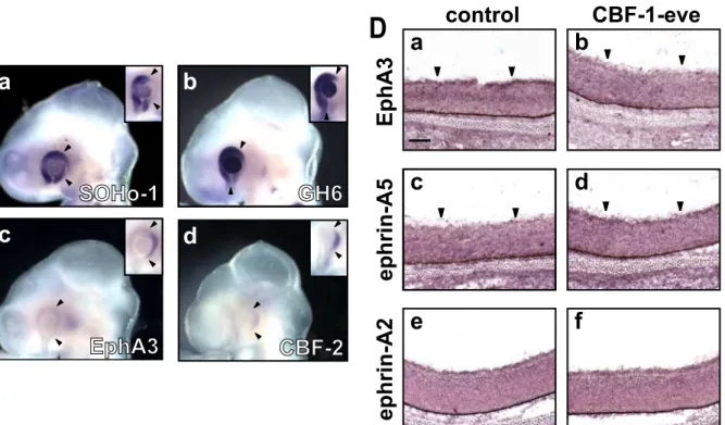
In ovo electroporation of CBF-1-eve/RCAS at HH stage 8 in the optic vesicle resulted in an expansion of SOHo-1 (Fig. 8-II Ca; 5 out of 9 embryos) and GH6 expression (Fig. 8-II Cb; 6 out of 11 embryos) into the temporal side of the retina at E3. On the other hand, expression of EphA3 (Fig. 8-II Cc; 7 out of 9 embryos) and CBF-2 (Fig. 8-II Cd; 3 out of 3 embryos) in the temporal retina was repressed by electroporation of CBF- 1-eve/RCAS. At E8, continuous repression of EphA3 expression (Fig. 8- II Db; 8 out of 8 embryos) and induction of ephrin-A5 expression (Fig. 8-II Dd; 6 out of 8 embryos) were observed in the temporal retina as expected. However, surprisingly, the expression of ephrin-A2 was not affected by the introduction of RCAS/CBF-1-eve (Fig. 8-II Df; 5 out of 5 embryos). These results suggest that CBF-1 functions as a transcriptional repressor for the regulation of expression of SOHo-1, GH6, EphA3, CBF-2, and ephrin- A5. On the other hand, expression of ephrin-A2 appeared to be regulated by CBF-1 through a different mechanism.
Mutant CBF-1 deficient in DNA binding ability exerts similar effects on expression of SOHo-1, GH-6, EphA3, CBF-2, and ephrin-A2 as the wild type CBF-1, but not on expression of ephrin-A5
I next tested whether DNA binding of CBF-1 is essential for the activity to regulate the expression of asymmetrically distributed molecules. Two
- 29 -
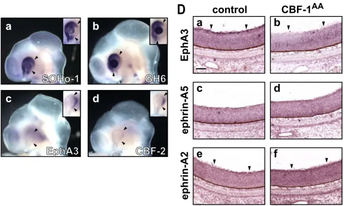
amino acid residues in the WH domain of murine BF-1, Asn219 and His223, are suggested to be important for binding to DNA, and it has been shown that the mutant generated by changing these two residues to alanine (AA mutant) is devoid of DNA binding ability (Dou et al., 2000). I prepared CBF-1AA/RCAS to express the mutant CBF-1 protein in which Asn189 and His193 were changed to alanine: These two are equivalent to Asn219 and His223 in murine BF-1, respectively (Fig. 9-I A). I examined the ability of the wild-type CBF-1 and CBF-1AA mutant proteins to bind to the BF-1 binding sequence by DNA pull-down assay beforehand. The wild-type protein was co-precipitated with the double-stranded DNA fragment containing the BF-1 binding sequence (Fig. 9-I Ba). On the other hand, CBF-1AA was not co-precipitated (Fig. 9-I Ba), although sufficient amounts of mutant proteins were expressed (Fig. 9-I Bb). Thus, the mutation at Asn189 and His193 abolishes the ability of CBF-1 to bind the BF-1 binding sequence.
Then, I misexpressed the CBF-1AA mutant in the developing retina by in ovo electroporation at HH stage 8. When the mutant protein was misexpressed, positive regions for SOHo-1 (Fig. 9-II Ca; 7 out of 13 embryos) and GH6 (Fig. 9-II Cb; 4 out of 11 embryos) expanded into the temporal sides in the E3 retina. On the other hand, expression of EphA3 (Fig. 9-II Cc; 5 out of 6 embryos) and CBF-2 (Fig. 9-II Cd; 4 out of 4 embryos) in the temporal retina was almost completely abolished. At E8, repression of the EphA3 expression was continuously observed in the temporal retina (Fig. 9-II Db; 4 out of 4 embryos). In addition, induction of ephrin-A2 expression (Fig. 9-II Df; 4 out of 5 embryos) was observed in the temporal retina. However, expression of ephrin-A5 was not affected by the introduction of CBF-1AA/RCAS (Fig. 9-II Dd; 6 out of 6 embryos). These results suggest that the DNA binding activity of CBF-1 is necessary for the regulation of the expression of ephrin-A5. On the other hand,
- 30 -
expression of SOHo-1, GH6, EphA3, CBF-2, and ephrin-A2 can be regulated by CBF-1 through a DNA binding-independent mechanism.
- 31 -
Discussion
In ovo electroporation of replication-competent retroviral vector allows immediate and sustained expression of transgenes
Our lab previously reported that misexpression of CBF-1 in the retina did not alter the distribution of EphA3 (Yuasa et al., 1996; Yamagata et al., 1999). Another group reported that the expression of SOHo-1 and GH6 was not affected by misexpression of CBF-1 (Schulte and Cepko, 2000) using our retroviral construct CBF-1/RCAS (B), a replication-competent retroviral vector carrying the CBF-1 gene (Yuasa et al., 1996). These experimental results were obtained through the infection of retroviral particles or in ovo electroporation of the pMiw expression plasmid vector to misexpress CBF-1 in the developing retina. By virus infection, it usually takes more than 12 h for the expression of transgenes to start, whereas by in ovo electroporation of the plasmid vector, the expression of transgenes begins immediately (within 3 h) but it stops in a few days. In the present study, the expression level of the CBF-1 transgene was improved by modification of the last RCAS construct (Yuasa et al., 1996): I replaced the long 5’-untranslated region of CBF-1 in the previous construct with the 5’-untranslated region of the src gene that confers efficient expression on heterologous coding sequences (Morgan and Fekete, 1996; Suzuki et al., 2000; see Materials and Methods). In addition, I employed in ovo electroporation of CBF-1/RCAS DNA instead of virus infection.
This strategy allowed the immediate and sustained expression of the transgene, because the mRNA is directly transcribed from the incorporated RCAS vector DNA shortly after the gene transfer, which is followed by the production of retroviral particles and widespread infection in the retina. When CBF-1/RCAS was introduced by in ovo electroporation into the
- 32 -
optic vesicle of HH stage 8 embryos, the transgene expression was detected at 6 h after the electroporation, as early as HH stage 10, just before the stage at which the polarity along the N-T axis appears to be determined by the endogenous CBF-1 expression (Dütting and Meyer, 1995; Dütting and Thanos, 1995; Thanos et al., 1996), indicating that this in ovo electroporation method fulfills the requirements for the functional study of CBF-1.
CBF-1 controls all of the asymmetrically distributed molecules along the N-T axis in the developing retina
Here, I show that misexpression of CBF-1 in the temporal retina results in induction of SOHo-1, GH6, ephrin-A2, and ephrin-A5, and down- regulation of EphA3 and CBF-2 expression (Fig. 13). These results indicate that all of the asymmetric molecules so far identified to be involved in the formation of the topographic map along the A-P axis (Yuasa et al. 1996; Drescher et al., 1995; Nakamoto et al., 1996; Schulte and Cepko, 2000; Dütting et al., 1999; Hornberger et al., 1999) are downstream genes of CBF-1. Endogenous expression of CBF-1 begins around HH stage 8-9 in the retina, and is confined to the nasal retina prior to HH stage 11 (Yuasa et al. 1996; Yamagata et al., 1999), preceding all other genes that are asymmetrically expressed along the N-T axis during development. CBF-2 is first asymmetrically expressed in the temporal retina at HH stage 11 (Yamagata et al., 1999). Expression of SOHo-1 and GH6 is restricted in the nasal retina from HH stage 12-14 (Yamagata et al., 1999; Schulte and Cepko, 2000). EphA3 expression is confined to the temporal retina at HH stage 15 (Yamagata et al., 1999; Schulte and Cepko, 2000). In addition, as shown in this report, graded distributions of ephrin-A5 and ephrin-A2 are observed after HH stage 18 (E3) and E5, respectively. The order of onset of these genes thus supports the view that CBF-1 determines the N-T
- 33 -
specificity, thereby controlling the expression of a series of asymmetrically distributed molecules in the developing retina. It is likely that misexpression of CBF-1 endows the temporal retinal ganglion cells with the character of nasal ganglion cells by inducing the expression of SOHo-1, GH6, ephrin-A5 and ephrin-A2, and inhibiting the expression of CBF-2 and EphA3.
In this study, I also misexpressed CBF-2 in parallel experiments. Unexpectedly, misexpression of CBF-2 had no effect on the expression of asymmetrically distributed molecules including CBF-1 (data not shown). Since misexpression of CBF-2 also results in retinotectal projection errors along the A-P axis (Yuasa et al., 1996), CBF-2 is also likely to be involved in the formation of the retinotectal map by regulating the expression of unknown topographic molecule(s). In previous experiments, misexpressed CBF-1 completely repressed the expression of CBF-2 (see also Yamagata et al., 1999), suggesting that CBF-1 can control the expression of these unknown topographic molecules through the regulation of CBF-2 expression. One potential candidate is a receptor for a membrane-associated glycoprotein, RGM, which was recently reported to be distributed in a gradient with increasing concentrations from the anterior to posterior pole of the embryonic tectum and collapse the temporal growth cones (Stahl et al., 1990; Müller et al., 1996; Monnier et al., 2002).
CBF-1 regulates the expression of asymmetrically distributed molecules through multiple mechanisms
BF-1 has been shown to act primarily as a transcriptional repressor (Li et al., 1995; Bourguignon et al., 1998). In addition to the functions which are dependent on their ability to bind to DNA, several transcription factors have been reported to function even when their DNA binding-ability is deficient (Beato et al., 1995; Um et al., 1995; Plaza et al., 1997; Schuur et
- 34 -
al., 2001; Zhao et al., 2001). This DNA binding-independent function is also observed in murine BF-1 (Dou et al., 2000; Rodriguez et al., 2001). To clarify the modes of action of CBF-1, I examined effects of misexpression of CBF-1-eve and CBF-1AA mutants. As a result, we found that the downstream asymmetric molecules can be classified into three distinct categories by the mode of action of CBF-1 (Fig. 13A): (1) those whose expression is affected only by the CBF-1-eve mutant (written in red), (2) those whose expression is affected only by the CBF-1AA mutant (in green), and (3) those whose expression is affected by both the CBF-1-eve and CBF-1AA mutants (in blue).
Ephrin-A5 belongs to the first category. Consistent with the reports that CBF-1 acts as a transcriptional repressor, misexpressed CBF-1-eve shows similar activity for the expression of ephrin-A5 as the wild-type CBF-1. Because misexpression of CBF-1AA mutant protein has no effect on the expression of ephrin-A5, the expression of this molecule is regulated only by the function of CBF-1 as a transcriptional repressor (Fig. 13A). How does CBF-1, a transcriptional repressor, induce the expression of ephrin-A5? The simplest explanation is that CBF-1 functions as a transcriptional repressor of some other putative intermediate repressor(s) which inhibits the expression of ephrin-A5 (indicated by ‘X’ in Fig. 13B). The onset of ephrin-A5 expression appears to be triggered first by a transcription activator(s) at stage 12 to yield the homogeneous expression in the retina (Fig. 5A). Subsequently, factor X may begin to be expressed in the temporal retina where CBF-1 is absent, and represses expression of ephrin-A5 in the temporal retina. In misexpressed embryos, ectopic CBF- 1 probably abrogates the repression by factor X from the temporal retina, and then expression of ephrin-A5 is re-induced by the transcriptional activator(s) which is ubiquitously expressed in the retina.
Ephrin-A2 belongs to the second category. Surprisingly, expression
- 35 -
of ephrin-A2 is not affected by misexpression of the CBF-1-eve fusion protein. In contrast, CBF-1AA exerted the same effect on the expression of ephrin-A2 as the wild-type protein. Therefore, expression of ephrin-A2 is regulated only by a DNA binding-independent mechanism, which clearly contrasts with ephrin-A5 whose expression is regulated only by a DNA binding-dependent mechanism (Fig. 13A). In the retina, both ephrin-A2 and ephrin-A5 are expressed in a nasal high-temporal low gradient. Although these ephrins commonly function as ligands for EphA receptors, their expression patterns are not identical. The gradient of ephrin-A2 expression in the GCL is gentle and wide along the N-T axis, as compared with that of ephrin-A5 expression. In addition, the onsets of their expression are also distinct: ephrin-A5 begins to be expressed at HH stage 12 (E2), but ephrin-A2 does not appear until E5. These differences may reflect the distinct regulatory mechanisms for their expression. I further investigated the molecular mechanism of the regulation of ephrin-A2 by a DNA binding-independent mechanism, and found that CBF-1 interferes with BMP signaling and ephrin-A2 is controlled by a BMP. See the next chapter for details.
SOHo-1, GH6, EphA3, and CBF-2 belong to the third category. The expression of these molecules is affected by misexpression of both the CBF-1-eve fusion protein and CBF-1AA mutant protein, indicating that the expression of these molecules is regulated by CBF-1 through not only a DNA binding-dependent but also a DNA binding-independent mechanism (Fig. 13A). Since SOHo-1 and GH6 repress the expression of EphA3 in the retina (Schulte and Cepko, 2000), repression of EphA3 expression by CBF-1 likely resulted from induction of SOHo-1 and GH6 expression by CBF-1. It may also be that CBF-1 directly down-regulates the expression of EphA3 not through SOHo-1 or GH6. This is, however, unlikely because EphA3 is homogenously expressed in the optic vesicle at HH stage
- 36 -
12, when CBF-1 is topographically expressed (Yamagata et al., 1999). In addition, CBF-1 and EphA3 are coexpressed in the same region of the chick forebrain (our unpublished observations). Thus, CBF-1 probably represses EphA3 expression through induction of SOHo-1 and GH6 expression. To explain the induction of SOHo-1 and GH6 expression by CBF-1 through a DNA binding-dependent mechanism, the existence of a putative intermediate repressor(s), which inhibits expression of SOHo-1 and GH6, must be postulated (indicated by ‘Y’ in Fig. 13B). As in the regulation of ephrin-A5 expression, misexpressed CBF-1 abrogates the repression by factor Y, and then the expression of SOHo-1 and GH6 is induced by a transcriptional activator(s) which is ubiquitously expressed in the retina.
Expression of SOHo-1, GH6, and CBF-2 is also regulated by CBF-1 through a DNA binding-independent mechanism (Fig. 13A). Several transcription factors are known to regulate the expression of their downstream target genes by interacting with other transcription factors (Beato et al., 1995; Um et al., 1995; Plaza et al., 1997; Schuur et al., 2001; Zhao et al., 2001). Thus, it is probable that CBF-1 regulates the expression of SOHo-1, GH6, and CBF-2 via interaction with other transcription factors which directly regulate the expression of SOHo-1, GH6, and CBF-2.
What is the meaning of using two distinct mechanisms, DNA binding-dependent and -independent (Fig. 13A), to regulate the expression of topographic molecules? It may be for security to maintain regulation of the EphA/ephrin-A system: For instance, if the DNA binding- dependent mechanism is lost by a mutation in the WH domain of CBF-1, or by a dysfunction of any factor which regulates this mechanism with CBF-1, CBF-1 can still regulate the asymmetrical distribution of EphA3 and ephrin-A2 through the remaining DNA binding-independent mechanism.
- 37 -
On the other hand, if the DNA binding-independent mechanism is lost, CBF-1 can still regulate the asymmetrical distribution of EphA3 and ephrin-A5 through the DNA binding-dependent mechanism. Gene knock- out of ephrin-A2 and ephrin-A5 in mice suggests the importance of this redundancy: In the single knock-out mice, a substantial part of the retinal axons normally projected onto the SC (Frisén et al., 1998; Feldheim et al., 2000). Since the total EphA/ephrin-A system plays an essential role in the formation of the retinotectal map, this dual regulatory system by CBF-1 might have evolved during the evolution of the visual system.
Fig. 4. Gene transfer by electroporation into the developing chick retina.
(A) Schematic representation of in ovo electroporation. Electroporation was performed on stage 8 embryos with electrodes arranged as shown in the drawing. A solution containing plasmid DNA and Fast Green was injected into the anterior neuropore (blue colored site). Upon charging the current, DNA molecules in the neuropore moved toward the anode side placed on the right side of the embryo, and the right eye was selectively transfected.
(B) An E3 (stage 19) embryo transfected with pEGFP-N1. Inset shows higher magnification of the right eye observed under UV light, visualizing the cells that took up the transgenes. Almost all the retinal cells were EGFP-positive.
(C) A stage 10 embryo transfected with CBF-1/RCAS. Retrovirus vector-derived CBF-1 signals were detected in the whole of the right optic vesicle (yellow arrowheads) at 6 h post-electroporation by in situ hybridization (note that signals in the anterior optic vesicle contain endogenous expression). The untransfected left side shows the endogenous expression of CBF-1 in the anterior optic vesicle (white arrow).
- +
electrodes
A B
C
DNA
GH6SOHo-1EphA3CBF-2
control CBF-1
F
D
B
H
E
C
A
G
Fig. 6. Misexpressed CBF-1 induces expression of SOHo-1 and GH6, and represses expression of EphA3 and CBF-2.
Whole-mount in situ hybridization of E3 chick embryos (HH stage 18 to 20) transfected with CBF-1/RCAS. Embryos were hybridized with antisense probes for SOHo-1 (A and B), GH6 (C and D), EphA3 (E and F), or CBF-2 (G and H). The untransfected sides (control) are shown in (A,C,E and G), and the contralateral transfected sides of the same embryo are shown in (B,D,F and H) as inverted images for ease of comparison. Arrowheads indicate the border of the endogenous expression area on the control side.
ephrin-A2 ephrin-A5
N
T
EphA3
CBF-2
GAPDH
Fig. 7. Misexpressed CBF-1 induces expression of ephrin-A5 and ephrin-A2, and repressed that of EphA3 and CBF-2.
(A,B,F,G,K,L,P and Q) Horizontal section in situ hybridization of E8 embryos electroporated with CBF-1/RCAS.
(D,E,I,J,N,O,S and T) Respective enlargements of the boxed temporal areas. Sections were hybridized with antisense probes for EphA3 (A,B,D and E), ephrin- A5 (F,G,I and J), ephrin-A2 (K,L,N and O), or CBF-2 (P,Q,S and T). The untransfected control eyes are shown in (A,D,F,I,K,N,P and S), and the contralateral transfected eyes of the same embryos are shown in (B,E,G,J,L,O,Q and T). Nasal (anterior) is up, temporal (posterior) is down. Scale bar, 600 µm.
(C,H,M,R and U) Northern blot analysis of E8 chick retina electroporated with CBF- 1/RCAS. Northern blot analysis was performed using 20 µg of total RNA prepared from the nasal (N) and temporal (T) thirds of E8 retinae transfected with CBF- 1/RCAS (CBF-1). RNA of control retinae (control) was prepared from the left eyes of the same embryos. Probes used for each panel are indicated on the right. A probe for GAPDH, glyceraldehyde phosphate dehydrogenase, was used for control of RNA amounts.
A
CBF-1-eve
myc-tagged CBF-1
DNA BD myc tag
B a b
eve RD myc tag DNA BD
Fig. 8-I. Misexpression of CBF-1 repressing form (CBF-1-eve) alters the expression of SOHo-1, GH6, EphA3, CBF-2, and ephrin-A5, but not ephrin-A2.
(A) Schematic representation of the wild-type and chimeric CBF-1. The top drawing represents the wild-type CBF-1 protein with myc tag. The repressing form of CBF-1 (CBF-1-eve) was constructed by fusion of the CBF-1 DNA binding domain (DNA BD) and even-skipped repression domain (eve RD).
(B) Nuclear localization of CBF-1 proteins. Chick embryonic fibroblasts were transfected with retroviral vectors for myc-tagged CBF-1 (Ba), or CBF-1-eve (Bb). Nuclear localization of the expressed proteins was visualized by immunofluorescence using anti-myc primary antibody (red). Transfected cells were detected by expression of the viral gag protein from RCAS vector using an anti-gag antibody (green). Scale bar, 20 µm.
Fig. 8-II. Misexpression of CBF-1 repressing form (CBF-1-eve) alters the expression of SOHo-1, GH6, EphA3, CBF-2, and ephrin-A5, but not ephrin-A2.
(C) Whole-mount in situ hybridization of E3 (stage 18 - 20) chick embryos transfected with CBF-1-eve/RCAS using antisense probes for SOHo-1 (Ca), GH6 (Cb), EphA3 (Cc), or CBF-2 (Cd). Insets show the normal expression of SOHo-1, GH6, EphA3, or CBF-2, in the control eyes. Arrowheads indicate the border of endogenous expression areas on the control side.
(D) Horizontal section in situ hybridization of E8 retina transfected with CBF-1- eve/RCAS using antisense probes for EphA3 (Da and Db), ephrin-A5 (Dc and Dd), or ephrin-A2 (De and Df). The temporal regions of the untransfected retinae (control) are shown in the left panels (Da, Dc, and De), and those of the contralateral transfected temporal retinae of the same embryos are shown in the right panels (Db, Dd, and Df). Scale bar, 100 µm.
Fig. 9-I. Misexpression of CBF-1AA mutant proteins alters expression of SOHo-1, GH6, EphA3, CBF-2, and ephrin-A2, but not ephrin-A5.
(A) Schematic representation of myc-tagged CBF-1AA mutant. The CBF-1AA mutant is deficient for DNA binding activity due to substitutions of asparagine 189 and histidine 193 with alanines.
(B) DNA pull-down assays using the nuclear extracts prepared from chick embryonic fibroblasts transfected with myc-tagged CBF-1 or CBF-1AA (Ba). Western blot analysis using anti-myc primary antibody indicated the amounts of nuclear extracts used in the DNA pull-down assays (Bb).
A
B
myc-tagged CBF-1AA
Fig. 9-II. Misexpression of CBF-1AA mutant proteins alters expression of SOHo-1, GH6, EphA3, CBF-2, and ephrin-A2, but not ephrin-A5.
(C) Whole-mount in situ hybridization of E3 (stage 18 - 20) chick embryos transfected with CBF-1AA/RCAS using antisense probes for SOHo-1 (Ca), GH6 (Cb), EphA3 (Cc), or CBF-2 (Cd). The normal expression of SOHo-1, GH6, EphA3, or CBF-2 in the control eyes, is shown in insets, respectively. Arrowheads indicate the border of the endogenous expression.
(D) Horizontal section in situ hybridization of E8 retina transfected with CBF- 1AA/RCAS using antisense probes for EphA3 (Da and Db), ephrin-A5 (Dc and Dd), or ephrin-A2 (De and Df). The temporal regions of untransfected retinas (control) are shown in the left panels (Da, Dc, and De), and those of the transfected ones in the same embryos are shown in the right panels (Db, Dd, and Df). Scale bar, 100 µm.
- 46 -
Chapter III
BMP signaling plays a pivotal role in the topographic
mapping along both axes
- 47 -
Introduction
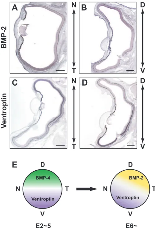
In Chapter II, I showed that CBF-1 regulates the expression of ephrin-A2 by a DNA binding-independent mechanism. Our lab recently identified a novel secreted molecule, Ventroptin, which also promotes expression of ephrin-A2. Ventroptin is an antagonist of BMP-4 in the retina, and implicated in the retinotectal topographic projection along both the D-V and A-P axes (Sakuta et al., 2001). At the early developmental stages of the eye, Ventroptin is specifically expressed in the ventral retina, with a complementary pattern to the dorsal-specific expression of BMP-4, and this counteraction between Ventroptin and BMP-4 governs the regional specification along the D-V axis (Sakuta et al., 2001). At the later stages (E6 onward), Ventroptin begins to be expressed in a nasal high-temporal low gradient, in addition to a ventral high-dorsal low gradient. Since expression of BMP-4 declines from E5, a member of the TGF-β family other than BMP-4 is supposed to appear in a temporal high-nasal low gradient along the N-T axis with a complementary pattern to the Ventroptin expression (Sakuta et al., 2001).
In this study, I found that a TGF-β family member, BMP-2, is expressed in a pattern complementary to that of Ventroptin, with a double- gradient along the N-T and D-V axes from E5 onward. Misexpression of BMP-2 in the developing retina represses expression of Ventroptin and ephrin-A2. Moreover, I demonstrate that CBF-1 perturbs BMP signaling through a DNA binding-independent mechanism, which leads to the induction of ephrin-A2 expression. These findings suggest that distinct BMP signals play pivotal roles in the topographic projection along both axes.
- 48 -
Materials and Methods Plasmids
To prepare the BMP-2/RCAS construct, the coding region of mouse BMP-2 was once inserted into SLAX-NS, and then subcloned into RCAS- NS. Ventroptin/RCAS was described previously (Sakuta et al., 2001).
Mouse Smad1, Smad4 and ALK-3 were cloned from a P0 mouse retina cDNA library by PCR and their sequences were verified by DNA sequence analysis. Smad1 and Smad4 were fused with double flag- epitope tags at the N terminus by PCR. A constitutively active mutant ALK-3 (ALK3-CA) was generated by substitution of glutamine 233 with aspartic acid by PCR as described previously (Hoodless et al., 1996). They were subcloned into pcDNA3.1(+) vector.
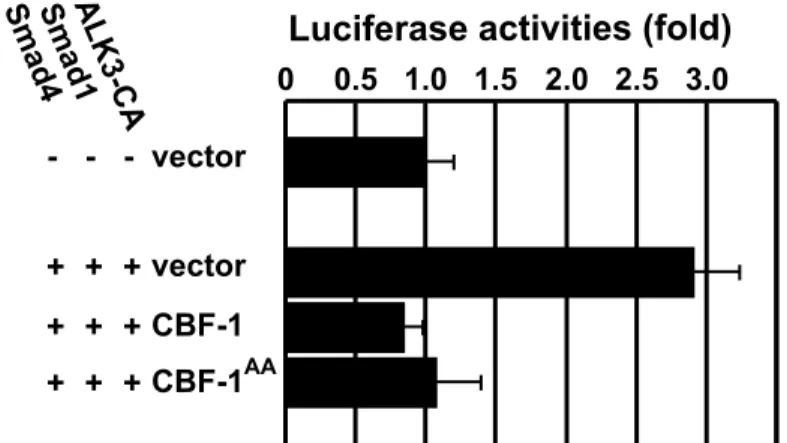
Luciferase assay
A minimal promoter construct (pGL3ti) was first made from pGL3-basic reporter vector (Promega) by inserting oligonucleotides carrying the adenovirus major-late promoter TATA box and the mouse terminal deoxynucleotidyl transferase gene initiator in the BglII site as described previously (Jonk et al., 1998). The BMP-responsive reporter (pGL3ti- 12GCCG) was constructed by further inserting four oligonucleotides, each containing three copies of the GCCG motif (Kusanagi et al., 2000), into the XhoI site of pGL3ti. Luciferase assays were carried out using human embryonic kidney 293 cells. Cells grown in 96-well microplates at a density of 4 X 10⁴ cells per well were transiently transfected with expression plasmids using a combination of the reporter (50 ng), receptor (10 ng), and Smads (10 ng each) together with or without the expression plasmid for CBF-1 (20 ng) or CBF-1AA (20 ng). Luciferase activity was measured with a Dual-Glo Luciferase Assay System (Promega) using