Panc r eat i c Endoc r i ne Cel l D
evel opm
ent
著者
Abdel l at i f Ahm
ed M
. , O
i s hi H
i s as hi , I t agaki
Takahi r o, J ung Yuns hi n, Shaw
ki H
os s am
H
. ,
O
ki t a Yukar i , H
as egaw
a Yos hi kaz u, Suz uki
H
i r oyuki , El - M
or s y Sal ah E. , El - Sayed M
es bah
A. , Shoai b M
ahm
oud B. , Sugi yam
a Fum
i hi r o,
Takahas hi Sat or u
j our nal or
publ i c at i on t i t l e
PLoS O
N
E
vol um
e
11
num
ber
2
page r ange
e0150010
year
2016- 02
権利
( C) 2016 Abdel l at i f et al . Thi s i s an open
ac c es s ar t i c l e di s t r i but ed under t he t er m
s of
t he Cr eat i ve Com
m
ons At t r i but i on Li c ens e,
w
hi c h per m
i t s unr es t r i c t ed us e, di s t r i but i on,
and r epr oduc t i on i n any m
edi um
, pr ovi ded t he
or i gi nal aut hor and s our c e ar e c r edi t ed.
U
RL
ht t p: / / hdl . handl e. net / 2241/ 00138655
β
-Cell-Specific
Mafk
Overexpression Impairs
Pancreatic Endocrine Cell Development
Ahmed M. Abdellatif1,2, Hisashi Oishi1,3,4
*, Takahiro Itagaki1,3, Yunshin Jung1,3,
Hossam H. Shawki1,3, Yukari Okita5, Yoshikazu Hasegawa3, Hiroyuki Suzuki5, Salah E. El-Morsy2, Mesbah A. El-Sayed2, Mahmoud B. Shoaib2, Fumihiro Sugiyama3,
Satoru Takahashi1,3,4,6*
1Department of Anatomy and Embryology, Faculty of Medicine, University of Tsukuba, Tsukuba, Ibaraki, Japan,2Department of Anatomy and Embryology, Faculty of Veterinary Medicine, Mansoura University, Mansoura, Egypt,3Laboratory of Animal Resource Center, Faculty of Medicine, University of Tsukuba, Tsukuba, Ibaraki, Japan,4Life Science Center, Tsukuba Advanced Research Alliance (TARA), University of Tsukuba, Tsukuba, Ibaraki, Japan,5Department of Experimental Pathology, Faculty of Medicine, University of Tsukuba, Tsukuba, Ibaraki, Japan,6International Institute for Integrative Sleep Medicine (WPI-IIIS), University of Tsukuba, Tsukuba, Ibaraki, Japan
*[email protected](ST);[email protected](HO)
Abstract
The MAF family transcription factors are homologs of v-Maf, the oncogenic component of the avian retrovirus AS42. They are subdivided into 2 groups, small and large MAF proteins, according to their structure, function, and molecular size. MAFK is a member of the small MAF family and acts as a dominant negative form of large MAFs. In previous research we generated transgenic mice that overexpress MAFK in order to suppress the function of large MAF proteins in pancreaticβ-cells. These mice developed hyperglycemia in adult-hood due to impairment of glucose-stimulated insulin secretion. The aim of the current study is to examine the effects ofβ-cell-specificMafkoverexpression in endocrine cell development. The developing islets ofMafk-transgenic embryos appeared to be disorga-nized with an inversion of total numbers of insulin+ and glucagon+ cells due to reducedβ -cell proliferation. Gene expression analysis by quantitative RT-PCR revealed decreased levels ofβ-cell-related genes whose expressions are known to be controlled by large MAF proteins. Additionally, these changes were accompanied with a significant increase in key β-cell transcription factors likely due to compensatory mechanisms that might have been activated in response to theβ-cell loss. Finally, microarray comparison of gene expression profiles between wild-type and transgenic pancreata revealed alteration of some uncharac-terized genes includingPcbd1,Fam132a,Cryba2, andNpy, which might play important roles during pancreatic endocrine development. Taken together, these results suggest that
Mafkoverexpression impairs endocrine development through a regulation of numerousβ -cell-related genes. The microarray analysis provided a unique data set of differentially expressed genes that might contribute to a better understanding of the molecular basis that governs the development and function of endocrine pancreas.
a11111
OPEN ACCESS
Citation:Abdellatif AM, Oishi H, Itagaki T, Jung Y, Shawki HH, Okita Y, et al. (2016)β-Cell-SpecificMafk
Overexpression Impairs Pancreatic Endocrine Cell Development. PLoS ONE 11(2): e0150010. doi:10.1371/journal.pone.0150010
Editor:Bridget Wagner, Broad Institute of Harvard and MIT, UNITED STATES
Received:December 30, 2015
Accepted:February 8, 2016
Published:February 22, 2016
Copyright:© 2016 Abdellatif et al. This is an open access article distributed under the terms of the
Creative Commons Attribution License, which permits unrestricted use, distribution, and reproduction in any medium, provided the original author and source are credited.
Data Availability Statement:All relevant data are within the paper and its Supporting Information files.
Funding:This work was supported by a Grant-in-Aid for Scientific Research (26221004) from the Japanese Ministry of Education, Culture, Sports, Science and Technology (MEXT) (to S.T.). The funders had no role in study design, data collection and analysis, decision to publish, or preparation of the manuscript.
Introduction
Diabetes mellitus is a group of metabolic diseases characterized by hyperglycemia resulting from defects in insulin secretion, insulin action, or both, which eventually leads to a series of complications in various organs [1]. Type 1 diabetes (T1D) results from the destruction ofβ -cells by aβ-cell-specific autoimmune reaction. In type 2 diabetes (T2D), the peripheral tissues are resistant to insulin action and the disease is often accompanied by obesity and hyperlipid-emia. For decades, several approaches have been developed for the treatment of diabetes including insulin-secretion stimulants, improving insulin preparations, and islet transplanta-tion, yet many unexamined avenues of research remain [2].
During pancreatic development, a subset of the pancreatic epithelial cells starts to express the proendocrine factorNgn3and gives rise to all types of endocrine cells [3–6]. The hormone-expressing cells are produced during 2 sequential stages, the primary and secondary transi-tions. The primary transition begins before E13.5, and is characterized by an appearance of hormone+ cells that are not fully functional. During the secondary transition starting from around E13.5, the differentiating endocrine cells expand markedly, then migrate into mesen-chyme, and eventually aggregate to form Langerhans islets [7]. The expression of a cascade of different transcription factors stimulates the differentiation into distinct endocrine lineages.α -cell-related transcription factors includeMafb,Nkx2.2,Pax6,Foxa2,Pou3f4, andArx, whereas β-cell differentiation is controlled byPdx1,Mafb,Pax4,Pax6,Nkx2.2, andNkx6.1[8].
The MAF family transcription factors belong to the activator protein 1 (AP1) superfamily of basic leucine zipper (bZIP) proteins. It derives its name from v-Maf—the oncogenic compo-nent of the avian retrovirus AS42 that was originally isolated from chicken musculoaponeuro-tic fibrosarcoma [9]. The MAF family is subdivided into 2 groups according to their molecular size: the small MAF proteins comprising MAFG, MAFF, and MAFK; and large MAF proteins, including MAFA, MAFB, c-MAF, and NRL. All MAF proteins contain basic leucine zipper domain that allows DNA binding. Compared to the other bZIP proteins, MAF proteins can recognize a longer palindromic sequence of DNA (Maf-recognition element, MARE) [10–12]. Increasing numbers of studies on endocrine development reveal that the expression of large MAF proteins is tightly regulated in a spatiotemporal manner [13–15].Mafbgene knockout (Mafb−/−) mice show around a 50% reduction ofα- andβ-cell numbers at E18.5. In contrast,
no developmental defects were observed inMafagene knockout (Mafa−/−) mice [16,17]. Small
MAF proteins are also found to display a complex expression pattern during embryogenesis [18]. They are able to form a homodimer or a heterodimer with other bZIP factors such as the cap’n’collar (CNC) family and play a role in many biological processes like hematopoiesis, neu-ronal function, and oxidative stress response [19–22]. Unlike the large MAF proteins, the small MAF proteins lack a transactivation domain and when they are expressed in large amounts, the homodimeric proteins compete with the binding of large MAF proteins to the cis-element of target genes at MARE sites, resulting in a dominant-negative effect [23].
Our previous studies demonstrated thatβ-cell-specificMafktransgenic (Mafk-Tg) mice exhibited hyperglycemia due to an impaired insulin secretion during early postnatal life [24]. When these mice are crossed withMafa−/−mice, the double mutants display destructiveβ-cell
development and an overt diabetic phenotype with typical characteristics of human diabetic nephropathy [25]. The aim of the current study was to characterize and evaluate the impact of Mafkoverexpression on the genetic pathways governingβ-cell development usingMafk-Tg andMafa−/−;Mafk-Tg mice embryos. In theMafk-Tg mutants we observed abnormalities inβ
contribute to the phenotypic alteration in theMafk-Tg mice at E15.5, to gain insights into the mechanisms controlling endocrine cell development and function.
Materials and Methods
Mice
The mice were maintained in specific pathogen-free conditions, in the Laboratory Animal Resource Center at the University of Tsukuba. This study was carried out in strict accordance with the recommendations in the Guide for the Care and Use of Laboratory Animals of the National Institutes of Health. The protocol was approved by the Committee on the Ethics of Animal Experiments of the University of Tsukuba (Permit Number: 14–049). All mice were euthanized with carbon dioxide gas, and all efforts were made to minimize suffering. The gen-erations ofMafaknockout (Mafa−/−) mice, transgenic mice expressingMafkunder the control
of ratInsulin 1promoter (Mafk-Tg), R26GRR mice, andIns1-Cre25 mice have previously been described [16,24,26,27]. Double mutantMafa−/−;Mafk-Tg mice were generated by mating
Mafa+/−females withMafa+/−;Mafk-Tg males. DNA extraction and embryo genotyping were
performed by NaOH extraction methods from tails as previously described [16,24].
Immunohistochemistry
The embryos were collected, washed in cold PBS, fixed in 4% PFA, and then embedded in par-affin. Immunohistochemistry was performed on 5-μm paraffin sections according to the stan-dard histological methods. The sections were blocked in appropriate serum for 1 hour and incubated overnight at 4°C with the following primary antibodies: guinea pig anti-insulin (ab7842, 1:1000, Abcam, Cambridge, UK), rabbit anti-glucagon (RAG-06P, 1:2000, Linco Research, St. Charles, MO, USA), guinea pig anti-glucagon (M182, 1:4000, Takara, Kyoto, Japan), rabbit anti-aristaless-related homeobox (gift from Drs. Kitamura and Morohashi; 1: 250) [28], rabbit Ki67 (NCL-Ki67p, 1:500, Novocastra, Newcastle, UK) and rabbit anti-PHH3 (ab5176, 1:500, Abcam). The antigens were visualized using the appropriate secondary antibodies conjugated with Alexa Fluor 350, 488, or 594 (1:1000, Life Technologies, Gaithers-burg, MD, USA). All sections were examined using a fluorescence microscope (BZ-9000, Key-ence, Tokyo, Japan). For cell counting experiments, serial sections spanning the entire pancreas were collected at a 100-μm intervals and immunostained. A total number of 25 sec-tions were used per pancreas. The total numbers of immunoreactive cells were quantified using ImageJ 1.48 software (NIH, Bethesda, Maryland, USA).
Measurement of total insulin contents
The whole pancreas was collected from embryos at both E15.5 and E18.5. The total insulin content was determined after extraction with acid-ethanol. Insulin levels were detected using a mouse insulin ELISA kit (Morinaga, Yokohama, Japan).
Luciferase assay
Chromatin Immunoprecipitation (ChIP) assay
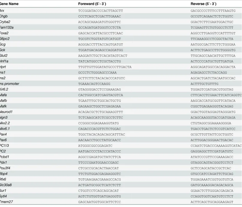
NMuMG cells expressing FLAG-MAFK were prepared as previously described [30]. In short, the NMuMG cells were transfected with pCAGIP-FLAG-MAFK or mock plasmids. The cells were cross-linked with 1% formaldehyde at 37°C for 15 minutes, suspended in 500μl of nuclear lysis buffer (1% SDS, 50 mM Tris-HCl, pH 8.1, 10 mM EDTA, 20,000 KIU/ml aprotinin, 1μg/ ml leupeptin), and sonicated. Soluble chromatin was diluted with 9 volumes of dilution buffer for immunoprecipitation (16.7 mM Tris-HCl, pH 8.1, 1.2 mM EDTA, 167 mM NaCl, 0.01% SDS, 1.1% Triton X-100, 20,000 KIU/ml aprotinin, 1μg/ml leupeptin) and incubated with anti-FLAG antibody (M2, Sigma, St Louis, MO, USA) with end-end rotation at 4°C over-night followed by incubation with 25μl of Dynabeads Protein A (Life Technologies) at 4°C for 1 hour. DNA was extracted from the Dynabeads by means of phenol-chloroform extraction. The PCR primers are described inTable 1.
Quantitative RT-PCR
The whole pancreas of E15.5 embryos were homogenized and total RNA isolated using the NucleoSpin RNA kit (Macherey-Nagel, Düeren, Germany). The cDNA was synthesized using a QuantiTect Reverse Transcription Kit (Qiagen, Hilden, Germany). Quantitative PCR reac-tions were carried out using a Thermal Cycler Dice Real Time System (Takara) with a SYBR Green PCR Master Mix (Takara). Expression ofHprtwas utilized to analyze the relative gene expression of other genes. All primer sequences are listed inTable 1.
Lineage tracing experiments
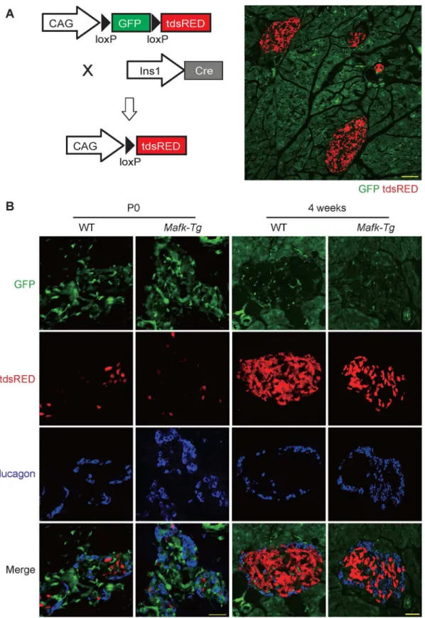
To test the possibility thatβ-cell specificMafkoverexpression inducedβ-cell transdifferentia-tion intoα-cells during early stages of development, we crossedMafk-Tg females with Ins1-Cre;R26GRR males, which express GFP ubiquitously before and tdsRed after Cre excision; thus we could label around 90% of theβ-cell lineage [26]. The pancreata ofMafk-Tg;Ins1-Cre; R26GRR and control WT;Ins1-Cre;R26GRR mice were collected at both P0 and 4 weeks of age, fixed overnight in 4% PFA, dehydrated in sucrose and embedded in OCT compound (Sakura, Tokyo, Japan). Frozen sections were stained using anti-glucagon antibody in order to deter-mine the cell fraction coexpressing both tdsRed and glucagon.
Microarray experiment
Total RNA was isolated from 3 pairs of WT andMafk-Tg pancreata at E15.5 using the
Statistical analysis
Data were expressed as the means ± standard errors of the means and compared using an unpairedttest. Probability values of less than 0.05 were considered significant.
Results
β
-cell-specific
Mafk
overexpression resulted in impaired endocrine cell
development and an abnormal islet structure
In order to study the impact ofβ-cell-specificMafkoverexpression on endocrine development, especially during the primary and secondary transitions, we performed immunohistochemical
Table 1. The primers used in this study.
Gene Name Foreward (5`- 3`) Reverse (5`- 3`)
Arx TCCGGATACCCCACTTAGCTT GACGCCCCTTTCCTTTAAGTG
Chgb CCCTCAGCTCGACTTGAAAC GCCGTCAGAACTCTCTGGTC
Cryba2 ACCAGCAAAGATGTGGGTTC GGACTCTTCGAATGGACTGC
Fam132a GCCAGATGATGGGTCCTCTA TCGAAGTTCTGTGGCCTCTT
Foxa2 GAGCACCATTACGCCTTCAAC AGGCCTTGAGGTCCATTTTGT
G6pc2 TGCGTCTGGTATGTCATGGT TTCAAAGGCCTCGGCTACTA
Gcg AGGGACCTTTACCAGTGATGT AATGGCGACTTCTTCTGGGAA
Gck TGGATGACAGAGCCAGGATGG ACTTCTGAGCCTTCTGGGGTG
Glut2 AAGGATCTGCTCACATAGTCACT TTGCAGCCAACATTGCTTTGA
Hnf1a TATCATGGCCTCGCTACCTG ACTCCCCATGCTGTTGATGA
Hprt TTGTTGTTGGATATGCCCTTGACTA AGGCAGATGGCCACAGGACTA
Ins1 GCCCTCTGGGAGCCCAAA AGAGAGCCTCTACCAGG
Ins2 GCTTCTTCTACACACCCATGTC AGCACTGATCTACAATGCCAC
Ins promoter TGAAACAGTCCAAGG ACTTTGCTGTTTG
Kir6.2 GTAGGGGACCTCCGAAAGAG TGGAGTCGATGACGTGGTAG
Mafa CACTGGCCATCGAGTACGTCA CTTCACCTCGAACTTCATCAGGTC
Mafb TGAATTTGCTGGCACTGCTG AAGCACCATGCGGTTCATACA
Mafk GAGAAGCTGGCTCGAGAGAA CGGCTGAGAAGGGTACAGAG
Neurod1 ACAGACGCTCTGCAAAGGTTT GGACTGGTAGGAGTAGGGATG
Ngn3 TCTCAAGCATCTCGCCTCTTC ACAGCAAGGGTACCGATGAGA
Nkx2.2 CCGGGCGGAGAAAGGTATG CTGTAGGCGGAAAAGGGGA
Nkx6.1 CAGACCCACGTTCTCTGGAC TGACCTGACTCTCCGTCATCC
Pax4 TGGCTACACAGACAGCATTTAC GCGCTTGTTATTCGCTGGTC
Pax6 AACAACCTGCCTATGCAACC ACTTGGACGGGAACTGACAC
PC1/3 ATGGGCGGCGGAGATC CCAATCTGACCCAAAAGGTCATAC
PC2 AATGACCCCTACCCATACCC GAGGAGGCTTCGATGATGTC
Pcbd1 AGGCCGAGATGCTATCTTCA ATATCCCGTTCCGAAAGACC
Pdx1 TTCCCGAATGGAACCGAGC GTAGGCAGTACGGGTCCTCT
Pou3f4 CTCGCCGCACACTAACCAT GCTCCAGCATACCGCTCAC
Rbp4 TTCTGTGGACGAGAAGGGTC GTGCCATCCAGATTCTGCAG
Rfx6 TGTGAAGAACGAAAGCCACG TGGAGAAATCGGTGGTGTCA
Slc30a8 ACTGATGCGGCTCATCTCTT GATGCAAAGGACAGACAGCA
Sur1 CTGGTCCTCAGCAGCACAT GGAACTCTTGGGACGAGACA
Sytl4 AGTCTGTGGTGATGAGGGTG CCAGGTGGTCAATGTCCTCT
Tmem27 GAGCAATGGTGGCATTCTCC ACTTCAGCTGCAGGAAGAGT
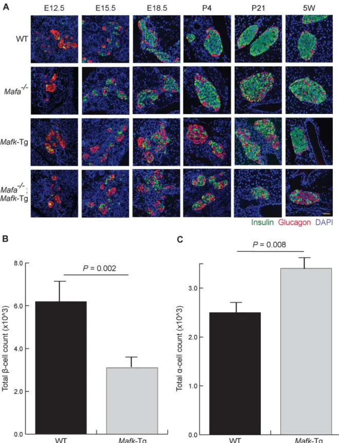
staining of pancreas sections from WT,Mafa−/−,Mafk-Tg, andMafa−/−;Mafk-Tg embryos
(Fig 1A). At E12.5, the point at which the primary cells emerge (the first wave of hormone-expressing cells), all groups of mice showed comparable manifestations. They contained a few insulin positive cells and a few insulin/glucagon double positive cells (which are considered progenitor cells), while the majority of cells were glucagon-positive [31,32]. At E15.5 and E18.5, the islets of WT andMafa−/−embryos displayed normal and comparable phenotypes,
whereasMafk-Tg andMafa−/−;Mafk-Tg embryos exhibited similar structural patterns with
reversed number of insulin+ and glucagon+ cells. These results indicate thatMafais not involved inβ-cell development either in theMafk-Tg background or in the WT background. We also stained pancreatic sections at postnatal day (P) 4, P21, and 5 weeks of age (5W). The islets from WT mice at all periods appeared with a clear central core of insulin+ cells sur-rounded by a peripheral layer of glucagon+ cells, whereasMafa−/−mice developed an
abnor-mal structure with many glucagon+ cells in the center of the islets in the early neonatal period [16]. The islets ofMafk-Tg mice were still displaying changes similar to their embryonic abnor-mal phenotype at P4 and P21, however at 5W the islet structure was apparently reverted to normal. As shown previously, in the islet ofMafa−/−;Mafk-Tg mice, the destructive changes
were more severe compared toMafa−/−mice [25]. These results suggest that the expression of
Mafaduring the neonatal period compensates for the effect ofMafkoverexpression in embryos, which is consistent with a previous report showing the functional significances of Mafaafter birth [33]. Hereafter, our experiment was focused on the comparison ofMafk-Tg and control WT mice in each experiment.
To clarify the extent of these changes, we quantified the changes in insulin+ and glucagon + cell populations based on their numbers. The total numbers of insulin+ and glucagon+ cells were counted in representative sections throughout the whole pancreas of embryos at E18.5. In Mafk-Tg embryos, the total count of insulin+ cells was decreased (3.1 ± 0.19 × 10^3 vs. 6.21 ± 0.72 × 10^3 in WT,P= 0.002) (Fig 1B). Conversely, we found the number glucagon + cells to be significantly increased (3.4 ± 0.14 × 10^3 vs. 2.4 ± 0.20 × 10^3 in WT,P= 0.008) (Fig 1C). The total insulin contents were also decreased inMafk-Tg embryos both at E15.5 and at E18.5, consistent with the immunohistochemistry results (Fig 2). The reduction in the total insulin contents was more dramatic at E15.5 (1.1 ± 0.1 ng in Tg vs. 5.6 ± 0.6 ng in WT, P<0.001) compared to E18.5 (10.0 ± 0.39 ng in Tg vs. 13.4 ± 0.33 ng in WT,P<0.001).
β
-cell-specific
Mafk
overexpression altered the gene expression of both
β
- and
α
-cell-related factors
Fig 1. TransgenicMafkoverexpression altered the normal islet structure.(A) Immunohistochemical analysis of insulin and glucagon in wild-type (WT),
Mafaknockout (Mafa−/−),Mafktransgenic (Mafk-Tg) andMafa−/−;Mafk-Tg pancreata at embryonic day (E) 12.5, E15.5, E18.5, postnatal day (P)4, P21 and 5
weeks of age (5W). Scale bar = 40μm. (B) The totalβ-cell count of WT (n = 3) andMafk-Tg (n = 5) embryos at E18.5. (C) Theα-cell count of WT (n = 3) and
Mafk-Tg (n = 5) embryos at E18.5. The error bars represent the standard errors of the means.
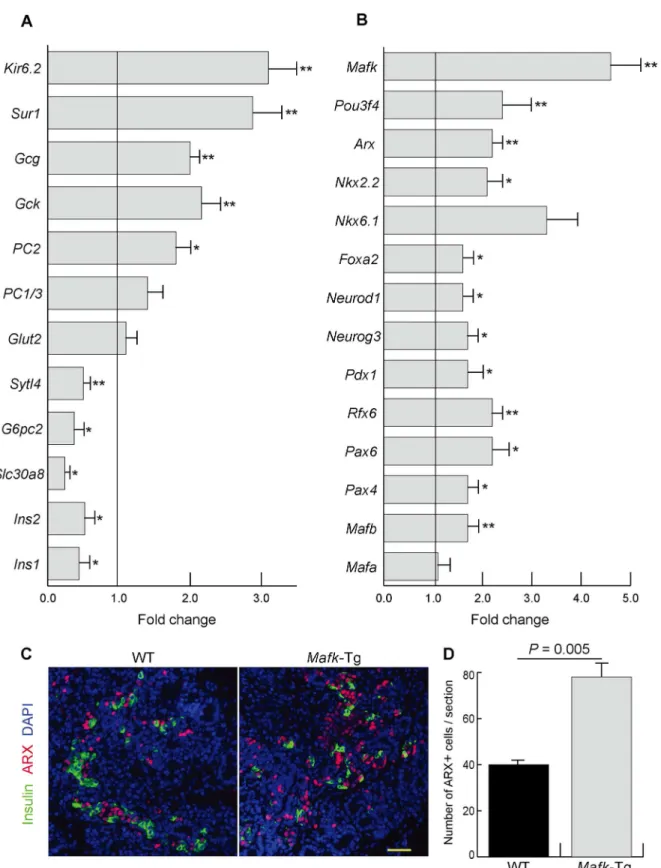
P= 0.01),Pdx1(1.7 ± 0.2 fold,P= 0.03),Foxa2(1.6 ± 0.1 fold,P= 0.03),Ngn3(1.7 ± 0.1 fold, P= 0.03),Neurod1(1.6 ± 0.1 fold,P= 0.05),Nkx2.2(2.1 ± 0.2 fold,P= 0.01),Nkx6.1(3.3 ± 0.6 fold,P= 0.09),Arx(2.1 ± 0.2 fold,P= 0.001), andPou3f4(2.4 ± 0.2 fold,P= 0.005). Further-more, immunohistochemistry showed that ARX expression, a master regulator ofα-cell devel-opment, was increased mainly in the noninsulin-positive cells ofMafk-Tg mice (Fig 3C). The latter finding was confirmed by quantitative assessment of the number of ARX+ cells per pan-creatic section in both WT (40 ± 1.2) andMafk-Tg mice (78 ± 6.6,P= 0.005) (Fig 3D).
MAFK inhibited the activation of insulin promoter
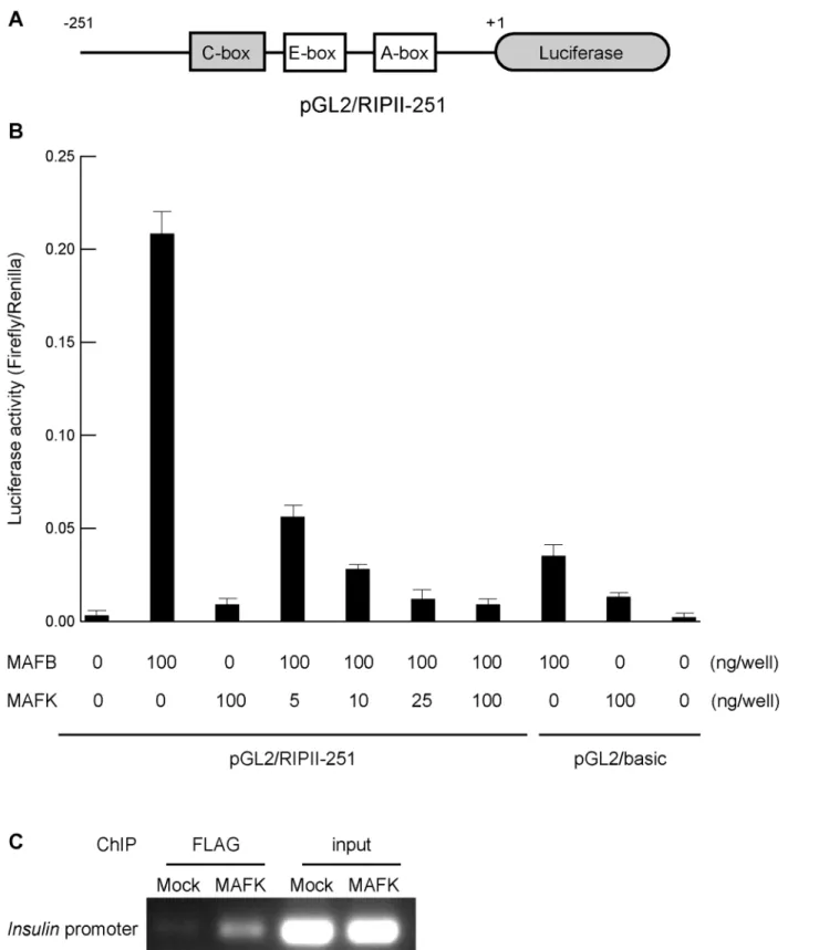
The ability of MAFK to suppress the activation of the insulin promoter was examined using luciferase assay. A reporter plasmid containing the luciferase gene under the control of the rat insulin 2 promoter (pGL2/RIPII-251) was used, as previously described [24](Fig 4A). NIH3T3 cells were transfected with MAFK and MAFB expression plasmids with a reporter plasmid, and the luciferase activity was monitored 48 hours after transfection (Fig 4B). MAFB activated the insulin promoter more than 100-fold. In the presence of increased amounts of MAFK, the enhanced activity of insulin promoter by MAFB showed a dose-dependent reduction. These findings indicate that MAFK inhibited MAFB binding to the C-box of the insulin promoter. Chromatin immunoprecipitation (ChIP) using FLAG antibody against NMuMG cells treated with either pCAGIP-FLAG-MAFK or mock plasmids further confirmed these observations
Fig 2. The changes in total pancreatic insulin content.The total insulin content of whole pancreata collected from embryos at E15.5 (A) (n: WT = 6;
Mafk-Tg = 7) and E18.5 (B) (n: WT = 4;Mafk-Tg = 7). The error bars represent the standard errors of the means.
Fig 3. Gene expression inMafk-Tg pancreases.(A, B) The mRNA expression of the indicated genes in the pancreas ofMafk-Tg relative to WT at E15.5 (n = 6 per each)*P<0.05,**P<0.01. The error bars represent the standard errors of the means. (A) Genes involved in hormone processing and secretion.
(B) Transcription factors related to the endocrine development. (C) Immunohistochemical staining of Insulin and ARX of WT andMafk-Tg mice at E15.5. Scale bars = 40μm. (D) The average number of cells that appeared positive for ARX immunostaining per pancreatic section at E15.5 (n: WT = 3,Mafk -Tg = 3).
Fig 4. MAFK inhibited the activation of insulin promoter.(A) Schema of the rat insulin promoter II reporter plasmid (pGL2/RIPII-251). (B)MafbandMafk
expression plasmids and the pGL2/RIPII-251 plasmid were transfected into NIH3T3 cells. The amount ofMafk-expression plasmid were serially increased from 0 to 100 ng. Three independent experiments were conducted and the error bars represent the standard errors of the means. (C) Chromatin
immunoprecipitation (ChIP) using anti-FLAG antibody detected binding of MAFK to theInsulinpromoter region including C-box in NMuMG cells transfected with pCAGIP-FLAG-MAFK. A representative figure of two independent experiments is shown.
(Fig 4C). The binding of MAFK directly to the insulin promoter was evident upon analysis of the immunoprecipitated protein-DNA complex with specific PCR primers.
No evidence of
α
-cell transdifferentiation in
Mafk
-Tg
β
-cells
The first observation of the reversion of the absolute number of insulin+ and glucagon+ cells inMafk-Tg mice raised the possibility thatβ-cell-specificMafkoverexpression could induce their transdifferentiation toα-cells during endocrine development. To answer this question, we labeled theβ-cell lineage usingIns1-Cre;R26GRR mice, which express GFP ubiquitously before and tdsRed exclusively inβcells after Cre recombination (Fig 5A). We collected 8 sections at 100-μm intervals apart fromMafk-Tg;Ins1-Cre;R26GRR and control WT;Ins1-Cre25;R26GRR (n = 3 per mouse) at P0 and 4 weeks of age and stained them with glucagon antibody in order to examine the adultα-cells that might be derived from theβ-cell lineage. Although higher numbers of glucagon+ cells were detected inMafk-Tg mice at 4 weeks of age, glucagon + tdsRed+ cells were observed in 0.52% of the total glucagon+ cells inMafk-Tg mice (n = 763) and these cells were 0.66% of the total glucagon+ cells in the controls (n = 752) (Fig 5B). These observations suggest that the increased number ofα-cells inMafk-Tg mice might be due to other causes rather thanβ-cell transdifferentiation.
Mafk
overexpression suppressed
β
-cell proliferation
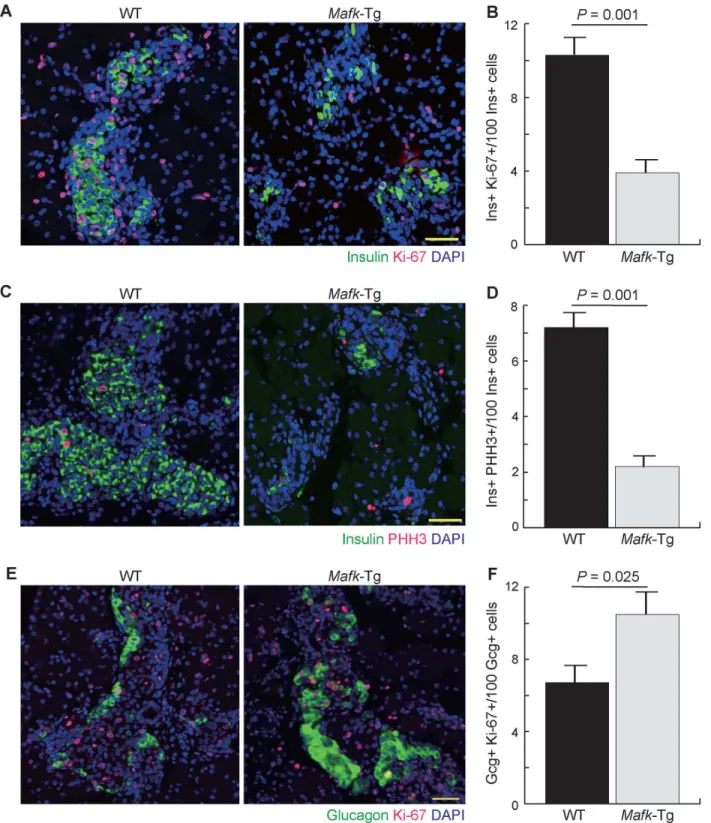
To examine the reason for reduction inβ-cell numbers during the prenatal and early postnatal periods, we performed immunohistochemical analysis using the cell cycle marker Ki-67 (Fig 6A). Six different sections from WT andMafk-Tg embryos (n = 3 per embryo) were collected at 100-μm intervals and stained using Ki-67 and insulin antibodies. The number of double pos-itive cells was counted and divided by the total number of insulin+ cells to estimate the per-centage of proliferatingβ-cells. The result showed that the percentage of proliferatingβ-cells was lower inMafk-Tg mice (3.9 ± 0.31%) than in WT mice (10.3 ± 0.91%,P= 0.001) at E18.5 (Fig 6B). This finding was further examined using another cell cycle marker, pHH3 (Fig 6C). Quantitative comparison of the percentage of the cells that appeared as double positive for both pHH3 and insulin (2.2 ± 0.4% in Tg vs. 7.2 ± 0.6% in WT,P= 0.001) was in line with that obtained from Ki-67 staining, suggesting thatβ-cell proliferation is reduced or delayed in Mafk-Tg mice (Fig 6D). Impaired proliferation of blood cells induced byMafkoverexpression is also reportedin vivo, suggesting that excess amounts of MAFK has an antiproliferative effect regardless of cell type [23,34].
We also examined the changes inα-cell proliferation using Ki-67 staining and found higher percentage of cells with double immunoreactivity for both glucagon and Ki-67 at E18.5 in Mafk-Tg (10.5 ± 0.9%) compared to WT embryos (6.7 ± 0.7%,P= 0.025)(n = 3 per embryo) (Fig 6E and 6F). This observation indicated that the increase in theα-cell numbers was mainly due to their proliferation rather thanβ-cell dedifferentiation.
Microarray analysis of pancreas from
Mafk
-Tg embryos revealed
differential expression of islet-related genes
Fig 5. Lineage tracing analysis ofβ-cell fate inMafk-Tg mice.(A) Strategy of the mice crossing. Forβ-cell labeling,Ins1-Cre;R26GRR male mice were mated withMafk-Tg females. The ability of the reporter mice to express tdsRED exclusively in pancreatic islets was confirmed by microscopic examination of unstained pancreas sections at 4 weeks of age. Scale bar = 100μm. (B) Higher number of glucagon+ cells was detected inMafk-Tg, however no cells coexpressing tdsRED and glucagon were observed at neither P0 nor 4W (n = 3 per each genotype). Scale bars = 40μm.
Fig 6. Cell proliferation assay inMafk-Tg mice.(A) Immunohistochemical analysis of pancreata from WT andMafk-Tg mice using Ki-67 and Insulin antibodies at E18.5. Scale bars = 40μm. (B) The percentage of cells that appeared double positive for both Ki-67 and insulin (n: WT = 4 (463/4135),Mafk -Tg = 5 (112/2893)). (C) Immunohistochemical analysis of pancreata from WT andMafk-Tg mice using pHH3 and Insulin antibodies at E18.5. Scale bars = 40μm. (D) The percentage of the cells that appeared as double positive for both pHH3 and Insulin (n: WT = 4 (158/2291),Mafk-Tg = 4 (21/957)). (E) Immunohistochemical analysis of pancreata from WT andMafk-Tg mice using Ki-67 and glucagon antibodies at E18.5. Scale bars = 40μm. (F) The percentage of proliferatingα-cell that appeared as double positive for both Ki-67 and Glucagon (n: WT = 3 (31/429),Mafk-Tg = 4 (76/746)). The error bars represent the standard errors of the means.
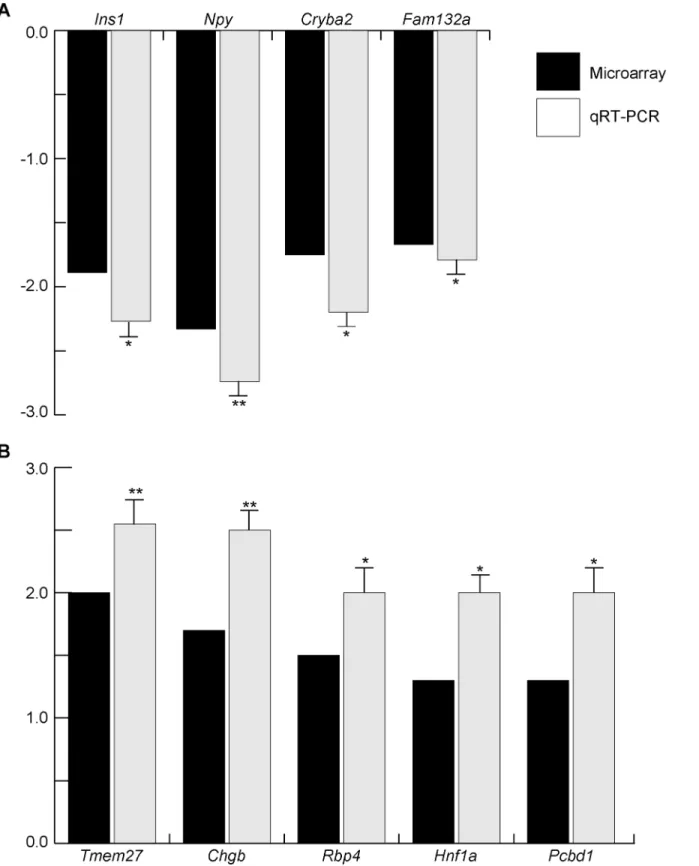
this stage of development, the cutoff was set at 1.2-fold change [35,36]. Sixteen genes (namely Ins1,Ins2,Slc30a8,G6pc2,Sytl4,Gcg,Mafb,Pax4,Pax6,Rfx6,Pdx1,Foxa2,Ngn3,Neurod1, Nkx2.2andPou3f4) from a total of 22 genes (72.7%) that show significant difference (Fig 3) were found to be included in the up- and down-regulated gene list, suggesting that microarray can be used for further analysis. In order to identify any uncharacterized genes that might be involved in endocrine development and function, we chose several candidate genes for qRT-PCR analyses. For that experiment we used individual biological samples (n = 4–8 per experiment) to confirm the changes observed with the technical replicates that were used for microarray analysis. For qRT-PCR, we chose several transcripts that we thought to be potential factors involved in endocrine development and function, some of which had not been fully characterized (Fig 7). In addition toIns1, we identified 3 novel candidate genes that showed a significant reduction of gene expression inMafk-Tg, namelyNpy,Cryba2, andFam132a(Fig 7A). Additionally, from the upregulated gene group, the expression ofTmem27,Chgb,Rbp4, Hnf1aandPcbd1was confirmed (Fig 7B). These results suggest that the gene expression profile ofMafk-Tg pancreata can provide a unique set of novel genes that possibly play various roles in endocrine development and function.
Discussion
In this study, we were able to detect insulin/glucagon double positive cells, which are consid-ered to be endocrine progenitors, inMafa−/−,Mafk-Tg, andMafa−/−;Mafk-Tg embryos as well
as WT embryos at E12.5. Our findings indicate that neitherMafadeficiency norβ-cell-specific Mafkoverexpression have any deleterious effect on the development of the early pancreatic progenitors. The islets of theMafa−/−mice also showed no marked structural changes when
compared to WT mice during the whole prenatal life, consistent with a previous study using MafaΔpancmice [17]. In contrast, as observed inMafk-Tg andMafa−/−;Mafk-Tg mice,
overex-pression ofMafkimpaired the islet morphogenesis at the late gestational stage, with up to 50% reduction in the number ofβ-cells and total insulin content. This finding corresponds with that of systemicMafb-deficient embryos in regard toβ-cell development [15]. The phenotype observed inMafk-Tg mice probably represents a part of conditional deletion ofMafbinβ-cells. On this point, we could not rule out the direct role of overexpressed MAFK on other differenti-ation factors in a large MAF independent manner. However, the increasing expression ofMafa in neonatalβ-cells probably restored the islet structure inMafk-Tg mice by 5 weeks of age, sug-gesting thatMafkoverexpression primarily blocked the large MAF function in embryos [37]. This idea is also supported by a recent report showing the opposing effect of MAFA and small MAFs on the insulin promoter [38].
Mafk-deficient mice do not exhibited any abnormalities [19].β-cell specific transgenic mice, which express a dominant negative form of MAFK (DN-MAFK), show normal glucose toler-ance under normal chow diet [38]. In contrast, under high fat diet conditions, DN-MAFK overexpression improves insulin secretion and glucose metabolism. Together with the observa-tion that high fat diet feeding increases small MAF expression inβ-cells, MAFK probably downregulates the expression of genes regulated by MAFA under stressed conditions partly due to the blocking of MAFA binding to MARE on the target [38].
Fig 7. Validation of microarray data by qRT-PCR in WT andMafk-Tg pancreata at E15.5.The expression levels of microarray and qRT-PCR are shown in black and gray columns, respectively. TheMafk-Tg expression (n = 8) of indicated genes was shown as relative to their expression in WT (n = 4). *P<0.05,**P<0.01. The error bars represent the standard errors of the means. (A) Downregulated genes. (B) Upregulated genes.
not able to find any cells coexpressing tdsRED and glucagon usingβ-cell lineage tracing, indi-cating that the increase inα-cells ofMafk-Tg mice is likely not to be due toβ- toα-cell transdif-ferentiation, but rather due to the increased proliferation ofα-cells. Recently, it is proposed that derepression ofMafbinβ-cells activatesβ- toα-cell reprogramming in the absence of Pdx1orMafa[43] [44]. Because overexpressed MAFK blocks MAFB function and probably does not initiate the reprogramming cascade inMafk-Tgβ-cells, the increase inα-cell numbers as well asα-cell transcription factors might be due to a loss of insulin action [45]. Alternatively, disruption of keyβ-cell transcription factors (MafsorPdx1) during the early postnatal period does not causeβ- toα- transdifferentiation (this manuscript and [46]), which occurs after 4 weeks of age [43] [44].
Finally, our analyses of microarray and subsequent qRT-PCR revealed that the downregu-lated gene group included a set of important transcripts forβ-cell function.Npy,Fam132a, and Cryba2, as well asIns1were encountered as potentialβ-cell-related genes. NPY (Neuropeptide Y) has pleiotropic functions in various tissues including hypothalamus, autonomic nervous system, and adipose tissue. In pancreas, its expression is observed inβ-cells during the second-ary transition to neonatal period [47,48]. A previous report showing that NPY treatment of mouse islets significantly enhancedβ-cell replication supports our finding of a reduction ofβ -cell proliferation inMafk-Tg mice [49].Fam132a(also calledAdipolin) is a novel adipokine associated with roles in glycemic control and insulin sensitization [50]. In addition to adipose tissue,Fam132ais thought to be secreted from mouse islets, according to the T1D base (https://www.t1dbase.org/page/Welcome/display). Notably,Cryba2is identified as an enriched gene in developing and adult pancreas and its expression is affected byNgn3-deficiency during development, although no clear phenotype in pancreas of ENU-inducedCryba2mutants has been reported [51–53].
From the upregulated gene group, TMEM27 plays a role in controlling insulin exocytosis by regulating the soluble N-ethylmaleimide-sensitive factor attachment protein receptor
(SNARE) complex assembly [54,55]. Retinol binding protein 4 (RBP4), which is a principle carrier of blood retinol, contributes insulin resistance in mice and humans [56]. Interestingly, bothTmem27andRbp4is upregulated in pancreas fromMafb-/-during late embryonic period, implying thatMafk-Tg mice andMafb-/-mice share common gene expression patterns as well as phenotypic similarities [17]. Chromogranin-B (CHGB) is a secretory glycoprotein co-stored with insulin and is found to control the rapid initial phase of insulin secretion [57]. Hepatocyte nuclear factor 1 alpha (HNF1A) controls many genes related toβ-cell differentiation, and gene mutations are the most common cause of maturity-onset diabetes of the young [58]. Pterin-4-alpha-carbinolamine dehydratase (PCBD1) is a novel protein that acts as a cofactor for HNF1A-dependent transcription protein, and it is reported thatPCBD1mutations cause early-onset nonautoimmune diabetes with features similar to dominantly inherited HNF1A-diabetes [59].
In conclusion,β-cell-specificMafkoverexpression resulted in impairment in endocrine development through alteration of the expression of many important genes for endocrine devel-opment and function. SinceMafkoverexpression can mimic the targeted disruption of many genes containing MARE sites in their regulatory regions to which MAFK homodimers can bind, our microarray analysis ofMafk-Tg embryos provides a unique data set for investigating novel factors that might have possible roles inβ-cell development, function, and survival.
Supporting Information
S1 Table. List of the up- and down-regulated genes in the pancreas ofMafk-Tg embryos at E15.5.
Acknowledgments
The authors thank Drs. Kitamura and Morohashi for ARX antibody, Masami Ojima and Gul-bekrem Xepket for technical assistance, and Thomas Mayers for grammatical revision of the manuscript.
Author Contributions
Conceived and designed the experiments: AMA HO SEE MAE MBS ST. Performed the experi-ments: AMA HO TI YJ HHS YO YH HS. Analyzed the data: AMA HO ST. Contributed reagents/materials/analysis tools: HS FS ST. Wrote the paper: AMA HO ST.
References
1. American Diabetes A. Diagnosis and classification of diabetes mellitus. Diabetes care. 2014; 37 Suppl 1:S81–90. doi:10.2337/dc14-S081PMID:24357215.
2. Vetere A, Choudhary A, Burns SM, Wagner BK. Targeting the pancreatic beta-cell to treat diabetes. Nature reviews Drug discovery. 2014; 13(4):278–89. doi:10.1038/nrd4231PMID:24525781.
3. Jensen J, Heller RS, Funder-Nielsen T, Pedersen EE, Lindsell C, Weinmaster G, et al. Independent development of pancreatic alpha- and beta-cells from neurogenin3-expressing precursors: a role for the notch pathway in repression of premature differentiation. Diabetes. 2000; 49(2):163–76. PMID:
10868931.
4. Gradwohl G, Dierich A, LeMeur M, Guillemot F. neurogenin3 is required for the development of the four endocrine cell lineages of the pancreas. Proceedings of the National Academy of Sciences of the United States of America. 2000; 97(4):1607–11. PMID:10677506; PubMed Central PMCID: PMC26482.
5. Gu G, Dubauskaite J, Melton DA. Direct evidence for the pancreatic lineage: NGN3+ cells are islet pro-genitors and are distinct from duct propro-genitors. Development. 2002; 129(10):2447–57. PMID:
11973276.
6. Desgraz R, Herrera PL. Pancreatic neurogenin 3-expressing cells are unipotent islet precursors. Devel-opment. 2009; 136(21):3567–74. doi:10.1242/dev.039214PMID:19793886; PubMed Central PMCID: PMC2761107.
7. Slack JM. Developmental biology of the pancreas. Development. 1995; 121(6):1569–80. PMID:
7600975.
8. Benitez CM, Goodyer WR, Kim SK. Deconstructing pancreas developmental biology. Cold Spring Har-bor perspectives in biology. 2012; 4(6). doi:10.1101/cshperspect.a012401PMID:22587935; PubMed Central PMCID: PMC3367550.
9. Nishizawa M, Kataoka K, Goto N, Fujiwara KT, Kawai S. v-maf, a viral oncogene that encodes a "leu-cine zipper" motif. Proceedings of the National Academy of Sciences of the United States of America. 1989; 86(20):7711–5. PMID:2554284; PubMed Central PMCID: PMC298140.
10. Kataoka K, Noda M, Nishizawa M. Maf nuclear oncoprotein recognizes sequences related to an AP-1 site and forms heterodimers with both Fos and Jun. Molecular and cellular biology. 1994; 14(1):700–12. PMID:8264639; PubMed Central PMCID: PMC358419.
11. Eychene A, Rocques N, Pouponnot C. A new MAFia in cancer. Nature reviews Cancer. 2008; 8(9): 683–93. doi:10.1038/nrc2460PMID:19143053.
12. Hang Y, Stein R. MafA and MafB activity in pancreatic beta cells. Trends in endocrinology and metabo-lism: TEM. 2011; 22(9):364–73. doi:10.1016/j.tem.2011.05.003PMID:21719305; PubMed Central PMCID: PMC3189696.
13. Matsuoka TA, Artner I, Henderson E, Means A, Sander M, Stein R. The MafA transcription factor appears to be responsible for tissue-specific expression of insulin. Proceedings of the National Acad-emy of Sciences of the United States of America. 2004; 101(9):2930–3. doi:10.1073/pnas.
0306233101PMID:14973194; PubMed Central PMCID: PMC365722.
14. Nishimura W, Kondo T, Salameh T, El Khattabi I, Dodge R, Bonner-Weir S, et al. A switch from MafB to MafA expression accompanies differentiation to pancreatic beta-cells. Developmental biology. 2006; 293(2):526–39. doi:10.1016/j.ydbio.2006.02.028PMID:16580660; PubMed Central PMCID: PMC2390934.
104(10):3853–8. doi:10.1073/pnas.0700013104PMID:17360442; PubMed Central PMCID: PMC1803762.
16. Zhang C, Moriguchi T, Kajihara M, Esaki R, Harada A, Shimohata H, et al. MafA is a key regulator of glucose-stimulated insulin secretion. Molecular and cellular biology. 2005; 25(12):4969–76. doi:10.
1128/MCB.25.12.4969-4976.2005PMID:15923615; PubMed Central PMCID: PMC1140590.
17. Artner I, Hang Y, Mazur M, Yamamoto T, Guo M, Lindner J, et al. MafA and MafB regulate genes critical to beta-cells in a unique temporal manner. Diabetes. 2010; 59(10):2530–9. doi:10.2337/db10-0190 PMID:20627934; PubMed Central PMCID: PMC3279542.
18. Lecoin L, Sii-Felice K, Pouponnot C, Eychene A, Felder-Schmittbuhl MP. Comparison of maf gene expression patterns during chick embryo development. Gene expression patterns: GEP. 2004; 4(1): 35–46. PMID:14678826.
19. Kotkow KJ, Orkin SH. Complexity of the erythroid transcription factor NF-E2 as revealed by gene target-ing of the mouse p18 NF-E2 locus. Proceedtarget-ings of the National Academy of Sciences of the United States of America. 1996; 93(8):3514–8. PMID:8622968; PubMed Central PMCID: PMC39641.
20. Shavit JA, Motohashi H, Onodera K, Akasaka J, Yamamoto M, Engel JD. Impaired megakaryopoiesis and behavioral defects in mafG-null mutant mice. Genes & development. 1998; 12(14):2164–74. PMID:9679061; PubMed Central PMCID: PMC317009.
21. Onodera K, Shavit JA, Motohashi H, Katsuoka F, Akasaka JE, Engel JD, et al. Characterization of the murine mafF gene. The Journal of biological chemistry. 1999; 274(30):21162–9. PMID:10409670.
22. Katsuoka F, Motohashi H, Ishii T, Aburatani H, Engel JD, Yamamoto M. Genetic evidence that small maf proteins are essential for the activation of antioxidant response element-dependent genes. Molecu-lar and celluMolecu-lar biology. 2005; 25(18):8044–51. doi:10.1128/MCB.25.18.8044-8051.2005PMID:
16135796; PubMed Central PMCID: PMC1234339.
23. Motohashi H, O'Connor T, Katsuoka F, Engel JD, Yamamoto M. Integration and diversity of the regula-tory network composed of Maf and CNC families of transcription factors. Gene. 2002; 294(1–2):1–12. PMID:12234662.
24. Shimohata H, Yoh K, Morito N, Shimano H, Kudo T, Takahashi S. MafK overexpression in pancreatic beta-cells caused impairment of glucose-stimulated insulin secretion. Biochemical and biophysical research communications. 2006; 346(3):671–80. doi:10.1016/j.bbrc.2006.05.184PMID:16780794.
25. Shimohata H, Yoh K, Fujita A, Morito N, Ojima M, Tanaka H, et al. MafA-deficient and beta cell-specific MafK-overexpressing hybrid transgenic mice develop human-like severe diabetic nephropathy. Bio-chemical and biophysical research communications. 2009; 389(2):235–40. doi:10.1016/j.bbrc.2009.
08.124PMID:19715672.
26. Hasegawa Y, Daitoku Y, Mizuno S, Tanimoto Y, Mizuno-Iijima S, Matsuo M, et al. Generation and char-acterization of Ins1-cre-driver C57BL/6N for exclusive pancreatic beta cell-specific Cre-loxP recombi-nation. Experimental animals / Japanese Association for Laboratory Animal Science. 2014; 63(2): 183–91. PMID:24770644.
27. Hasegawa Y, Daitoku Y, Sekiguchi K, Tanimoto Y, Mizuno-Iijima S, Mizuno S, et al. Novel ROSA26 reporter knock-in C57BL/6N mice exhibiting green emission before and red emission after Cre-mediated recombination. Experimental animals / Japanese Association for Laboratory Animal Science. 2013; 62(4):295–304. PMID:24172193; PubMed Central PMCID: PMC4160954.
28. Kitamura K, Yanazawa M, Sugiyama N, Miura H, Iizuka-Kogo A, Kusaka M, et al. Mutation of ARX causes abnormal development of forebrain and testes in mice and X-linked lissencephaly with abnor-mal genitalia in humans. Nature genetics. 2002; 32(3):359–69. doi:10.1038/ng1009PMID:12379852.
29. Kajihara M, Sone H, Amemiya M, Katoh Y, Isogai M, Shimano H, et al. Mouse MafA, homologue of zeb-rafish somite Maf 1, contributes to the specific transcriptional activity through the insulin promoter. Bio-chemical and biophysical research communications. 2003; 312(3):831–42. doi:10.1016/j.bbrc.2003.
10.196PMID:14680841.
30. Okita Y, Kamoshida A, Suzuki H, Itoh K, Motohashi H, Igarashi K, et al. Transforming growth factor-beta induces transcription factors MafK and Bach1 to suppress expression of the heme oxygenase-1 gene. The Journal of biological chemistry. 2013; 288(28):20658–67. doi:10.1074/jbc.M113.450478 PMID:23737527; PubMed Central PMCID: PMC3711329.
31. Hashimoto T, Kawano H, Daikoku S, Shima K, Taniguchi H, Baba S. Transient coappearance of gluca-gon and insulin in the progenitor cells of the rat pancreatic islets. Anatomy and embryology. 1988; 178(6):489–97. PMID:2464956.
33. Eto K, Nishimura W, Oishi H, Udagawa H, Kawaguchi M, Hiramoto M, et al. MafA is required for postna-tal proliferation of pancreatic beta-cells. PloS one. 2014; 9(8):e104184. doi:10.1371/journal.pone. 0104184PMID:25126749; PubMed Central PMCID: PMC4134197.
34. Yoh K, Sugawara T, Motohashi H, Takahama Y, Koyama A, Yamamoto M, et al. Transgenic over-expression of MafK suppresses T cell proliferation and function in vivo. Genes to cells: devoted to molecular & cellular mechanisms. 2001; 6(12):1055–66. PMID:11737266.
35. Wilding Crawford L, Tweedie Ables E, Oh YA, Boone B, Levy S, Gannon M. Gene expression profiling of a mouse model of pancreatic islet dysmorphogenesis. PloS one. 2008; 3(2):e1611. doi:10.1371/ journal.pone.0001611PMID:18297134; PubMed Central PMCID: PMC2249940.
36. Rubel CA, Lanz RB, Kommagani R, Franco HL, Lydon JP, DeMayo FJ. Research resource: Genome-wide profiling of progesterone receptor binding in the mouse uterus. Molecular endocrinology. 2012; 26(8):1428–42. doi:10.1210/me.2011-1355PMID:22638070; PubMed Central PMCID: PMC3404303.
37. Aguayo-Mazzucato C, Koh A, El Khattabi I, Li WC, Toschi E, Jermendy A, et al. Mafa expression enhances glucose-responsive insulin secretion in neonatal rat beta cells. Diabetologia. 2011; 54(3): 583–93. doi:10.1007/s00125-010-2026-zPMID:21190012; PubMed Central PMCID: PMC3047400.
38. Nomoto H, Kondo T, Miyoshi H, Nakamura A, Hida Y, Yamashita K, et al. Inhibition of Small Maf Func-tion in Pancreatic beta-Cells Improves Glucose Tolerance Through the Enhancement of Insulin Gene Transcription and Insulin Secretion. Endocrinology. 2015; 156(10):3570–80. doi:
10.1210/en.2014-1906PMID:25763640; PubMed Central PMCID: PMC4588816.
39. Thorel F, Nepote V, Avril I, Kohno K, Desgraz R, Chera S, et al. Conversion of adult pancreatic alpha-cells to beta-alpha-cells after extreme beta-cell loss. Nature. 2010; 464(7292):1149–54. doi:10.1038/
nature08894PMID:20364121; PubMed Central PMCID: PMC2877635.
40. Collombat P, Hecksher-Sorensen J, Krull J, Berger J, Riedel D, Herrera PL, et al. Embryonic endocrine pancreas and mature beta cells acquire alpha and PP cell phenotypes upon Arx misexpression. The Journal of clinical investigation. 2007; 117(4):961–70. doi:10.1172/JCI29115PMID:17404619; PubMed Central PMCID: PMC1839241.
41. Dhawan S, Georgia S, Tschen SI, Fan G, Bhushan A. Pancreatic beta cell identity is maintained by DNA methylation-mediated repression of Arx. Developmental cell. 2011; 20(4):419–29. doi:10.1016/j.
devcel.2011.03.012PMID:21497756; PubMed Central PMCID: PMC3086024.
42. Talchai C, Xuan S, Lin HV, Sussel L, Accili D. Pancreatic beta cell dedifferentiation as a mechanism of diabetic beta cell failure. Cell. 2012; 150(6):1223–34. doi:10.1016/j.cell.2012.07.029PMID:22980982; PubMed Central PMCID: PMC3445031.
43. Gao T, McKenna B, Li C, Reichert M, Nguyen J, Singh T, et al. Pdx1 maintains beta cell identity and function by repressing an alpha cell program. Cell metabolism. 2014; 19(2):259–71. doi:10.1016/j.
cmet.2013.12.002PMID:24506867; PubMed Central PMCID: PMC3950964.
44. Nishimura W, Takahashi S, Yasuda K. MafA is critical for maintenance of the mature beta cell pheno-type in mice. Diabetologia. 2015; 58(3):566–74. doi:10.1007/s00125-014-3464-9PMID:25500951.
45. Liu Z, Kim W, Chen Z, Shin YK, Carlson OD, Fiori JL, et al. Insulin and glucagon regulate pancreatic alpha-cell proliferation. PloS one. 2011; 6(1):e16096. doi:10.1371/journal.pone.0016096PMID:
21283589; PubMed Central PMCID: PMC3026810.
46. Gannon M, Ables ET, Crawford L, Lowe D, Offield MF, Magnuson MA, et al. pdx-1 function is specifi-cally required in embryonic beta cells to generate appropriate numbers of endocrine cell types and maintain glucose homeostasis. Developmental biology. 2008; 314(2):406–17. doi:10.1016/j.ydbio.
2007.10.038PMID:18155690; PubMed Central PMCID: PMC2269701.
47. Myrsen-Axcrona U, Ekblad E, Sundler F. Developmental expression of NPY, PYY and PP in the rat pancreas and their coexistence with islet hormones. Regulatory peptides. 1997; 68(3):165–75. PMID:
9100283.
48. Whim MD. Pancreatic beta cells synthesize neuropeptide Y and can rapidly release peptide co-transmitters. PloS one. 2011; 6(4):e19478. doi:10.1371/journal.pone.0019478PMID:21559341; PubMed Central PMCID: PMC3084883.
49. Cho YR, Kim CW. Neuropeptide Y promotes beta-cell replication via extracellular signal-regulated kinase activation. Biochemical and biophysical research communications. 2004; 314(3):773–80. PMID:
14741702.
50. Enomoto T, Ohashi K, Shibata R, Higuchi A, Maruyama S, Izumiya Y, et al. Adipolin/C1qdc2/CTRP12 protein functions as an adipokine that improves glucose metabolism. The Journal of biological chemis-try. 2011; 286(40):34552–8. doi:10.1074/jbc.M111.277319PMID:21849507; PubMed Central PMCID: PMC3186379.
52. Hoffman BG, Zavaglia B, Witzsche J, Ruiz de Algara T, Beach M, Hoodless PA, et al. Identification of transcripts with enriched expression in the developing and adult pancreas. Genome biology. 2008; 9(6):R99. doi:10.1186/gb-2008-9-6-r99PMID:18554416; PubMed Central PMCID: PMC2481431.
53. Puk O, Ahmad N, Wagner S, Hrabe de Angelis M, Graw J. First mutation in the betaA2-crystallin encod-ing gene is associated with small lenses and age-related cataracts. Investigative ophthalmology & visual science. 2011; 52(5):2571–6. doi:10.1167/iovs.10-6443PMID:21212184.
54. Fukui K, Yang Q, Cao Y, Takahashi N, Hatakeyama H, Wang H, et al. The HNF-1 target collectrin con-trols insulin exocytosis by SNARE complex formation. Cell metabolism. 2005; 2(6):373–84. doi:10.
1016/j.cmet.2005.11.003PMID:16330323.
55. Akpinar P, Kuwajima S, Krutzfeldt J, Stoffel M. Tmem27: a cleaved and shed plasma membrane protein that stimulates pancreatic beta cell proliferation. Cell metabolism. 2005; 2(6):385–97. doi:10.1016/j.
cmet.2005.11.001PMID:16330324.
56. Yang Q, Graham TE, Mody N, Preitner F, Peroni OD, Zabolotny JM, et al. Serum retinol binding protein 4 contributes to insulin resistance in obesity and type 2 diabetes. Nature. 2005; 436(7049):356–62. doi:
10.1038/nature03711PMID:16034410.
57. Obermuller S, Calegari F, King A, Lindqvist A, Lundquist I, Salehi A, et al. Defective secretion of islet hormones in chromogranin-B deficient mice. PloS one. 2010; 5(1):e8936. doi:10.1371/journal.pone. 0008936PMID:20126668; PubMed Central PMCID: PMC2812483.
58. Shields BM, Hicks S, Shepherd MH, Colclough K, Hattersley AT, Ellard S. Maturity-onset diabetes of the young (MODY): how many cases are we missing? Diabetologia. 2010; 53(12):2504–8. doi:10.
1007/s00125-010-1799-4PMID:20499044.
59. Simaite D, Kofent J, Gong M, Ruschendorf F, Jia S, Arn P, et al. Recessive mutations in PCBD1 cause a new type of early-onset diabetes. Diabetes. 2014; 63(10):3557–64. doi:10.2337/db13-1784PMID: