Frontal theta activation during
motor synchronization in autism
Masahiro Kawasaki
1,2,3, Keiichi
Kitajo
2,3, Kenjiro
Fukao
4,5, Toshiya
Murai
4, Yoko Y
amaguchi
6&
Yasuko
Funabiki
4,7Autism is characterized by two primary characteristics: deicits in social interaction and repetitive behavioral patterns. Because interpersonal communication is extremely complicated, its underlying brain mechanisms remain unclear. Here we showed that both characteristics can be explained by a unifying underlying mechanism related to diiculties with irregularities. To address the issues, we measured electroencephalographm during a cooperative tapping task, which required participants to tap a key alternately and synchronously with constant rhythmic a PC program, a variable rhythmic PC program, or a human partner. We found that people with autism had great diiculty synchronizing tapping behavior with others, and exhibited greater than normal theta-wave (6 Hz) activity in the frontal cortex during the task, especially when their partner behaved somewhat irregularly (i.e. a variable rhythmic PC program or a human partner). Importantly, the higher theta-wave activity was related to the severity of autism, not the performance on the task. This indicates that people with autism need to use intense cognition when trying to adapt to irregular behavior and can easily become overtaxed. Diiculty adapting to irregular behavior in others is likely related to their own tendencies for repetitive and regular behaviors. Thus, while the two characteristics of autism have been comprehended separately, our unifying theory makes understanding the condition and developing therapeutic strategies more tractable.
Autism spectrum disorder (ASD) is characterized by two features in the Diagnostic and Statistical Manual of Mental Disorders (DSM-5) (American Psychiatric Association, 2013): persistent deicits in social interaction, and restricted, repetitive patterns of behavior, interests, or activities. Most ASD studies have focused on the irst component, for aspects including imitation, language development, theory of mind, joint attention, and eye con-tact1–8. Some studies have shown cognitive inlexibility relevant to the second component, but these executive
function studies did not include reciprocal interactive tasks9–12. To understand these two features of ASD in
terms of a single underlying mechanism, we designed a rhythm-synchronization task featuring simple reciprocal communication with limited social demands. Previous studies have shown the behavioral synchronization in the nods during conversation13, the hand clapping14, and the tapping timing15, and so on. hese synchronization plays
important roles in facilitating and improving human relationship16.
Moreover, we included conditions that necessitated cognitive lexibility, because some studies have shown that the ASD people have inlexibility through executive functioning tasks10–12. Although we found no similar studies
that investigated interpersonal behavioral synchronization in ASD during communication, recent studies have shown tight interpersonal synchronizations both in electroencephalograms (EEG) and behavioral rhythms in typical development (TD) individuals17,18.
Previous human neuroimaging studies have identiied diferent mechanisms in the brain functions between ASD and TD, such as the emotion-related amygdala and the empathy-related mirror neuron systems (i.e.,
1Department of intelligent interaction technology, Graduate School of Systems and information engineering, University of Tsukuba, 1-1-1, Tennodai, Tsukuba-shi, Ibaraki 305-8573, Japan. 2Rhythm-based Brain Information Processing Unit, RIKEN BSI-TOYOTA Collaboration Center, 2-1, Hirosawa, Wako-shi, Saitama, 351-0198, Japan. 3Laboratory for Advanced Brain Signal Processing, RIKEN Brain Science Institute, 2-1, Hirosawa, Wako-shi, Saitama, 351-0198, Japan. 4Department of Psychiatry, Graduate School of Medicine, Kyoto University, Yoshida-nihonmatsu-cho, Sakyo-ku, Kyoto, 606-8501, Japan. 5Department of Psychology, Faculty of Human Sciences, Tezukayamagakuin University, 2-1823, Imakuma, Sayama-shi, Osaka, 589-8585, Japan. 6Neuroinformatics Japan Center, RIKEN Brain Science Institute, 2-1, Hirosawa, Wako-shi, Saitama, 351-0198, Japan. 7Department of Cognitive and Behavioral Sciences, Graduate School of Human and Environmental Studies, Kyoto University, Yoshida-nihonmatsu-cho, Sakyo-ku, Kyoto 606-8501, Japan. Correspondence and requests for materials should be addressed to M.K. (email:
kawasaki@iit.tsukuba.ac.jp) or Y.F. (email: funabiki.yasuko.8a@kyoto-u.ac.jp) Received: 29 June 2016
Accepted: 11 October 2017 Published: xx xx xxxx
www.nature.com/scientificreports/
superior temporal sulcus)19–22. Moreover, the EEG studies showed the enhancements of theta synchronization
(4-8 Hz) in ASD at both resting and cognitive states in comparison with that in TD participants23,24. However, no
studies have examined how to modulate the brain activity with inter-person behavioral synchronization in ASD during communication.
Here, we found that synchronizing with the behavioral rhythms of others led to diferent behaviors and brain activities in ASD and TD individuals. We used the DSM-525 for diagnosing ASD, and the Autism Diagnostic
Observation Schedule (ADOS)26 and the Multi-dimensional Scale for Pervasive developmental disorder and
Attention deicit/hyperactivity disorder (MSPA)27 for assessing its features and degree. We administered a task that
required two participants (divided by a partition and unseen by each other) to alternate tapping a key back and forth. he participant pairs were either ASD-TD or TD-TD. During the task, we recorded electroencephalograms.
Materials and Methods
participants.
We recruited 24 ASD adults (14 men, 10 women; Age: 29.2 ± 7.2 years), Mean intelligence quo-tients were: full-scale IQ (FIQ) = 111 ± 11, Verbal IQ (VIQ) = 114 ± 12, and Performance IQ (PIQ) = 105 ± 13. ASD participants were without physical complications, psychosis or medication and were recruited from outpa-tient clinics or colleges in our city. TD controls were 24 adults (12 men and 12 women) who were age, sex, and IQ-matched to the ASD group (Age = 25.5 ± 6.6 years, FIQ = 111 ± 12, VIQ = 109 ± 14, PIQ = 110 ± 11). Other inclusion criteria were: FIQs ≥ 80 (for suicient understanding of the study directions) and no medication (to avoid afecting the electroencephalogram (EEG). All participants gave written informed consent. Trained psychi-atrists conirmed diagnosis of ASD according to the criteria in DSM-5. IQ was assessed with the Wechsler Adult Intelligent Scale-hird Edition28. We used data from the Wechsler Intelligence Scale for Children-hird Edition29for a 20-year-old patient and the Wechsler Adult Intelligent Scale-Revised30 for another patient because the data
were already available within the last 5 years. his study was approved by the RIKEN Ethical Committee and Kyoto University Ethical Committee in accordance with the Declaration of Helsinki. All participants gave written informed consent before participation in the study.
AsD Assessment.
We administered the following assessment batteries in order to grasp the degree and individual diferences of autistic and comorbid features. Trained staf conducted the ADOS with supervision by specialists with research licenses, and the MSPA. he ADOS is a well-known assessment tool for autism. It con-sists of semi-structured tasks and interviews that involve communication and social interaction. We used module 4, which is for older adolescents and adults and takes 40–60 min to administer. he MSPA is a comprehensive assessment tool encompassing not only social communication of core ASD features but also other features such as restricted interests/behavior, sensory, inattention and motor problems that may relate to the present study. While the ADOS focuses on the behavior of social communication within the session, the MSPA includes the develop-mental history gathered from several informants and the objective behavior. he MSPA consists of 14 domains: ive core domains (communication, social adaptation, empathy, restricted interests/behavior, stereotyped/repet-itive motion), gross and ine motor, three attention-deicit/hyperactivity (ADHD) domains (inattention, hyper-activity and impulsivity), sensory, sleep cycle, learning, and language development. Each domain is assessed on a scale of one to ive with half-point steps, according to the severity: one (no sign), two (somewhat but no need to support), three (clinical level: mild), four (moderate), ive (severe).We also used the autism-spectrum quotient (AQ) to assess the severity of ASD. Independent of the above assessments, participants illed out the form by themselves. hus, we scored the severity of ASD in multiple ways of self-report, observation and interviews.
experimental procedure.
The tapping task included three alternate tapping conditions: Human, Constant-PC, and Variable-PC. hroughout the task, participants wore headphones, closed their eyes, and sat in a chair alone in an electric- and sound-shielded room.In the Human condition, two participants tapped their respective keys back and forth with their right index ingers. When a participant tapped, a sound (“do” or “mi”) was presented through both right and let headphones. When the partner tapped, the other sound (“mi” or “do”) was presented through both right and let headphones. If the diference between the previous tapping interval (e.g., from participant A to participant B) and the current one (e.g., from participant B to participant A) was within 50 ms, the one-octave higher sound was presented. Participants were instructed to tap the key at a time interval equal to that of the partner’s. he tapping rhythms were not predetermined or directed. Each participant was required to tap 250 times per session (total 500 taps/ session).
In the Constant-PC condition, a participant performed the same task with a virtual person (a PC program) that they thought was the human partner. he PC generated taps at a constant interval (600 ms ater the partic-ipants’ taps). In the Variable-PC condition, the tapping interval of the PC program varied from 400 to 800 ms in two patterns. For both patterns, the intervals changed ater blocks of 50 trials. Pattern 1 was 600 ms → 400 ms → 600 ms → 800 ms → 600 ms and pattern 2 was 600 ms → 800 ms → 600 ms → 400 ms → 600 ms. Each per-son completed 250 trials for both of these conditions.
Each participant in the ASD and TD groups completed three or four separate sessions, respectively (ASD group: one session each for the Constant-PC, Variable-PC, and Human conditions; TD group: one session each for the Constant-PC, and Variable-PC conditions, and two sessions for the Human condition). In the Human condition, the partner for an ASD participant was always from the TD group, whereas the partner of a TD partic-ipant was either from the ASD or TD group. he results of ASD and TD particpartic-ipants were equally compared with each other under the same condition (with the same TD partner). he order of conditions was random.
EEG measurement session. he stimulus was generated on a Windows computer using Matlab (Mathworks, Inc., Natick, MA, USA) with the Psychophysics Toolbox extension. Each sound was highly distinctive.
eeG recordings and analyses.
Individual EEG data were recorded continuously with 27 active scalp elec-trodes embedded in an electro cap (actiCAP) (Brain Products, Germany) in accordance with the international 10/10 system. EEG signals were re-referenced digitally according to the averaged recordings from the right and let earlobes. Electrode impedance was maintained below 14.6 kΩ. he vertical electrooculography (EOG) was recorded from electrodes that were placed above and below the let eye to monitor eye blinks or vertical eye move-ments. he horizontal EOG was recorded from electrodes that were placed 1 cm lateral from the right and let eyes. he EEG and EOG signals were ampliied by BrainAmp ExG MR equipment (Brain Products, Germany). he sampling rate was 1000 Hz.To reduce or eliminate artefacts, we conducted infomax independent components analysis (ICA) on the EEG data31. he ICA components with the most signiicant correlations with the vertical and horizontal EOGs were
rejected, and the ICA-corrected data were recalculated by regression of the remaining components.
Next, to identify cortical activity with reduced efects of volume conduction, we applied a current source den-sity transformation to the voltage distribution on the surface of the scalp using the spherical Laplace operator32.
Finally, to identify the time-frequency phases, wavelet transforms using Morlet’s wavelet function were applied33. We conducted the wavelet analyses for EEG data for each time epoch which was segmented into 3-sec
epochs around the onset of button press (i.e. from −1.5 to 1.5 sec from button press). Ater that, we segmented the EEG data during the 0.5-sec epochs around the onset of button press (i.e. from −0.25 to 0.25 sec from button press), to minimize the edge artifacts of the low-frequency wavelets and to assure the epoch durations which are suicient for the low-frequency analyses. he total number of epochs was 1000 epochs per one session for each participant (i.e. 500 taps × 2 participants). he number of epochs were almost same in all participants in both groups.
Morlet’s wavelets were used for the high time and frequency resolutions, which allowed a better observation of transitions in both low- and high-frequency oscillations. he amplitude for each time point during the tapping and observation periods was the arctangent of the result of the convolution of the original EEG signal s(t) with a complex Morlet’s wavelet function w(t, f):
σ
π
=
−
w t f( , ) f exp t exp i ft
2 t ( 2 )
2
2
where σt is the standard deviation of the Gaussian window (the number of cycles = 6), with f ranging from 1 to 20 Hz in 1-Hz steps. We used the same wavelets on diferent time and frequency points.
statistical analyses.
The behavioral performance was evaluated by the Two-way analysis of variance (ANOVA) for groups (ASD vs. TD) and conditions (constant PC, variable PC, and Human). Furthermore, we analysed the Pearson’s correlations (N = 48 for total; N = 24 for each ASD and TD group; two-tailed) of the per-formance with 3 IQ scores and 18 ASD assessment scores (3 ADOS scores, 14 MSPA scores, and AQ score). We calculated the false discovery rate (FDR) corrected q-values34, instead of the uncorrected p-values. In the sameway, we calculated the FDR q-values in the correlations of the EEG amplitudes with synchronization rate, ID scores and ASD scores.
Results and Discussions
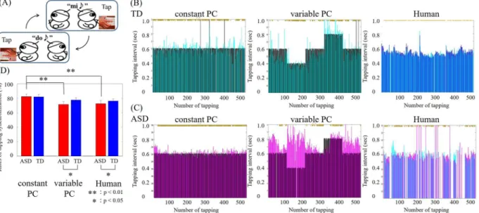
Behavioral performance was evaluated by the rates of synchronized tapping. Examples of tapping intervals for the ASD and TD groups are shown in Fig. 1. Two-way analysis of variance (ANOVA) for groups (ASD vs. TD) and conditions (constant PC, variable PC, and Human) showed the main efects of conditions (F(2,138) = 3.07;
p< 0.05) but no main efect of groups (F(1,138) = 1.19; p= 0.28) and interaction (F(2,138) = 0.42; p= 0.65). he rates of synchronization in the ASD group were signiicantly lower than those in the TD group under the Human condition (mean ± sd: ASD: 72.50% ± 4.39%; TD: 76.18% ± 3.00%; Z = 1.85, p< 0.05, Mann–Whitney U test) and the Variable-PC condition (ASD: 71.38% ± 4.22%; TD: 77.61% ± 3.04%; Z = 2.20, p< 0.05, efect size = 0.45). In contrast, no signiicant diferences between groups were observed in the Constant-PC condition (ASD: 82.26% ± 3.14%; TD: 81.94% ± 3.44%; Z =−0.28, p= 0.39, efect size = 0.06). Moreover, only in the ASD group did the performance signiicantly difer between the Constant-PC and Variable-PC conditions (Z = 2.96,
p< 0.01, efect size = 0.60) and between the Constant-PC and Human conditions (Z = 3.18, p< 0.01, efect size = 0.65). hese results show that individuals with ASD had greater diiculty in adapting to sudden luctua-tions in partner behaviors, which are usual in daily communication.
To investigate which feature of ASD contributed to the synchronization rate, we calculated Pearson’s correla-tions between the rate and the severity of each ASD feature taken from the 3 main domains of the ADOS and all 14 domains of the MSPA. he FDR corrected q-values of correlations with IQ scores and ASD assessment scores were shown at Table 1A and B, respectively. Analysis showed that rates of synchronization were signiicantly correlated with ADOS and MSPA scores under Variable-PC and Human conditions (Table 1A). Synchronization rates in ASD participants correlated with MSPA scores for only restricted interests/behaviors in the Variable-PC and Human conditions (q< 0.05, sample size = 48). Additionally, synchronization rates correlated with MSPA scores for communication skill and empathy, and ADOS scores for reciprocal social interaction skill in the Variable-PC condition (all qs< 0.01, sample size = 48). In human condition, synchronization rates correlated
www.nature.com/scientificreports/
Figure 1. (A) Schematic illustration of the alternate tapping task. (B) Sample performance of a TD participant in the three conditions. Cyan, TD participant. Black, virtual partner. Blue, human partner. he vertical axis is the interval between one partner’s tap and the other’s. he horizontal axis represents the number of taps over time. (C) Sample performance of an ASD participant on the three conditions. Purple, ASD participant. Blue, same human participant as in (B). (D) Averaged rates of tapping synchronization for all participants within each group. Error bars denote the s.e.m. Signiicant diferences between conditions were evaluated by Mann– Whitney U test.
ASD TD Total correlation Total correlation TD correlation
scores scores con.PC vari.PC Human con.PC vari.PC Human con.PC vari.PC Human
(A)
IQ 111.33 ± 2.29 111.00 ± 2.49 0.544 0.984 1 1 0.931 0.986 1 0.893 0.529 verbal IQ 113.96 ± 2.35 109.75 ± 2.79 0.655 0.970 1 0.750 0.544 0.892 0.648 1 1 performance IQ 105.29 ± 2.50 110.04 ± 2.29 0.728 1 0.883 0.826 0.432 1 0.562 0.953 0.526 (B)
communication 3.13 ± 0.16 * 1.40 ± 0.07 0.910 0.079 0.462 0.413 0.034* 0.170 1 1 0.959 social adaptation 3.23 ± 0.11 * 1.40 ± 0.08 0.897 0.068 0.549 0.724 0.061 0.038* 1 1 0.887 empathy 2.98 ± 0.12 * 1.42 ± 0.07 0.939 0.065 0.602 0.672 0.047* 0.145 1 0.988 0.847 restricted interests/behavior 3.38 ± 0.13 * 1.79 ± 0.09 0.921 0.089 0.361 0.556 0.034* 0.045* 1 0.964 0.993 Sensory 2.33 ± 0.16 * 1.42 ± 0.10 1 0.725 0.875 0.643 1 0.944 1 1 0.977 stereotyped/repetitive motion 1.58 ± 0.15 * 1.02 ± 0.02 0.850 0.682 0.938 1 0.973 0.885 0.993 1 0.659 gross motor 1.94 ± 0.16 * 1.25 ± 0.07 1 0.249 0.400 0.711 0.830 0.318 0.981 0.722 0.871 ine motor 1.65 ± 0.15 1.46 ± 0.09 0.803 0.517 0.676 0.812 0.839 0.420 1 0.934 0.839 Inattention 2.92 ± 0.19 * 2.23 ± 0.14 0.905 0.958 0.737 1 0.996 0.917 0.962 0.981 0.972 hyperactivity 1.85 ± 0.15 1.46 ± 0.09 0.861 0.449 0.946 0.939 0.961 0.920 1 0.692 0.872 Impulsivity 2.19 ± 0.16 * 1.50 ± 0.11 1 0.180 0.832 0.652 1 1 1 0.736 1 sleep cycle 2.23 ± 0.18 * 1.46 ± 0.15 1 1 0.896 0.984 0.475 0.930 0.491 0.717 0.904 learning 1.73 ± 0.17 * 1.17 ± 0.08 1 0.117 0.831 0.616 0.087 0.395 0.413 0.554 0.564 language development 1.41 ± 0.15 * 1.30 ± 0.11 0.929 1 0.929 0.936 0.987 0.991 1 1 0.916 ADOS (communication) 2.17 ± 0.30 * 0.63 ± 0.14 0.793 0.076 0.549 0.777 0.120 0.209 0.908 0.995 1 ADOS (reciprocal social interaction) 4.67 ± 0.70 * 0.92 ± 0.25 1 0.007* 0.272 0.492 0.028* 0.153 0.599 0.607 0.844 ADOS (imagination and creativity) 0.75 ± 0.17 * 0.13 ± 0.07 0.915 0.249 0.924 0.942 0.513 0.924 1 1 0.888 AQ 17.09 ± 0.85 ** 9.62 ± 0.89 1 0.066 0.625 1 0.494 1 0.620 0.711 0.826
To examine the relationship between behavior and instantaneous brain activity, we conducted time-frequency analyses on the electroencephalogram data collected during tapping (tapping period) and while listening to part-ner tapping (observation period). We compared these data with those recorded during the inter-session interval
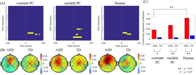
Figure 2. (A) Colour maps showing the channels and the time during the observation period in which signiicantly higher theta amplitudes were observed in the ASD group (yellow; p< 0.05, Mann-Whitney U-test with Bonferroni corrected for multiple comparisons). (B) Topographical maps for the averaged theta (6 Hz) amplitude modulations during the 0–50 ms ater onset of the other’s tapping for TD and ASD groups. (C) Averaged frontal (Fz electrode) theta amplitude modulations during the same 0–50 ms. Error bars denote s.e.m. Signiicant diferences between conditions were evaluated by Mann–Whitney U test.
Total correlation
con.PC vari.PC Human
(A)
Behavior 0.121 0.850 0.295 (B)
IQ 0.702 0.894 1
verbal IQ 0.690 1 4.390
performance IQ 1 4.502 0.849 (C)
communication 0.203 0.149 0.020*
social adaptation 0.171 0.160 0.017*
empathy 0.042* 0.177 0.042*
restricted interests/behavior 0.043* 0.088 0.011*
Sensory 0.969 0.204 0.156 stereotyped/repetitive motion 0.834 0.146 0.106 gross motor 0.865 0.881 0.488 ine motor 0.863 0.559 0.322 Inattention 0.847 0.290 0.097 hyperactivity 0.897 0.481 0.215 Impulsivity 0.645 0.516 0.093 sleep cycle 0.893 0.518 0.319 learning 0.803 0.497 0.202 language development 0.799 0.546 0.195 ADOS (communication) 0.198 0.129 0.102 ADOS (reciprocal social interaction) 0.482 0.144 0.055 ADOS (imagination and creativity) 0.165 0.470 0.100
AQ 0.110 0.142 0.056
www.nature.com/scientificreports/
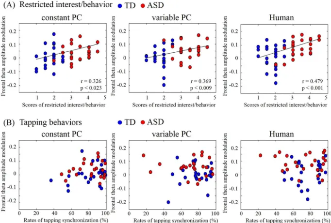
(baseline period). In all three conditions, the ASD group showed increased theta activity in the midline prefron-tal regions just ater the onset of the observation period (from onset to 50 ms). Two-way ANOVA for groups and conditions showed the main efects of groups (F(1,138) = 20.20; p< 0.001) but no main efect of conditions (F(2,138) = 2.04; p= 0.13) and interaction (F(2,138) = 1.06; p= 0.35). No such increase was detected during the tapping period for either group, or during the observation period for the TD group. he theta activity in the ASD group was signiicantly higher than that during the same time window in the TD group (Fig. 2; diferent peak frequency: 6 Hz; p< 0.05, Bonferroni corrected for multiple comparisons). No other regions were signiicantly diferent between the groups. We calculated Pearson’s correlations between the frontal activity and ASD assess-ment scores. he FDR corrected q-values of correlations with synchronization rate, ID scores and ASD scores were shown at Table 2A,B, and C respectively. We found that the amount of theta activity positively correlated with MSPA communication skills, social adaptation, empathy, and restricted behaviors in the Human condition and with empathy and restricted behaviors in the constant PC condition (all qs< 0.01). he sensory domain in
the MSPA, which shows atypical sensory reactivity, did not relate the EEG activity. Figure 3 shows the relationship between theta activity and the restricted behavior with which it correlated the most. In contrast, no signiicant correlations were found between the frontal activity and synchronization rates.
Decreased beta amplitudes and increased theta amplitude in the central electrodes which is closed to the motor area were commonly observed in all conditions and in both groups around the onset of the tapping period. hese results were consistent with the inding that beta desynchronization is related to several motor movements, such as the voluntary hand movements35 and the synchronous hand movements36. hus, the simple
sensory-motor systems in ASD appear similar to those in TD for behaviors related to synchronization, which do not require empathy-like processes.
Notably, we also found large individual diferences in the synchronization rate under the Variable-PC and Human conditions. he variance in the ASD group was particularly large; the best and the worst performers in the Human condition and the best two and the worst three performers (n = 48) in the Variable-PC condition were all from the ASD group. Indeed, not all ASD participants performed poorly on our synchronization task. his is unsurprising because ASD has a wide variety of features and comorbidities, and individual diferences are large. However, interpretation of the variance is not complicated, because our task related only to speciic cognitive functions (i.e., task switching, sensory-motor adaptation, cooperation with the behaviors of others). Our MSPA data showed that only communication, social adaptation, and restricted interests/behavior inluenced the results, while other features such as ine motor coordination, stereotyped movement, sensory, attention, hyperactivity, impulsivity, had no efect on either behavioral performance or frontal theta activity.
Increased frontal theta activity is known as one of the brain areas which correlate with executive processing capacity37–39. herefore, the theta elevation that we observed suggests that this communication might need cognitive
resources on individuals with ASD. Interestingly, the increase in frontal theta activity was correlated with the severity of ASD, rather than with the behavioral performance, meaning that the frontal activation cannot reveal how much they struggled with the task. Instead, the results suggest that individuals with ASD must put efort into thinking and planning their communicative behaviors, while TD individuals synchronize their behaviors with others automatically.
Also, the frontal theta enhancements in the ASD group might relect the diiculties in motor planning charac-teristic of ASD40–43. Although this study could not identify the exact brain regions from only the EEG recordings
and analyses due to low spatial resolution, the theta-enhanced areas were close to the motor areas, especially the supplementary motor areas. he supplementary motor areas are thought to be associated with motor planning as well as motor execution44. herefore, the ASD participants would be expected to have diiculty with motor
planning as well as executive function.
he frontal theta activation was seen even when the tapping rhythm was regular. Because participants were not informed that the rhythm was regular, they might have consciously planned their tapping in response to the regular PC and were thus able to cope with it behaviorally. In the irregular conditions, simultaneously with further frontal activation, behavioral performance dropped. In addition to not being able to keep up with unex-pected changes in rhythm made by the PC, ASD participants could not even cope with natural luctuations made by a human partner or with changes made by the PC partner. Even natural human luctuations have a irregular rhythm that might be diicult for ASD people to follow. hus, communication diiculties of people with ASD might result from their inability to adapt to irregularity. ASD includes persistent deicits in social communication, as well as restricted and repetitive patterns of behavior, interests, or activities (deine in DSM-5). Further, people with ASD are known to prefer objects to people to a greater extent than TD individuals do45,46. Assuming that
human behaviors are essentially irregular, the irst component of ASD (deicits in social communication) could be explained by this diiculty with irregularity. he second component (restricted and repetitive behavior) itself is a preference for the regular and can be viewed as intolerance of irregularity. hus, we can see how this single underlying symptom might explain a fundamental mechanism of ASD.
Furthermore, because the frontal theta activity occurred just ater the onset of the partner’s tapping, ASD participants might have been consciously conirming the other’s behavior and planning their own in response. he increased activity in the irregular conditions would suggest that individuals with ASD need more cognitive resources to make such strategies in unpatterned situations. In clinical practice, some ASD patients receive social skill training or behavioral interventions47,48. hese therapies might boost cognitive compensation for diiculty
in communication. In daily-life situations, individuals with ASD oten need experience simulations to adapt to new or irregular environments. hese indings increase our understanding of the mechanisms underlying ASD and the diiculties these patients have in daily life.
References
1. Frith, U. Mind blindness and the brain in autism. Neuron 32, 969–979 (2001).
2. Klin, A., Jones, W., Schultz, R., Volkmar, F. & Cohen, D. Deining and quantifying the social phenotype in autism. Am. J. Psychiatry.
159, 895–908 (2002).
3. Baron-Cohen, S. Autism: the empathizing-systemizing (E-S) theory. Ann. N.Y. Acad. Sci. 1156, 68–80 (2009).
4. Senju, A., Southgate, V., White, S. & Frith, U. Mindblind eyes: an absence of spontaneous theory of mind in Asperger syndrome.
Science. 325, 883–885 (2009).
5. Jones, W. & Klin, A. Attention to eyes is present but in decline in 2-6-month-old infants later diagnosed with autism. Nature. 504, 427–431 (2013).
6. Happe, F. & Frith, U. Annual research review: Towards a developmental neuroscience of atypical social cognition. J. Child Psychol. Psychiatry. 55, 553–557 (2014).
7. Kennedy, D. P. & Adolphs, R. Violations of personal space by individuals with autism spectrum disorder. PLoS One. 9, e103369 (2014). 8. Soto-Icaza, P., Aboitiz, F. & Billeke, P. Development of social skills in children: neural and behavioral evidence for the elaboration of
cognitive models. Front. Neurosci. 9, 333 (2015).
9. Hill, E. L. Executive dysfunction in autism. Trends Cogn. Sci. 8, 26–32 (2004).
10. Van Eylen, L. et al. Cognitive lexibility in autism spectrum disorder: Explaining the inconsistencies? Res Autism Spectr Disord. 5, 1390–1401 (2011).
11. Yasuda, Y. et al. Cognitive inlexibility in Japanese adolescents and adults with autism spectrum disorders. World J Psychiatry. 22, 42–48 (2014).
12. Ambery, F. Z., Russell, A. J., Perry, K., Morris, R. & Murphy, D. G. M. Neuropsychological functioning in adults with Asperger syndrome. Autism 10, 551–564 (2006).
13. Kwon, J., Ogawa, K., Ono, E. & Miyake, Y. Detection of Nonverbal Synchronization through Phase Difference in Human Communication. PLos One e0133881 (2015).
14. Neda, Z. et al. he sound of many hands clapping. Nature 403, 849–850 (2000).
15. Tognoli, E., Lagarde, J., de Guzman, G. C. & Kelso, J. A. he phi complex as a neuromarker of human social coordination. Proc. Natl. Acad. Sci. USA 104, 8190–8195 (2007).
16. Feldstein, S. & Welkowitz, J. Conversational congruence: Correlates and concerns. In Siegman, A., Feldstein, S. (eds.) Nonverbal Behavior and Communication. (Hillsdale, NJ: Lawrence Erlbaum Associates, 1978).
17. Kawasaki, M. et al. Inter-brain synchronization during coordination of speech rhythm in human-to-human social interaction. Sci. Rep. 3, 1692 (2013).
18. Yun, K., Watanabe, K. & Shimojo, S. Interpersonal body and neural synchronization as a marker of implicit social interaction. Sci. Rep. 2, 959 (2012).
19. Baron-Cohen, S. et al. he amygdala theory of autism. Neuroscience and Biobehavioral Reviews 24, 355–364 (2000).
20. Schultz, R. T. et al. Abnormal ventral temporal cortical activity during face discrimination among individuals with autism and Asperger syndrome. Arch Gen Psychiatry 57, 331–340 (2000).
21. Walsh, P., Elsabbagh, M., Bolton, P. & Singh, I. In search of biomarkers for autism: scientiic, social and ethical challenges. Nat Rev Neurosci. 12, 603–612 (2011).
22. Hamilton, A. F. C. Relecting on the mirror neuron system in autism: a systematic review of current theories. Dev Cogn Neurosci. 3, 91–105 (2013).
www.nature.com/scientificreports/
24. Tierney, A. L., Gabard-Durnam, L., Vogel-Farley, V., Tager-Flusberg, H. & Nelson, C. A. Developmental Trajectories of Resting EEG Power: An Endophenotype of Autism Spectrum Disorder. PLoS ONE 7(6), e39127 (2012).
25. American Psychiatric Association. Diagnostic and Statistical manual of Mental Disorders (American Psychiatric Press, Washington, DC, ed. 5) (2013).
26. Lord, C. et al. Autism diagnostic observation schedule: a standardized observation of communicative and social behavior. J. Autism Dev. Disord. 19, 185–212 (1989).
27. Funabiki, Y., Kawagishi, H., Uwatoko, T., Yoshimura, S. & Murai, T. Development of a multi-dimensional scale for PDD and ADHD.
Res. Dev. Disabil. 32, 995–1003 (2011).
28. Wechsler, D. he Wechsler adult intelligence scale (Harcourt, San Antonio, Texas, ed. 3) (1997).
29. Wechsler, D. Manual for the Wechsler adult intelligence scale-revised (he Psychological Corporation, New York, New York) (1981). 30. Wechsler, D. he Wechsler intelligence scale for children (he Psychological Corporation, San Antonio, Texas, ed. 3) (1991). 31. Kawasaki, M., Kitajo, K. & Yamaguchi, Y. Dynamic links between theta executive functions and alpha storage bufers in auditory and
visual working memory. Eur. J. Neurosci. 31, 1683–1689 (2010).
32. Kayser, J. & Tenke, C. E. Principal components analysis of Laplacian waveforms as a generic method for identifying ERP generator patterns: I. Evaluation with auditory oddball tasks. Clin. Neurophysiol. 117, 348–368 (2006).
33. Tallon-Baudry, C., Bertrand, O., Delpuech, C. & Pernier, J. Stimulus speciicity of phase-locked and non-phase-locked 40 Hz visual responses in human. J. Neurosci. 16, 4240–4249 (1996).
34. Benjamini, Y. & Hochberg, Y. Controlling the false discovery rate: a practical and powerful approach to multiple testing. Journal of the Royal Statistical Society, Series B., 58, 289–300.
35. Pfurtscheller, G. & Lopes da Silva, F. H. Event-related EEG/MEG synchronization and desynchronization: basic principles. Clin. Neurophysiol. 110, 1842–1857 (1999).
36. Ritter, P., Moosmann, M. & Villringer, A. Rolandic alpha and beta EEG rhythms’ strengths are inversely related to fMRI-BOLD signal in primary somatosensory and motor cortex. Hum. Brain Mapp. 30, 1168–1187 (2009).
37. Gevins, A. S. et al. Electroencephalogram correlates of higher cortical functions. Science 203, 665–668 (1979).
38. Jensen, O. & Tesche, C. D. Frontal theta activity in humans increases with memory load in a working memory task. Eur. J. Neurosci.
15, 1395–1399 (2002).
39. Onton, J., Delorme, A. & Makeig, S. Frontal midline EEG dynamics during working memory. Neuroimage. 27, 341–356 (2005). 40. Dewey, D., Cantell, M. & Crawford, S. G. Motor and gestural performance in children with autism spectrum disorders,
developmental coordination disorder, and/or attention deicit hyperactivity disorder. J Int Neuropsych Soc 13, 246–256 (2007). 41. Matson, M. L., Matson, J. L. & Beighley, J. S. Comorbidity of physical and motor problems in children with autism. Res Dev Disabil
32, 2304–2308 (2011).
42. Sipes, M., Matson, J. L. & Horovitz, M. Autism spectrum disorders and motor skills: he efect on socialization as measured by the Baby And Infant Screen For Children with a Utism Traits (BISCUIT). Developmental Neurorehabilitation 14, 290–296 (2011). 43. Funabiki, Y., Mizutani, T. & Murai, T. Fine motor skills relate to visual memory in autism spectrum disorder. Journal of Educational
and Developmental Psychology. 5, 88–96 (2015).
44. Nachev, P., Kennard, C. & Husain, M. Functional role of the supplementary and pre-supplementary motor areas. Nat Rev Neurosci
9, 856–869 (2008).
45. Sasson, N. J. & Touchstone, E. W. Visual attention to competing social and object images by preschool children with autism spectrum disorder. J. Autism Dev. Disord. 44, 584–592 (2014).
46. Pierce et al. Eye Tracking Reveals Abnormal Visual Preference for Geometric Images as an Early Biomarker of an Autism Spectrum Disorder Subtype Associated with Increased Symptom Severity. Bio. Psychiatry. 79, 657–666 (2015).
47. Couteur, A.L. & Szatmari, P. in Rutter’s Child and Adolescent Psychiatry, (John Wiley & Sons Ltd., Oxford, UK., ed. 6), 665–82 (2015). 48. Jonsson, U., Choque Olsson, N. & Bolte, S. Can indings from randomized controlled trials of social skills training in autism
spectrum disorder be generalized? he neglected dimension of external validity. Autism. 20, 295–305 (2016).
Acknowledgements
his research was supported by Grant-in-Aid for Scientiic Research on Innovative areas (21120005, 24120706, 25119512 and 15H01576), JST PRESTO, and Program to Disseminate Tenure Tracking System MEXT. We would like to thank Mr. Yosuke Ushiku, Mr. Yohei Yamada, and Ms. Eri Miyauchi for assisting with data analysis and Mr. Tadao Mizutani and Ms. Miho Yoshizumi for their support in data acquisition. We also thank Prof. Shigetada Nakanishi, Prof. Ichiro Tsuda, and Dr. Kazuo Funabiki for careful reading and fruitful suggestions.
Author Contributions
M.K. designed the study, performed EEG experiments, analyzed behavioral and EEG data, and wrote the paper; K.K. and Y.Y. performed EEG experiments and analyzed behavioral and EEG data; K.F. and T.M. supported experiments and assessments, and discussed the data; Y.F. designed the study, performed ASD assessments, analyzed behavioral and ASD data, and wrote the paper.
Additional Information
Competing Interests: he authors declare that they have no competing interests.
Publisher's note: Springer Nature remains neutral with regard to jurisdictional claims in published maps and institutional ailiations.
Open Access This article is licensed under a Creative Commons Attribution 4.0 International License, which permits use, sharing, adaptation, distribution and reproduction in any medium or format, as long as you give appropriate credit to the original author(s) and the source, provide a link to the Cre-ative Commons license, and indicate if changes were made. he images or other third party material in this article are included in the article’s Creative Commons license, unless indicated otherwise in a credit line to the material. If material is not included in the article’s Creative Commons license and your intended use is not per-mitted by statutory regulation or exceeds the perper-mitted use, you will need to obtain permission directly from the copyright holder. To view a copy of this license, visit http://creativecommons.org/licenses/by/4.0/.