Functional analysis of G protein-coupled receptors
Flop1 and Flop2 in Xenopus development
(アフリカツメガエル胚の発生における
G タンパク質共役受容体 Flop1 と Flop2 の機能解析)
Miyagi, Asuka
DOCTOR OF PHILOSOPHY
Department of Basic Biology, School of Life Science,
SOKENDAI (The Graduate University for Advanced Studies)
2015
Table of Contents ...01
Introduction ...02
Chapter 1: General Features of Flop1 and Flop2 1-1. Background ...09
1-2. Results .....11
1-3. Discussion ...19
1-4. Materials and Methods .....22
Chapter 2: Functional Role of Flop1 and Flop2 during Embryogenesis 2-1. Background ...28
2-2. Results .....29
2-3. Discussion ...36
2-4. Materials and Methods .....40
Conclusion .....42
Acknowledgments .....43
References ......44
Figures and Tables ......55
Introduction
Head formation is one of the most critical and complex steps of animal embryogenesis, involving the interplay of a multitude of signaling pathways that impact neural fate and development of the central nervous system (CNS). The molecular mechanism underlying the elaborated cell-to-cell interactions and tissue dynamics has been intensively studied with model organisms such as the mouse, chick, Xenopus laevis, and zebrafish. In particular, Xenopus laevis has been serving as an excellent model because of its convenience in embryo manipulation, microinjection, etc., due to its relatively large size of eggs and embryos. Head formation in Xenopus is achieved by a series of cell-to-cell communications, as first demonstrated by the landmark experiment performed by Spemann and Mangold (1924). In that experiment, the organizer, a small region on the dorsal lip of the early gastrula, was transferred from a host embryo to the ventral side of a recipient embryo and shown to induce a secondary body axis with completely developed head structures (Spemann and Mangold, 1924; Spemann and Mangold, 2001). It is now well understood that the functions of growth factors secreted during early embryogenesis must be suppressed to allow for the induction of neural fate in the ectoderm as well as head formation.
I. Signals essential for dorsal specification and neural development
First, I summarize general knowledge about the molecular mechanisms for the dorso–ventral (D–V) patterning during early embryogenesis in Xenopus, which is
thought to be conserved, in principle, in all vertebrates. During early Xenopus embryo- genesis, it is known that finely tuned regulation of maternally deposited factors and sequentially activated growth factors establishes the vertebrate body plan. After sperm entry, oocyte-deposited maternal factors are activated by cortical rotation. The maternal signals of Vg1/VegT at the vegetal hemisphere and of β-catenin at the dorso–vegetal side of embryo are activated, and the region in which both signals are highly intersected induces the Nieuwkoop center. At the Nieuwkoop center, Nodal-related genes are activated and the gradient of Nodal signal from dorsal to ventral side in vegetal hemi- sphere is generated. Nodal signal induces mesodermal tissues, and the region in which high β-catenin signal and low Nodal signal are received induces the blastula Chordin- and Noggin-expressing (BCNE) center (Introduction Fig. A). Dorsal mesoderms, endomesoderm and chordamesoderm, chordamesoderm contains a part of the BCNE center, express many secreted antagonists and act as head organizer and trunk organizer, respectively, and then these two organizers are also known as "Spemann organizer" (De Robertis and Kuroda, 2004; Hikasa and Sokol, 2013) (Introduction Fig. B).
During dorsal specification, growth factors such as bone morphogenetic proteins (BMPs) and Wnts are negatively regulated by antagonists secreted from Spemann organizer. Neural tissues are developed from the dorsal ectoderm that is lined by the involuting dorsal mesoderms. To establish neural fate in the ectoderm, BMP signaling, which plays crucial roles in establishing epidermal fate, must be inhibited by antagonists such as Chordin, Noggin, and Follistatin, all of which are secreted from the organizers (Fainsod et al., 1997; Iemura et al., 1998; Sasai et al., 1994; Smith and
Harland, 1992). Induced neural ectoderm then patterned along with the antero–posterior (A–P) axis. During this patterning event, zygotic Wnt/β-catenin signaling acts in posterior specification. Head is the most anterior neural structure and head formation is one of the key steps during animal embryogenesis. To induce head formation, therefore, zygotic Wnt/β-catenin signaling must be inhibited by sFRP, Dkk, and Cerberus, which are secreted Wnt antagonists produced from head organizer (Glinka et al., 1998; Leyns et al., 1997; Pera and De Robertis, 2000; Piccolo et al., 1999). Thus, the graded expression of secreted Wnt antagonists from anterior to posterior generated by head organizer characterizes neural tissue properties along with the A–P axis (Introduction Fig. B).
Although priciple moleclular mechanisms described above are now accepted and the current model appears to be well established to explain early dorsal specification and neural development, new molecules are still emerging to be integrated into the pathways.
II. Recently identified molecules antagonizing Wnt signals
As mentioned above, many secreted antagonists against growth factors have been identified and investigated so far and they are considered to be critical regulators of early patterning events. Recently, several molecules were identified as Wnt signal inhibitors. Wise and Notum were reported as secteted inhibitors, which bind to Wnt receptor LRP and Wnt ligand, respectively, and inhibited signal transduction (Guidato and Itasaki, 2007; Zhang et al., 2015). Shisa1, Waif1, Apcdd1, and Tiki1 were identified as transmembrane molecules and inhibited Wnt signaling by prevending the
maturation of Wnt receptor Frizzled, by binding LRP, by binding both Wnt ligand and LRP, and by inactivating Wnt ligand, respectively (Kagermeier-Schenk et al., 2011; Shimomura et al., 2010; Yamamoto et al., 2005; Zhang et al., 2012). In particular, since Shisa1 and Apcdd1 were confirmed to be expressed in the neural ectoderm at the early gastrula stage and their loss-of-function caused abnormal embryos with defective head morphology, suggesting that these moleclules have important roles in head fomation.
These findings suggested that in addition to the previously known secreted antagonists with pivotal roles in head formation, there may be as-yet-unidentified regulators that employs novel mechanisms for head formation.
III. Crosstalk between Wnt and RhoA pathways
RhoA, one of the small GTPases, is also a candidate for such Wnt antagonistic molecule because it has the capacity to induce ectopic head structures (Wünnenberg-Stapleton et al., 1999). RhoA has been known as an important mediator of non-canonical Wnt/planar cell polarity (PCP) pathway. In the Wnt/PCP pathway, RhoA transduces Wnt-Frizzled signal to cytoskeletal regulation and controls cell morphology and migration. On the other hand, it has been considering that RhoA does not interact with β-catenin-Tcf/Lef cascade and the relationship between RhoA and canonical Wnt/β-catenin pathway are largely unknown. Therefore, the study of RhoA in head induction has raised the possibility of a functional connection between RhoA and Wnt/β-catenin pathway.
A head-inducing activity of RhoA was demonstrated by the head induction
assay. To validate functional activity of Wnt inhibitor, a versatile method was well established in earlier studies, which is known as the head induction assay. Since the inhibition of BMP signaling results in the neural fate induction, expression of BMP signal inhibitor such as Chordin or dominant-negative BMP receptor (tBR) at the ventral side of embryo induces a secondary dorsal axis. However, the induced secondary axis has no head structures due to the existence of zygotic Wnt/β-catenin signaling. Therefore, by the co-expression of Wnt signal inhibitor such as Dkk or dominant-negative Wnt (dnWnt), the embryos can form complete head structures in the secondary axis (Glinka et al., 1998). RhoA also showed the head-inducing activity in this assay system, while it was still unclear how RhoA is integrated into the regulatory pathway of head formation and Wnt/β-catenin signaling.
IV. The purpose of the present study
From the background described above, I thought that there are still undiscovered molecules and mechanisms during head formation. In this study, to further understand head formation processes, I explored molecules potentially serve as a novel inhibitor, and found Flop1 and Flop2 that are G protein-coupled receptors (GPCRs) related to Gpr4 (Chung et al., 2004; Tao et al., 2005). Flop1 and Flop2 contributed to head formation during Xenopus embryogenesis by inhibiting Wnt/β-catenin signaling. Furthermore, Flops activated RhoA and functioned through both RhoA-dependent and -independent pathways. The Flops-activated RhoA- independent pathway induced the phosphorylation and proteasome-dependent
degradation of β-catenin, disabling β-catenin-mediated transcription. In contrast, the Flops-activated RhoA-dependent pathway inhibited Wnt/β-catenin signaling upstream of Dishevelled (Dvl). Consistent with the reported mechanism for transmembrane Wnt signal inhibitors, such as Shisa1 and Apcdd1, Flops inhibited Wnt/β-catenin signaling in a cell-autonomous manner. This is the first report demonstrating that GPCRs have essential functions in head formation and act as novel negative regulators of the Wnt/β-catenin pathway.
Chapter 1
General Features of Flop1 and Flop2
1-1. Background
Neural development which is essential for the establishment of the central nervous system (CNS) of animals is comprised of several intricate processes such as organizer formation, sequential neural induction and antero–posterior (A–P) as well as dorso–ventral (D–V) patterning. Although intensive studies have clarified basic principles of their molecular mechanisms by using several excellent model organisms such as Xenopus laevis (X. laevis) and zebrafish (Danio rerio), new molecules such as a transmembrane protein Shisa1 that serves as an important regulator of the anterior specification of the neural ectoderm are still emerging in the filed, highlighting the complexity of the regulatory system. This encouraged me to think that there are still undiscovered molecules and hidden mechanisms in these processes.
Thus, I explored new genes that have a potential to play a key role in neural development, particularly focusing on head development during Xenopus early embryogenesis. By screening genes that are dominantly expressed in the neural tissues throughout neural development from databases of Xenopus gene expression, I identified a gene whose function appears to be less well understood, but possibly involved in neural tissue development. By whole-mount in situ hybridization (WISH), I confirmed that this gene, provisionally designated as xgpcr4 by an earlier study (Chung et al., 2004), had an interesting spatiotemporal expression pattern and its transcripts were detected in the neural ectoderm at early gastrulae. Concomitantly, I found that another gene provisionally designated as xflop (Tao et al., 2005), which is phylogenetically
closed to xgpcr4, showed a similar expression pattern. These genes were separately reported and suggested to play roles in the regulation of gastrulation, but their functions in the regulation of neural development were unknown.
Therefore, I investigated their functions in the neural tissue development. In this study, I renamed these two genes as flop1 and flop2, respectively, based on their structural similarity and to clarify their nomenclature. In Chapter 1, I firstly introduce the general features of Flop1 and Flop2 in Xenopus embryogenesis.
1-2. Results
I. Phylogenetic positions of Flop1 and Flop2
The primary structure of Flop1 and Flop2 (Flop1/2 or Flops) indicated that they are typical G protein-coupled receptors (GPCRs) of Rhodopsin-like receptor family (Class A), which have seven transmembrane domains and G protein-binding motif (DRY motif), and transduce the extracellular signals to the intracellularly by mediating the activation of G protein cascades. Both Flop1 and Flop2 have been reported to be phylogenetically related to human GPR4 (Chung et al., 2004; Tao et al., 2005) that is a proton-sensing receptor, and phylogenetic tree showed that Flops are also included in the proton-sensing receptor family (Fig. 1A). However, since a recent search of the Xenopus genomic DNA database identified a gene that exhibits significantly higher homology to human GPR4 than either flop1 or flop2, Flops are thought not to be the orthologs of human GPR4. Moreover, I confirmed that each respective Flop exists in X. tropicalis genome, which suggests that they evolved as independent genes and were not derived by the allotetraploidization of the X. laevis genome. In amino acid sequence homology, human GPR4 exhibits 47% identity with either Xenopus Gpr4 and 32–3% identity with Flops (Fig. 1B). The identity between Xenopus Gpr4 and Flops is 26–9%. Flop1 and Flop2 share 60% amino acid sequence identity. Comparison of Flop1 and Flop2 with Gpr4 revealed that Flop1 displays a slightly higher homology with Gpr4 than does Flop2.
I searched for the homologs of Flops in human, model animals (mouse, chick,
zebrafish, medakafish, sea squirt, fly, and worm), reptiles (snake and lizard), and other amphibians Caudata (newt and salamander), but was unable to identify the homologs of Flops in these organisms. However, very interestingly, I found that coelacanth (Latimeria chalumnae) has a Flop ortholog in its genome (Fig. 1A). The coelacanth ortholog has 56% and 50% identity with X. laevis Flop1 and Flop2, respectively, at the amino acid level.
II. Differences from proton-sensing receptor
From the phylogenetic positions, Flop1 and Flop2 were thought to function as proton-sensing receptors similar to human GPR4. In human GPR4, five histidine residues distributed over the protein have been implicated in proton-sensing (Ludwig et al., 2003), but most of them are not conserved and either replaced to another amino acid or deleted in Flop1/2 (Fig. 2). As human GPR4 elicits the formation of cyclic AMP (cAMP) (Ludwig et al., 2003), I compared the roles of X. laevis Gpr4, Flop1, and Flop2 in cAMP formation in animal caps, ectodermal explants from St. 9 Xenopus embryos. Each mRNA was injected into the animal pole of two-cell-stage embryos and then animal caps were excised from the embryo and subjected to the cAMP assay. As a result, while Gpr4 overexpression promoted cAMP production under these conditions, the overexpression of neither Flop1 nor Flop2 affected cAMP production (Fig. 3). Thus, although Xenopus Gpr4 retains the capacity to function as a proton-sensing receptor, Flop1/2 may have different functions of yet unidentified.
III. Expression patterns during embryogenesis
To investigate the fundamental properties of Flops, I examined the temporal and spatial expression patterns of Flops during early Xenopus development. Quantitative RT-PCR (QRT-PCR) analysis revealed that flop1/2 mRNAs were maternally encoded and that their expression levels gradually declined as development proceeded (Fig. 4A). On the other hand, related gene gpr4 was expressed zygotically after the mid-blastula transition. Then, by WISH analysis, I found that maternal flop1/2 mRNAs localized to the animal hemisphere (Fig. 4B), whereas early zygotic flop1/2 transcripts were detected in the prospective neural ectoderm and organizer region of the blastula, known as the blastula Chordin- and Noggin-expressing (BCNE) center (Kuroda et al., 2004) (Fig. 4C and G, arrow). In particular, during early gastrulation, flop1/2 were detected in the bottle cells and neural ectoderm, and in the endoderm in a salt-and-pepper pattern (Fig. 4D-F and H-J). The flop1/2 transcripts were detected in the deep layers of the neural ectoderm, and a profound flop2 expression was detected in the anterior region of the ectoderm.
At the early neurula stage, flop1, but not flop2, was detected in the anterior and posterior neural plate and in the prechordal plate (Fig. 5A-E and O). During the late neurula stage, flop1/2 transcripts were detected in the eye primordia and the future brain; flop1 expression was restricted to the hindbrain, while flop2 expression extended from the fore- to hindbrain areas (Fig. 5F, G, P, and Q). Sagittal sections of tailbud stage embryos revealed that the flop1 expression domain corresponded to the region encompassing rhombomere 5/6, and was not detected in the notochord (Fig. 5H-J).
From the late tailbud stage on, flop1/2 were expressed in the somites (Fig. 5K, L, R, and S) in a non-overlapping pattern; while flop1 was expressed at the center of each somite segment, flop2 was expressed at their edges. The expression of flop1 was also detected in the lens (Fig. 5K, M, and N) and flop2 was expressed weakly in the mouth primordium (Fig. 5R, arrow).
In contrast, gpr4 expression was not detectable until the late tailbud stage and was subsequently detected in the heart (Fig. 5T-W, arrow), consistent with previous studies in which human and mouse GPR4 were shown to be highly expressed in cardiovascular tissues, including the heart (Mahadevan et al., 1995; Yang et al., 2007).
IV. Initial cue of zygotic induction
Flop1 has been reported to be a target of both FGF (Chung et al., 2004) and TGF-β signaling (gpr-4, Dickinson et al., 2006). I confirmed that Flop1/2 were more responsive to Smad2, a cytoplasmic signaling component of the TGF-β signaling pathway, than to a TGF-β ligand Nodal (Fig. 6), possibly due to the much higher signal transmission efficacy of Smad2. Likewise, eFGF failed to induce Flops expression significantly; a previous study reported that only 1.3- to 1.4-fold expression was induced by eFGF (Chung et al., 2004). These results suggested that the initial cue inducing zygotic Flop1/2 expression at the BCNE center may be TGF-β-related ligands. In addition, Flops expression was also reduced by BMP4 suggesting the possibility that endogenous Flops expression is under the regulation of BMP signaling. Taken together, it is likely that the transcription of Flop1/2 is properly tuned by the balance of the
two opposing TGF-β activities, Nodal/activin and BMP.
V. Gain- and loss-of-function effects
Previous studies reported that both the overexpression and depletion of either Flop led to gastrulation defects (Chung et al., 2004; Tao et al., 2005). As shown above, I found that Flop1/2 were expressed in the neural ectoderm and their transcripts were particularly abundant in the presumptive head region (Fig 4E, F, I, and J). Therefore, I examined the possibility that they are also involved in the neural tissue development, especially in the patterning of the CNS, separately from the regulation of gastrulation.
In gain-of-function experiments, the injection of Flop1 or Flop2 mRNA into the dorsal side of four-cell-stage embryos caused a severe defect in blastopore closure and led to embryonic phenotypes including spina bifida, as previously described (Fig. 7A and B) (Chung et al., 2004; Tao et al., 2005). In addition, the embryos with relatively mild phenotypes such as shortened body axis exhibited micro- or anencephaly (Fig 8A, arrows), which was not emphasized in the previous study. To further examine the effect of Flops overexpression on head development, I injected Flop1 or Flop2 mRNA into two dorso–animal blastomeres of 16-cell-stage embryos to avoid causing spina bifida phenotypes (Fig. 8B). The embryos treated with either mRNA underwent normal gastrulation as expected, but displayed microcephalic phenotypes, consistent with the prediction that Flops play a role in head formation (Fig. 8C and D). Expression of sonic hedgehog (shh), which marks axial structures including prechordal plate, notochord, and floor plate (Ekker et al., 1995), and is required for establishing bilateral
symmetry during head formation (Chiang et al., 1996), was attenuated in Flops mRNA-injected regions (Fig. 8E, arrow). In addition, most embryos overexpressing either Flop exhibited a reduction of body pigmentation (Fig. 8C, arrows). Expression of neural crest marker slug disappeared from the injected regions (Fig. 8F, arrow), suggesting that the perturbation of Flops also affected neural crest formation.
In loss-of-function experiments, knockdown of Flop1 and/or Flop2 was achieved by injecting specific morpholino antisense oligonucleotides (MOs) (Fig. 9) into the dorsal side of four-cell-stage embryos. Similar to Flops-overexpressing embryos, Flops-depleted embryos exhibited shortened axis with microcephaly (Fig. 10A). In contrast to the results of previous studies (Chung et al., 2004; Tao et al., 2005), the effects in blastopore closure were milder and the spina bifida phenotype was barely detected (Fig. 7C and D). Differences in experimental procedures between the current and earlier studies such as the sequence of antisense oligonucleotides could account for the inconsistent results. Notably, the microcephalic phenotypes of Flops morphants were rescued by co-injection with Flops rescue constructs, which contained mutations in MO recognition sequence (Fig. 9 and 10B). Further analysis of Flops morphants by QRT-PCR showed that the expression of forebrain (bf1) and midbrain (en2) markers and the expression of fore- to anterior hindbrain (otx2) marker were considerably and modestly reduced, respectively (Fig. 11A). In contrast, the expression of hindbrain marker krox20 was unchanged. This indicated that Flops are required for head development especially in anterior region. Reduction of bf1 and en2 expression in MO-injected region was also observed by WISH (Fig. 11B). In addition, Flops
morphants exhibited delayed body pigmentation, which in most cases was restored by St. 40. The expression of slug was retained, although its expression pattern was slightly altered, possibly due to the delay in neural folding (Fig. 12). Taken together, these data indicated that Flop1/2 are involved in neural patterning and head development, in addition to the regulation of gastrulation.
VI. RhoA activation and induction of bottle cell-like cells by Flops
Flop2 was implicated in cortical actin assembly during early embryogenesis (Tao et al., 2005). Flop2 overexpression increases the assembly of cortical actin, whereas the depletion of its maternal mRNA does the reverse. I confirmed this initial finding of Flop2 and demonstrated that Flop1 exhibited a similar function (Fig. 13A). I also found that both Flop1 and Flop2 were capable of activating RhoA, raising the possibility that the cortical actin regulation by Flops is at least partly mediated by RhoA (Fig. 13B).
In addition, the blastocoel roof (BCR) phenotypes of Flops-overexpressing embryos were strikingly similar to those of RhoA-overexpressing embryos. Normally, at the blastula stage, BCR is composed of 2–3 cell layers, while Flops- or RhoA- overexpressing embryos had approximately 3-times more BCR cell layers and enhanced pigment accumulation (Fig. 14A and B). The abnormal morphology of BCR by Flops overexpression was invalidated by co-injection with their specific MOs (Fig. 14C). Considering our finding that Smad2 could induce the expression of Flops, these observations suggest that TGF-β-mediated bottle cell formation (Kurth and Hausen, 2000) was due to the ectopic expression of Flops or RhoA. BCR thickening induced by
ectopic bottle cell formation is associated with the ectopic expression of chordin (chrd), goosecoid (gsc), and brachyury (xbra) (Kurth et al., 2005). In QRT-PCR analysis using animal cap system, I found that both chrd and xbra were induced by Flop1/2 and RhoA overexpression, and that gsc was induced by Flop2 and RhoA overexpression (Fig. 15A). Ectopic expression of chrd was also confirmed by WISH (Fig. 15B). To identify the layer of cells that contributed to BCR thickening, I examined the expression of two ectodermal markers, claudin4 (marker of the superficial layer) and hyaluronan synthase1 (has1, marker of the deep layer) (Chalmers et al., 2006). I found that the has1-positive cell layer was multi-layered in both Flop1/2- and RhoA-overexpressing embryos (Fig. 14B). Moreover, multi-layered cells contained bottle cell-like cells with an apical constriction-like morphology and actin accumulation (Fig. 16). As the endogenous expression of Flop1/2 was normally observed in bottle cells (Fig 4E, F, I, and J), and both their overexpression and knockdown caused gastrulation defects (Fig. 7A-D) (Chung et al., 2004; Tao et al., 2005), Flops and RhoA may be involved in mediating TGF-β signal-induced bottle cell formation and gastrulation.
In addition, Flops have a DRY motif, a conserved region in GPCRs that interacts with G proteins (Rovati et al., 2007), and I confirmed that Flops' expression was not affected by DRY motif mutation using western blotting (data not shown). However, Flops DRY motif mutants failed to exhibit BCR thickening and RhoA activation (Fig. 17A and B). This finding was consistent with the possibility that RhoA activation occurs downstream of Flops-mediated G protein signaling.
1-3. Discussion
Flop1 and Flop2 were independently reported to be human GPR4-related molecules (Chung et al., 2004; Tao et al., 2005). However, I revealed that authentic Gpr4 existed in Xenopus laevis genome and Flop1/2 were not orthologs of human GPR4. While Flop1/2 are included in the proton-sensing receptor family phylogenetically, they might play different roles from proton-sensing receptors with the ability of cAMP production. I found that both Flop1/2 were capable of regulating the cortical actin assembly, which is thought to be in part mediated by RhoA activation. As DRY motif mutants of Flops failed to activate RhoA, RhoA activation by Flops mediates G protein signaling. Since G12/13-RhoA activation pathway is well known as one of G protein signaling cascades of GPCR (Dhanasekaran and Dermott, 1996; Suzuki et al., 2009), I speculate that Flops have G12/13 signaling cascade as second messenger pathway and then activate RhoA.
From WISH analyses, I found that Flop1 and Flop2 have unique expression patterns in the BCNE center at the blastula stage and in the neural ectoderm at the early gastrula stage. Both gain- and loss-of-functioned embryos resulted in the abnormal head development, suggesting that they function in the neural tissue development, in addition to the regulation of gastrulation as previously reported (Chung et al., 2004; Tao et al., 2005). Furthermore, I found that both Flops and RhoA have the inducing ability of several developmental markers including organizer genes. These results also suggested that Flop1/2 have important roles in neural tissue development mediating RhoA
activation. Therefore, I further investigated their functions in neural tissue development and those data are shown and discussed in Chapter 2. WISH data further suggested that Flop1/2 might be involved in the somitogenesis as observed in their alternating expression in somitic tissues, and that Flop1 and Flop2 might also be associated with the development of lens and mouth, respectively.
Several GPCRs form functionally important dimers. In particular, human proton-sensing receptors and lysophosphatidic acid (LPA) receptors, which are phylogenetically related to proton-sensing receptors (Fig. 1A), have been reported to form both homo- and heterodimers (Zaslavsky et al., 2006). It is also possible that Flop1 and Flop2 form functional dimers, because their single knockdown phenotypes were indistinguishable from that of their double knockdown. My study demonstrated more functional similarities between Flop1/2 and LPA receptors, both of which regulate RhoA-mediated cytoskeletal rearrangement (Lloyd et al., 2005) and anterior head development (Geach et al., 2014) than between Flop1/2 and proton-sensing receptors. Originally, proton-sensing receptors were reported to be receptors for lipid mediators similar to LPA receptors, while more recent studies have redefined them as multi-functional receptors capable of mediating signals from protons and/or lysolipids (Im, 2005; Tomura et al., 2005). Although the ligands for Flop1/2 remain to be identified, I speculate that they could be analogs of lipid mediators. Lipid mediators have multiple biological functions and play important roles in several phases of embryonic development by modulating cell proliferation and differentiation. Thus it is possible that Flop1/2 evolved in Xenopus (amphibians) as a mechanism for mediating
the transduction of such important signals in developing embryos and allowing them to adapt to the unstable aquatic circumstances.
At present, Flops' ortholog is only found in the coelacanth genome, and therefore, Flop1/2 are thought to be following an interesting evolution. Coelacanth has been supposed to be a "missing link" between fish and amphibians (tetrapods) (Nikaido et al., 2013). On the other hand, the Pipidae family including Xenopus is completely aquatic animals different from other frogs and is considered to be primitive species of amphibians. Taken together, the fact that Flops are only found in these two animals could suggest that Flops may be the key molecules to understand the evolution and development of tetrapods.
1-4. Materials and Methods
Embryo handling and microinjection
Xenopus laevis embryos were obtained by standard methods (Morita et al., 2010). mRNAs or morpholino antisense oligonucleotides (MOs) were injected into the appropriate region of two-, four-, or 16-cell-stage embryos. The injected embryos were cultured in 3% Ficoll/0.1x Steinberg's Solution until stage (St.) 9 and then cultured in 0.3x Marc's Modified Ringer's Solution until the desired stage (Nieuwkoop and Faber, 1967).
DNA constructs, mRNA preparation, and MOs
The following Xenopus laevis (X. laevis) cDNA clones were obtained from the EST database (XDB3, http://xenopus.nibb.ac.jp) and cloned into the pCS2p+ vector: XL072l08 (gpr4), XL506k06ex (flop1), XL155m04 (flop2), XL255j14ex (rhoA), XL280g21ex (smad2), XL011e23 (claudin4), XL109a23 (hyaluronan synthase1), XL085m19 (slug), and XL250b20ex (sonic hedgehog). bf1 was subcloned into pCS2p+ vector from the X. laevis cDNA and en2 construct was used as reported previously (Hemmati-Brivanlou et al., 1991). Expression constructs for Wnt8 (Christian et al., 1991), Xnr1 (Jones et al., 1995), eFGF (Lombardo and Slack, 1997), and BMP4 (Nishimatsu et al., 1992) were reported previously. The dnRhoA (T19N) and Flops DRY motif mutants (Flop1; R113N, Flop2; R112N) were generated by PCR. Flop1 and Flop2 rescue constructs were generated by changing the nucleotides at the MO
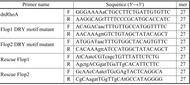
target site as follows (changed nucleotides are indicated in small letters): Flop1 (-2) 5′-GAATGTGcAAtCAatcCGTcagcTG-3′ (+23) and Flop2 (-1) 5′-TATGGCaTGcAA cCAatctTGcGAg-3′ (+24). Capped mRNAs were synthesized using the mMESSAGE mMACHINE® SP6 kit (AM1340; Ambion) and purified on NICK columns (17-0855; GE Healthcare). MOs were purchased from Gene Tools. PCR primers for expression constructs and MO sequences are shown in Tables 1 and 2.
Whole-mount in situ hybridization (WISH) and quantitative RT-PCR (QRT-PCR) WISH was performed as described previously (Goda et al., 2009). As the endogenous expression signals of flop1/2 were very weak, I used albino embryos to avoid the bleaching process, which might further reduce their signals. Pigmented embryos used in other experiments were bleached before WISH. To visualize the injected regions, a GFP tracer was injected and immunostained with an anti-GFP antibody after WISH. For the temporal expression analysis of gpr4 and flops, the total RNA from one whole embryo at each stage of interest was isolated as described previously (Yamamoto et al., 2001). For marker gene analysis, the total RNA was isolated from five anterior or posterior halves of St. 20 embryos, and five animal caps from St. 9 embryos. QRT-PCR was performed with the SYBR® premix ExTaq (Tli RNaseH plus) (RR420; TaKaRa) on a Thermal Cycler Dice Real Time System Single (TP850; TaKaRa) according to the manufacturer's instructions, and the relative gene expression was calculated with the ΔΔCt method. The primer sets used in this experiment are shown in Table 3.
Immunostaining
Embryos or animal caps excised from St. 9 embryos were fixed in MEMFA for 2 h at room temperature and then placed in PBS. The fixed embryos were embedded in fish gelatin, and 16-µm cryosections were generated (Morita et al., 2010; Suzuki et al., 2010). The sections, fixed animal caps, or embryos previously analyzed by WISH were incubated with the primary antibody rabbit anti-GFP (1:500, #598; MBL) or with Alexa Fluor® 546 phalloidin (1:50, A22283; Molecular Probes) for F-actin staining. The secondary antibody was Alexa Fluor® 488 goat anti-rabbit IgG (1:1,000, A11017; Molecular Probes).
Western blotting and RhoA pull-down assay
Prior to western blotting, mRNAs and/or MOs were injected into the animal pole of two-cell-stage embryos. Subsequently, 30 animal caps were excised from St. 9 embryos and lysed in 50 µl PBST (PBS with 0.3% TritonX-100) containing a protease inhibitor cocktail (1:50, 25955-11; Nacalai Tesque). After incubating on ice for 5 min, the lysates were centrifuged at 17,400 xg for 5 min at 4oC, and the supernatants were collected, diluted in 2x sample buffer, and boiled for 5 min. The samples were then subjected to SDS-PAGE (13.5% gel for Histone and 10% gel for all other proteins) and then blotted onto a PVDF membrane (162-0177; Bio-Rad). The membranes were incubated with the following primary antibodies: rabbit anti-GFP (1:2,000, #598; MBL), mouse anti-Histone H2B (1:1,000, #2934; Cell Signaling Technology), and mouse anti-Flag (1:1,000, F3165; Sigma). The following secondary antibodies were used: HRP-
conjugated sheep anti-mouse IgG (1:10,000, NA931; GE Healthcare) and HRP- conjugated donkey anti-rabbit IgG (1:10,000, NA934; GE Healthcare), and the signals were detected using ECL™ Prime or ECL™ Select Western Blotting Detection Reagent (RPN2232, RPN2235; GE Healthcare).
The RhoA pull-down assay was performed as described previously (Hara et al., 2013). Prior to the assay, mRNAs were injected into the animal pole of two-cell- stage embryos, and 100 animal caps were excised from the St. 9 embryos and analyzed.
cAMP assay
The cAMP assay was performed with the Cyclic AMP XP® Assay Kit (#4339; Cell Signaling Technology). mRNAs were injected into the animal pole of two-cell-stage embryos, and the animal caps were excised from St. 9 embryos. As a positive control, four- to eight-cell-stage embryos were incubated with 100 µM Forskolin (F6886; Sigma) overnight at 13oC until reaching St. 9, and then the animal caps were excised. Twenty animal caps were lysed in 50 µl 1x lysis buffer from the assay kit with 1 mM PMSF. After incubating on ice for 5 min, the lysates were centrifuged at 17,400 xg, for 5 min at 4oC. The supernatants were collected, and their protein concentrations were determined using the Pierce® BCA Protein Assay Kit (#23227; Thermo Scientific). The protein concentrations were adjusted to 1.2 µg/µl and used in the cAMP assay according to the manufacturer's instructions. The optical density of the samples was measured on an SH-9000Lab (Corona Electric). The cAMP assays were performed using three batches of embryos.
Image acquisition, processing, and statistical analysis
Images of whole embryos were captured using a fluorescence stereomicroscope SZX16 (Olympus) equipped with a CCD camera DP71 (Olympus), and images of sections were captured using a fluorescence microscope BX63 (Olympus) equipped with a CCD camera DP72 (Olympus). Immunostained sections or animal caps were observed using an A1R laser scanning confocal microscope (Nikon). Confocal images were processed with NIS-Elements software (Nikon) to generate the maximum projection intensity of the acquired z-stacks. Pixel intensity was measured using ImageJ. For statistical analysis, data presented by 100% stacked column chart were calculated by the Pearson's chi-square test with adjusted standardized residuals (α=0.05, critical value=±1.96; α=0.01, critical value=±2.58; α=0.001, critical value=±3.29). Statistical analyses for other data were performed using the Student's t-test.
Chapter 2
Functional Role of Flop1 and Flop2
during Embryogenesis
2-1. Background
During early embryogenesis, bone morphogenetic proteins (BMPs) play essential roles as ventral-inducing signals in the D–V patterning. In the ectoderm, since BMPs act as signals of epidermal induction, the inhibition of BMP signaling is required for neural induction and dorsal development. Notably, the inhibition of BMP signaling by expressing a dominant-negative (truncated) BMP receptor (tBR) at the ventral side of embryo induced the formation of ectopic dorsal structures, including neural tissues, but excluding the head (partial secondary axis) (Suzuki et al., 1994), suggesting that BMP suppression is not sufficient for inducing head formation. However, the simultaneous inhibition of BMP and Wnt signaling by expressing both tBR and a dominant-negative Wnt8 (dnWnt8) frequently induced the formation of a secondary axis with head structures (complete secondary axis), suggesting that the inhibition of both BMP and Wnt signals is required for head formation (Glinka et al., 1997). Interestingly, ventrally expressed RhoA also induced ectopic head formation when BMP signaling on the ventral side is inhibited, suggesting that RhoA also plays a role in head induction, similar to that of Wnt antagonists (Wünnenberg-Stapleton et al., 1999).
In Chapter 1, I revealed that Flop1/2 are required for normal head development and have the RhoA activation activity. Thus, I hypothesized that Flops also have the head-inducing activity via RhoA activation and contribute to head development. In Chapter 2, I investigated how Flops regulate head development focusing on this point, and elucidated a part of their functional roles in Xenopus embryogenesis.
2-2. Results
I. Flops have head-inducing activity as well as RhoA
To investigate how Flops are involved in the head development, I focused on the unique function of RhoA. RhoA has a head-inducing activity when BMP signaling is inhibited at the ventral region (Wünnenberg-Stapleton et al., 1999). In addition to the similar BCR phenotypes of the Flops- and RhoA-overexpressing embryos, the endogenous expression of RhoA overlapped with that of Flops in the neural ectoderm (Fig. 4K and L). Therefore, I speculated that Flops also induce head structure formation in the absence of BMP signals through the RhoA activity. To examine this hypothesis, Flops mRNAs were injected along with tBR mRNA into the ventral side of four-cell-stage embryos. As expected, the injection of either Flops or RhoA mRNA induced the development of a complete secondary axis (Fig. 18). Notably, the eye structures of the secondary axis in Flops- or RhoA-injected embryos exhibited a fused eye morphology, suggesting that from the point of spacing of eyes, the head-forming activity of Flops and RhoA is not as complete as that of dnWnt8 (Fig. 18C, arrows and E-G, arrowheads). In contrast, the ventral injection of Gpr4 mRNA failed to induce head structures (Fig. 18D). These results suggested that the head-inducing activity is rather specific to Flop1/2 among the family members.
Since DRY motif mutants could not induce a complete secondary axis (Fig. 19A), Flops' head-inducing activity is thought to mediate G protein signaling. Furthermore, I confirmed that the co-injection of Flop1/2 with dominant-negative RhoA
(dnRhoA) mRNA partially prevented the induction of a complete secondary axis (Fig. 19B), suggesting that RhoA, at least in part, acts downstream of the Flops-mediated head induction.
II. Flops and RhoA inhibit Wnt/β-catenin signaling
To investigate the mechanism by which Flops and RhoA influence head formation, I next examined whether they serve as Wnt antagonists by performing the TOP-flash assay, which measures β-catenin-dependent transcriptional activity (Wnt/β-catenin signaling). I injected the TOP-flash reporters alone or together with Wnt8, Flops, or RhoA mRNA into the animal pole of two-cell-stage embryos, and then measured the luciferase activity of excised animal caps corresponding to St. 12. Notably, the overexpression of either Flops or RhoA inhibited Wnt/β-catenin signaling in a dose- dependent manner (Fig. 20A). Conversely, the knockdown of Flop1 and/or Flop2 or the expression of dnRhoA upregulated the endogenous Wnt/β-catenin signaling (Fig. 20B). I also confirmed that DRY motif mutants of Flops failed to inhibit Wnt/β-catenin signaling (Fig. 20C), suggesting that Flops' inhibitory activity of Wnt/β-catenin signaling depends on G protein signaling.
Ectopic activation of Wnt/β-catenin signaling in the ventral region of the early stage embryo results in the induction of complete secondary axis (early Wnt signal in the D–V patterning, see the review by Hikasa and Sokol, 2013). Therefore, I tested whether Flops and RhoA can inhibit Wnt/β-catenin signaling by observing the complete secondary axis formation by early Wnt signal. As a result, the induction of a complete
secondary axis by Wnt8 mRNA injection on the ventral side of four-cell-stage embryos was efficiently blocked by co-injection with Flop1, Flop2, or RhoA mRNA in a dose- dependent manner (Fig. 21). These findings suggested that Flops and RhoA contribute to head induction by inhibiting Wnt/β-catenin signaling, in contrast to the results of a previous study, which showed that RhoA ectopic expression was unable to block the Wnt8-mediated induction of a complete secondary axis (Wünnenberg-Stapleton et al., 1999). This discrepancy may be due to a difference in the experimental conditions used in the two studies, such as the amount of Wnt8 and RhoA mRNA injected. In fact, one pg of Wnt8 mRNA was sufficient to induce a complete secondary axis and higher doses of Wnt8 mRNA often made rescue by RhoA difficult. Moreover, excess RhoA affected blastopore closure even by ventral expression, complicating the interpretation of its effects on secondary axis formation.
III. Cell-autonomous inhibition of Wnt/β-catenin signaling by Flops and RhoA During head induction, Wnt/β-catenin signaling in the anterior side of the neural ectoderm is inhibited by Wnt antagonists secreted from the endomesoderm (head organizer). I found that cerberus (cer) was highly induced by Flop2 and RhoA, while frzb2 was slightly induced by Flop1 in St. 9 animal caps (Fig. 15A and B). Thus, I speculated that Flops and RhoA might inhibit Wnt/β-catenin signaling by mediating the expression of Wnt antagonists. To explore this possibility, I injected the TOP-flash reporters alone or together with Wnt8 mRNA into one blastomere of two-cell-stage embryos (Fig. 22A; green), and simultaneously injected mRNA encoding Flops, RhoA,
or a secreted Wnt antagonist (Cer or Frzb2) into the other blastomere (Fig. 22A; blue). At St. 9, the animal caps were excised and cultured until St. 12, and then the luciferase activity was analyzed. Although the expression of Cer or Frzb2 significantly inhibited Wnt under these experimental conditions, I did not observe any suppression of Wnt signaling by the expression of Flops or RhoA (Fig. 22B), suggesting that Flops and RhoA inhibited Wnt/β-catenin signaling cell-autonomously.
IV. The effects on developmental marker genes expression
Why did both Flops and RhoA act cell-autonomously whereas they have inducing ability of the expression of secreted Wnt antagonists? I further examined the effects of Flops and RhoA on the expression of genes, which seemed to be markedly affected by Flops or RhoA overexpression in animal cap system (Fig. 15A): organizer genes cer, gsc, chrd, and siamois (sia), which are Wnt antagonist, ventral signaling inhibitor, BMP antagonist, and organizer-inducing factor, respectively, and mesoderm- specifying factor xbra. I found that the overexpression of Flops or RhoA on the ventral side of four-cell-stage embryos was almost unable to induce the expression of these genes in ventral marginal zone (VMZ) explants (Fig. 23A). Consistent with this, I could not observe the induction of any types of secondary axis by Flops or RhoA over- expression in the ventral side (data not shown). Furthermore, the Flops- or RhoA- upregulated expression of these genes in the animal cap was considerably lower at St. 12 than at St. 9 (Fig. 23B). These data suggested that the Flops or RhoA induction of these developmental markers was restricted spatially and temporally. Since endogenous
cer and frzb2 are not expressed in the neural ectoderm throughout the gastrula stage (Bouwmeester et al., 1996; Pera and De Robertis, 2000), I believe that endogenous Flops and RhoA act cell-autonomously, not mediate the expression of secreted Wnt antagonists, in Wnt signal inhibition.
In addition to that, I also investigated whether the expression of endogenous organizer genes is affected by Flops or RhoA depletion. Flops MOs or dnRhoA mRNAs were injected into the dorsal side of four-cell-stage embryos and the expression of organizer genes in dorsal marginal zone (DMZ) explants was analyzed at St. 10. As a result, the expression of cer and gsc was reduced by Flop2 MO and dnRhoA, and that of chrd was reduced by Flops MOs (Fig. 23C). Flop1 depletion did not affect the gsc expression, which is consistent with previous study (Chung et al., 2004). Thus, these data suggested that Flops and RhoA could be involved in the regulation of endogenous organizer genes expression, as expected from the Flops expression observed in the BCNE center at the blastula stage (Fig. 4C and G).
V. Flops and RhoA function in different steps of Wnt/β-catenin signaling
To determine which step(s) of Wnt/β-catenin signaling were regulated by Flops and RhoA, mRNAs encoding the intracellular components Dishevelled (Dvl) and β-catenin were co-injected with Flops or RhoA mRNAs followed by the TOP-flash assay. When the embryos expressed Flop1 or Flop2, the Dvl or β-catenin-mediated enhancement of Wnt/β-catenin signaling was significantly suppressed (Fig. 24A and B), suggesting that Flops inhibit Wnt/β-catenin signaling downstream of β-catenin, possibly
impacting β-catenin's degradation or β-catenin-mediated transcription. Surprisingly, no suppressive activity was observed when RhoA mRNA was co-injected with Dvl or β-catenin mRNA (Fig. 24A and B), suggesting that RhoA can employ another pathway to inhibit Wnt/β-catenin signaling upstream of Dvl.
In the earlier head induction experiments, the injection of dnRhoA mRNA only partially suppressed the Flops-induced head formation (Fig. 19B). Taken together, these data suggested that Flops inhibit Wnt/β-catenin signaling via both RhoA- dependent and -independent pathways.
VI. Flops promote β-catenin degradation by both RhoA-independent and -dependent pathways
To further clarify the mechanism by which Flops inhibit Wnt/β-catenin signaling, I examined the intracellular localization of β-catenin in the animal cap cells of Flops-overexpressing embryos. β-catenin binds the cytoplasmic domain of cadherins to promote the formation of cell-cell junctions, and the balance between the cytoplasmic β-catenin and cadherin-binding β-catenin levels is important in the activation of Wnt/β-catenin signaling (Heuberger and Birchmeier, 2010; Nelson and Nusse, 2004). Because Flop2 upregulated the C-cadherin protein expression (Tao et al., 2007), I speculated that Flops overexpression might trap β-catenin at the cell membrane through the induction of C-cadherin expression. However, contrary to my expectation, β-catenin localization was almost unchanged in Flops-overexpressing animal cap cells compared with control cells (Fig. 25). In addition, I could not confirm that Flops overexpression
upregulates the C-cadherin expression at either the transcriptional or translational level (Fig. 26A and B). Therefore, I next examined the β-catenin expression by western blotting, and found that Flops overexpression reduced the β-catenin protein levels (Fig. 27A). In addition, I performed the TOP-flash assay using animal cap cells that co-expressed Flops with a constitutively active β-catenin (caβ-catenin) mutant, in which the Gsk3β and Ck1 phosphorylation sites were deleted (Fig. 27B). In these cells, the Wnt/β-catenin signaling was resistant to inhibition by Flops (Fig. 27C). These data strongly suggested that Flops inhibited Wnt/β-catenin signaling by promoting β-catenin's phosphorylation and degradation.
Interestingly, although the injection of RhoA mRNA also promoted β-catenin degradation (Fig. 27A), the mechanism was different from that of RhoA-independent pathway (Fig. 28B). As the inhibitory effect of RhoA on Wnt/β-catenin signaling was shown to function upstream of Dvl, I speculate the involvement of the extracellular matrix (ECM) as discussed in 2-3. Discussion.
2-3. Discussion
Here, I found that both Flop1 and Flop2 exhibited head-inducing activity, which was mediated by the inhibition of Wnt/β-catenin signaling, and that the knockdown of Flop1 and/or Flop2 led to a small head phenotype, similar to the knockdown of secreted Wnt antagonists (Glinka et al., 1998; Kazanskaya et al., 2000; Kuroda et al., 2004; Shibata et al., 2005). The overexpression of some secreted Wnt antagonists such as Dkk1 and Frzb1 promotes an enlarged head phenotype (Bradley et al., 2000; Glinka et al., 1998; Pera and De Robertis, 2000). In contrast, interestingly, Flops overexpression resulted in the reduced formation of head structures. This observation was similar to the phenotype induced by ectopically expressed Frzb2, another Wnt antagonist (Bradley et al., 2000; Pera and De Robertis, 2000). The expression of the anterior region of the dorsal midline (axial mesoderm) marker shh is suppressed in Frzb2-overexpressing embryos, indicating that the small head phenotype associated with Frzb2 overexpression results from the failure in the proper positioning and formation of the prechordal plate (Bradley et al., 2000; Pera and De Robertis, 2000). I found that the expression of shh in the anterior region of Flops mRNA-injected embryos was compromised, and that endogenous flop1 and flop2 were expressed in the prechordal plate and mouth primordium (a prechordal plate derivative), respectively, similar to frzb2's expression pattern (Dickinson and Sive, 2009; Pera and De Robertis, 2000). Since head marker otx2 expression was ectopically induced in the ectoderm by Flops overexpression (Fig. 23D), Flops have the potential to enlarge head structures.
Similar to Frzb2, however, Flops are thought to be important for the proper development of the prechordal plate and the failure of this process results in the insufficient head development. The transcription factor Xbra was reported to inhibit the migration of prechordal cells, and the misexpression of Xbra in the prechordal mesoderm resulted in embryos with head truncations (Kwan and Kirschner, 2003). Thus, it is possible that the Flops-induced Xbra expression causes the small head phenotype, by disrupting prechordal cell migration. Besides, Flops also affected the neural crest formation as observed in the expression of slug. Neural crest is important for patterning of head structures and the neural crest formation depends on Wnt/β-catenin signaling (Wu et al., 2003). Therefore, Flops' inhibitory activity to Wnt/β-catenin signaling could affect the neural crest development and impaired head structures.
I also demonstrated that both Flops and RhoA inhibited Wnt/β-catenin signaling in a cell-autonomous manner, and that Flops used both RhoA-independent and -dependent mechanisms (Fig. 28B). Flops regulated β-catenin phosphorylation and degradation through a RhoA-independent signaling. LPA receptors, which are phylo- genetically related GPCRs to Flops, have been known to activate Gsk3β being mediated by a Gαi-PLC-Pyk2 pathway (Sayas et al., 2006). Therefore, I speculate that, like LPA receptors, Flops may activate Gsk3β, thereby promoting β-catenin degradation. In addition, several GPCRs, identified as upstream signaling molecules, have been shown to both positively and negatively regulate the Yap/Taz pathway (Yu et al., 2012), which regulates Wnt/β-catenin signaling through physical interactions with Dvl and β-catenin (Imajo et al., 2012; Varelas et al., 2010). Thus, the Yap/Taz pathway may also be
involved in mediating the Flops-induced degradation of β-catenin.
In the RhoA-dependent pathway, RhoA inhibited Wnt signal upstream of Dvl. Thus, I speculate the involvement of ECM, which impacts signal transduction by sequestering secreted morphogens (Dityatev et al., 2010; Kim et al., 2011). RhoA was previously shown to regulate the configuration of fibronectin (FN), to promote fibrillogenesis (Mao and Schwarzbauer, 2005; Zhong et al., 1998), which in turn controls morphogen diffusion (Kenny et al., 2012). In fact, recent study demonstrated that FN has the potential to inhibit Wnt/β-catenin signaling (Astudillo et al., 2014). In Xenopus embryo, FN is well known to overlying the inner side of BCR and extends to Brachet's cleft, which is the narrow cavity that separates the endomesoderm from the ectoderm, and is essential for the mesodermal cell migration during gastrulation. This FN distribution coincides with RhoA expression patterns and raises the possibility that RhoA regulates FN layer formation that serves as a barrier against morphogens (Fig. 28C). Although the mechanism by which Flops inhibit Wnt/β-catenin through RhoA remains to be solved, further investigations of the interplay between RhoA, ECM, and Wnt/β-catenin signaling would provide a cue to elucidate this mechanism.
From these observations, I propose that Flop1/2 function in several critical steps during early embryogenesis (Fig. 28A). First, at the blastula stage, they are expressed at the BCNE center and regulate the induction of organizer genes such as gsc and chrd, which are essential for dorsal specification and neural induction, respectively. Second, Flops induce the formation of bottle cells at the invaginating region of early gastrula and contribute to gastrulation. Third, Flops are expressed in the neural
ectoderm of early gastrula and regulate the head induction event by inhibiting Wnt/β-catenin signaling. Loss of Flops' functions particularly in the first and third steps contributes to the microcephaly of Flops morphants. I believe that microcephaly of the morphants could be attributable mainly to the failure of the third step, because Flops morphants retained the definite trunk structures whereas the body length is shortened due to the mild defect of gastrulation.
Related with the first step, Flops are unlikely to inhibit the maternal Wnt signaling because the expression level of sia, which is a target gene of maternal Wnt/β-catenin signaling and is an organizer-inducing factor expressed in BCNE center, was almost unchanged by Flops depletion (Fig. 23C). On the other hand, Flops overexpression could induce organizer genes such as cer in animal caps as observed by QRT-PCR analysis. It has been known that cer can be induced in the ectodermal cells by TGF-β signaling but not by Wnt/β-catenin signaling (Zorn et al., 1999; Engleka and Kessler, 2001). In addition, chrd, gsc, and xbra are the common genes induced by both TGF-β and Flops, suggesting that Flops might be involved in the specification of BCNE center and/or organizers through the regulation of TGF-β signaling, not of maternal Wnt/β-catenin signaling (Crease et al., 1998) (Fig. 28A).
2-4. Materials and Methods
Additional materials and methods from Chapter 1-4. are the following:
DNA constructs
The following X. laevis cDNA clones were obtained from the XDB3, and cloned into the pCS2p+ vector: XL281d07ex (frzb2), XL330o07ex (cerberus), XL301p20ex (dishevelled3), and XL285b09ex (β-catenin). Expression constructs for dnWnt8 (Hoppler et al., 1996) and tBR (Suzuki et al., 1995) were reported previously. Gsk3β, 6xMyc-β-catenin, and 6xMyc-caβ-catenin (Δaa 1–53) expression constructs were kindly provided by Dr. Noriyuki Kinoshita.
TOP-flash assay
To analyze Wnt/β-catenin signaling activity, the TOP-flash reporter (Super 8x TOPFlash; provided by Dr. Randall T. Moon) and an internal control reporter pRL-TK (E2241; Promega) were used. The two reporters were injected alone (TOP-flash reporter 50 pg and control reporter 5 pg) or together with mRNAs or MOs into the animal pole of two-cell-stage embryos or the dorsal side of four-cell-stage embryos. Animal caps or DMZ explants were excised from St. 9 or 10 embryos, respectively, and cultured in 1x Steinberg's Solution until reaching St. 12 to allow for sufficient reporter expression. Five explants were lysed in 100 µl 1x lysis buffer from the Dual-Luciferase® Reporter Assay System (E1910; Promega), and the activities of Firefly luciferase (TOP-flash)
and Renilla luciferase (pRL-TK) were measured on a Luminescencer-PSN (AB-2200; ATTO) according to the manufacturer's instructions. Firefly luciferase activity was normalized to Renilla luciferase activity. The TOP-flash assays were performed on three batches of embryos.
QRT-PCR
For marker gene analysis, the total RNA was isolated from five animal caps from St. 9 embryos, or five St. 9 animal caps cultured until reaching St. 12, and five explants of the dorsal or ventral marginal zone (DMZ, VMZ) of St. 10 embryos. QRT-PCR was performed as described in Chapter 1-4.
Immunostaining
Immunostaining was performed as described in Chapter 1-4. I also used a primary antibody rabbit anti-β-catenin (1:500, C2206; Sigma). The nuclei were stained with 4′,6-diamidino-2-phenylindole (DAPI) (5 µg/ml, 049-18801; Wako).
Western blotting
Western blotting was performed as described in Chapter 1-4. I also used following primary antibodies: mouse anti-cMyc (1:1,000, 9E10 #sc-40; Santa Cruz) and mouse anti-C-cadherin (1:50, 6B6 supernatant; DSHB).
Conclusion
In this study, I demonstrated novel functions for G protein-coupled receptors Flop1 and Flop2 in early Xenopus development, particularly in head development. Through overexpression and knockdown approaches, I found that Flops regulate head induction by inhibiting Wnt/β-catenin signaling during the A–P axis patterning of the neural ectoderm via RhoA-independent and -dependent pathways. In addition, analysis of the endogenous expression of Flops indicated that they may have additional, unknown functions in both early and late Xenopus development and organogenesis. Further studies to fully understand the precise molecular mechanism and their variations among animal species would help unveil the divergent function and evolution of Flops.
Acknowledgments
First of all, I sincerely thank my advisor Prof. Naoto Ueno for helpful advices and encouragements for my research life. Secondly, I would like to thank my colleagues for data acquisition, technical help, and constructive comments. I would also like to thank Dr. Noriyuki Kinoshita for providing constructs and experimental advices. I also thank the Spectrography and Bioimaging Facility, NIBB Core Research Facilities for technical support.
Finally, I would like to thank all laboratory members of the Division of Morphogenesis, for technical assistance, discussions, and fruitful comments.
References
Agius, E., Oelgeschläger, M., Wessely, O., Kemp, C., De Robertis, E.M., 2000. Endodermal Nodal-related signals and mesoderm induction in Xenopus. Development 127, 1173-1183.
Astudillo, P., Carrasco, H., Larraín, J., 2014. Syndecan-4 inhibits Wnt/beta-catenin signaling through regulation of low-density-lipoprotein receptor-related protein (LRP6) and R-spondin 3. Int. J. Biochem. Cell Biol. 46, 103-112.
Bouwmeester, T., Kim, S., Sasai, Y., Lu, B., De Robertis, E.M., 1996. Cerberus is a head-inducing secreted factor expressed in the anterior endoderm of Spemann's organizer. Nature 382, 595-601.
Bradley, L., Sun, B., Collins-Racie, L., LaVallie, E., McCoy, J., Sive, H., 2000. Different activities of the frizzled-related proteins frzb2 and sizzled2 during Xenopus anteroposterior patterning. Dev. Biol. 227, 118-132.
Chalmers, A.D., Lachani, K., Shin, Y., Sherwood, V., Cho, K.W., Papalopulu, N., 2006. Grainyhead-like 3, a transcription factor identified in a microarray screen, promotes the specification of the superficial layer of the embryonic epidermis. Mech. Dev. 123, 702-718.
Chiang, C., Litingtung, Y., Lee, E., Young, K.E., Corden, J.L., Westphal, H., Beachy, P.A., 1996. Cyclopia and defective axial patterning in mice lacking Sonic hedgehog gene function. Nature 383, 407-413.
Christian, J.L., McMahon, J.A., McMahon, A.P., Moon, R.T., 1991. Xwnt-8, a Xenopus Wnt-1/int-1-related gene responsive to mesoderm-inducing growth factors, may play a role in ventral mesodermal patterning during embryogenesis. Development 111, 1045-1055.
Chung, H.A., Hyodo-Miura, J., Kitayama, A., Terasaka, C., Nagamune, T., Ueno, N., 2004. Screening of FGF target genes in Xenopus by microarray: temporal dissection of the signalling pathway using a chemical inhibitor. Genes Cells 9, 749-761. Crease, D.J., Dyson, S., Gurdon, J.B., 1998. Cooperation between the activin and Wnt
pathways in the spatial control of organizer gene expression. Proc. Natl. Acad. Sci. USA 95, 4398-4403.
De Robertis, E.M., Kuroda, H., 2004. Dorsal-ventral patterning and neural induction in Xenopus embryos. Annu. Rev. Cell Dev. Biol. 20, 285-308.
Dhanasekaran, N., Dermott, J.M., 1996. Signaling by the G12 class of G proteins. Cell. Signal. 8, 235-245.
Dickinson, A.J., Sive, H.L., 2009. The Wnt antagonists Frzb-1 and Crescent locally regulate basement membrane dissolution in the developing primary mouth. Development 136, 1071-1081.
Dickinson, K., Leonard, J., Baker, J.C., 2006. Genomic profiling of mixer and Sox17 beta targets during Xenopus endoderm development. Dev. Dyn. 235, 368-381. Dityatev, A., Seidenbecher, C.I., Schachner, M., 2010. Compartmentalization from the
outside: the extracellular matrix and functional microdomains in the brain. Trends Neurosci. 33, 503-512.
Ekker, S.C., McGrew, L.L., Lai, C.J., Lee, J.J., von Kessler, D.P., Moon, R.T., Beachy, P.A., 1995. Distinct expression and shared activities of members of the hedgehog gene family of Xenopus laevis. Development 121, 2337-2347.
Engleka, M.J., Kessler, D.S., 2001. Siamois cooperates with TGF-beta signals to induce the complete function of the Spemann-Mangold organizer. Int. J. Dev. Biol. 45, 241-250. Fainsod, A., Deißler, K., Yelin, R., Marom, K., Epstein, M., Pillemer, G., Steinbeisser,
H., Blum, M., 1997. The dorsalizing and neural inducing gene follistatin is an antagonist of BMP-4. Mech. Dev. 63, 39-50.
Guidato, S., Itasaki, N., 2007. Wise retained in the endoplasmic reticulum inhibits Wnt signaling by reducing cell surface LRP6. Dev. Biol. 310, 250-263.
Geach, T.J., Faas, L., Devader, C., Gonzalez-Cordero, A., Tabler, J.M., Brunsdon, H., Isaacs, H.V., Dale, L., 2014. An essential role for LPA signalling in telencephalon development. Development 141, 940-949.
Glinka, A., Wu, W., Delius, H., Monaghan, A.P., Blumenstock, C., Niehrs, C., 1998. Dickkopf-1 is a member of a new family of secreted proteins and functions in head induction. Nature 391, 357-362.
Glinka, A., Wu, W., Onichtchouk, D., Blumenstock, C., Niehrs, C., 1997. Head induction by simultaneous repression of Bmp and Wnt signalling in Xenopus. Nature 389, 517-519.
Goda, T., Takagi, C., Ueno, N., 2009. Xenopus Rnd1 and Rnd3 GTP-binding proteins are expressed under the control of segmentation clock and required for somite formation. Dev. Dyn. 238, 2867-2876.
Hara, Y., Nagayama, K., Yamamoto, T.S., Matsumoto, T., Suzuki, M., Ueno, N., 2013. Directional migration of leading-edge mesoderm generates physical forces: Implication in Xenopus notochord formation during gastrulation. Dev. Biol. 382, 482-495.
Heuberger, J., Birchmeier, W., 2010. Interplay of cadherin-mediated cell adhesion and canonical Wnt signaling. Cold Spring Harb. Perspect. Biol. 2, a002915.
Hemmati-Brivanlou, A., de la Torre, J.R., Holt, C., Harland, R.M., 1991. Cephalic expression and molecular characterization of Xenopus En-2. Development 111, 715-724.
Hikasa, H., Sokol, S.Y., 2013. Wnt signaling in vertebrate axis specification. Cold Spring Harb. Perspect. Biol. 5, a007955.
Hoppler, S., Brown, J.D., Moon, R.T., 1996. Expression of a dominant-negative Wnt blocks induction of MyoD in Xenopus embryos. Genes Dev. 10, 2805-2817.
Iemura, S., Yamamoto, T.S., Takagi, C., Uchiyama, H., Natsume, T., Shimasaki, S., Sugino, H., Ueno, N., 1998. Direct binding of follistatin to a complex of bone-morphogenetic protein and its receptor inhibits ventral and epidermal cell fates in early Xenopus embryo. Proc. Natl. Acad. Sci. USA 95, 9337-9342.
Im, D.S., 2005. Two ligands for a GPCR, proton vs lysolipid. Acta Pharmacol. Sin. 26, 1435-1441.
Imajo, M., Miyatake, K., Iimura, A., Miyamoto, A., Nishida, E., 2012. A molecular mechanism that links Hippo signalling to the inhibition of Wnt/beta-catenin signalling. EMBO J. 31, 1109-1122.