Contents lists available atScienceDirect
Interdisciplinary Neurosurgery
journal homepage:www.elsevier.com/locate/inat
Technical Note & Surgical Technique
Musculo-cutaneous
fl
ap for reconstruction surgery for deep surgical site
infection after total en bloc spondylectomy: A technical note
Masao Koda, MD, PhD
a,⁎, Takeo Furuya, MD, PhD
a, Tomoe Kira, MD, PhD
b,
Satoshi Maki, MD, PhD
a, Masashi Yamazaki, MD, PhD
c, Seiji Ohtori, MD, PhD
aaDepartment of Orthopedic Surgery, Chiba University Graduate School of Medicine, Chiba, Japan
bDepartment of Plastic and Reconstructive Surgery, Kimitsu Chuo Hospital, Kisarazu, Japan
cDepartment of Orthopedic Surgery, University of Tsukuba, Tsukuba, Japan
A R T I C L E I N F O
Keywords:
Total en bloc spondylectomy Surgical site infection Flap
A B S T R A C T
Background: Total en bloc spondylectomy (TES) is potential radical resection surgery for spinal tumors. Surgical procedure of TES includes extremely wide detachment of surrounding soft tissue from pathological vertebra, resulting in impairment of blood supply. Moreover, massive dead space inevitably is made after vertebral body resection. Therefore deep surgical site infection (SSI) after TES could be intractable. To date, suitable treatment for deep SSI after TES has not been established.
Case description:A 72 years old man underwent TES of 12th thoracic level via single posterior approach for primary leiomyosarcoma. Postoperative additional irradiation was performed. One year after surgery, late in-fection around the cage occurred. We removed the cage followed by autologous iliac bone grafting, we treat the wound by open therapy and daily irrigation, followed by negative pressure wound therapy. Four-month later, we performed musculo-cutaneousflap using latissimus dorsi muscle with plastic surgeons. At the follow-up visit one year afterflap surgery, no evidence of recurrence of infection was observed.
Conclusion:Musculo-cutaneousflap is one of treatment options tofill the dead space and to control deep SSI after TES.
1. Introduction
Total en bloc spondylectomy (TES) is potential radical resection surgery for spinal tumors including primary and secondary ones [1]. Surgical procedure of TES includes extremely wide detachment of surrounding soft tissue from pathological vertebra, resulting in im-pairment of blood supply[2]. Moreover, massive dead space inevitably is made after vertebral body resection. Therefore deep surgical site infection (SSI) after TES could be intractable [3]. To date, suitable treatment for deep SSI after TES has not been established.
Here we report a case that was successfully treated by musculocu-taneousflap using latissimus dorsi muscle for reconstruction surgery after deep SSI after TES.
2. Case presentation
A 72 years old man complained back pain and referred to our in-stitute in suspicion of spinal tumor at T12 level. On examination, the
patient complained back and lateral abdominal pain. Neurological ex-amination revealed no apparent abnormalities except for hypoalgesia around the navel. Plain X-ray revealed lucency at T12 vertebral body. CT scan showed osteolytic lesion at T12 vertebral level (Fig. 1A), which partially includes right pedicle. MRI showed isolow intensity change in T1-weighted image and isointensity change lesion in T2-weighted image at T12 vertebral level (Fig. 1B).
We performed needle biopsy to determine the pathology of the le-sion. Pathologist diagnosed the lesion as leiomyosarcoma. Therefore we performed total en bloc spondylectomy of T12 via single posterior ap-proach (Fig. 1C, D). Postoperative additional irradiation (36 Gy) was performed to prevent tumor recurrence from possible remnants. Post-operative course was uneventful.
One year after surgery, follow-up MRI showed high intensity mass lesion around the cage in T2-weighted image (Fig. 1E) and positron emission tomography-CT scan showed apparently increased uptake at the primary surgical site (Fig. 1F). Therefore we performed revision surgery for possible recurrence of the tumor. During surgery, there was
http://dx.doi.org/10.1016/j.inat.2017.10.004
Received 18 March 2017; Received in revised form 25 April 2017; Accepted 15 October 2017
⁎Corresponding author at: Department of Orthopedic Surgery, Chiba University Graduate School of Medicine, 1-8-1 Inohana, Chuo-Ku, Chiba 2608670, Japan. E-mail address:masaokod@md.tsukuba.ac.jp(M. Koda).
Abbreviations:CT, (computed tomography); MRI, (magnetic resonance imaging); SSI, (surgical site infection); TES, (total en bloc spondylectomy); T, (thoracic vertebra)
Interdisciplinary Neurosurgery 11 (2018) 8–10
2214-7519/ © 2017 The Authors. Published by Elsevier B.V. This is an open access article under the CC BY-NC-ND license (http://creativecommons.org/licenses/BY-NC-ND/4.0/).
no apparent tumor mass. Instead of the tumor, pus discharge was ob-served. Thus we irrigated and curetted around the cage. However, pus discharge continued. Then we removed the cage followed by auto-logous iliac bone grafting (Fig. 1G, H). After that, we treat the wound by open therapy and daily irrigation, followed by negative pressure wound therapy (Fig. 2A). Four-month wound treatment resulted in granulation tissue formation around the grafted bone, we performed musculo-cutaneousflap using latissimus dorsi muscle with plastic sur-geons (Fig. 2B). Postoperative course was uneventful. At the follow-up visit one year afterflap surgery, no evidence of recurrence of infection was observed (Fig. 2C). Follow-up MRI and CT scan also revealed that there was nofluid collection around the instruments andflap, showing no evidence of infection (not shown).
3. Discussion
Surgical procedure of TES includes massive soft tissue detachment and complete abruption of blood vessels attached to the respective vertebra, potentially leading ischemia, for which one of major obstacles to heal infection[3]. Moreover, TES results in production of massive dead space, which is one of inhibitory factors for infection healing. Bulky implants including mesh cage substituting for resected vertebral body and pedicle screws and rods tofix the unstable spinal column after resection is one of the major risk factors for deep surgical site infection. Additionally, radiation therapy is frequently performed as an adjuvant therapy after TES, resulting in more severe soft tissue and bone ischemia, also possibly resulting in late instrumentation failure[4]. All those mechanisms could make deep SSI more intractable.
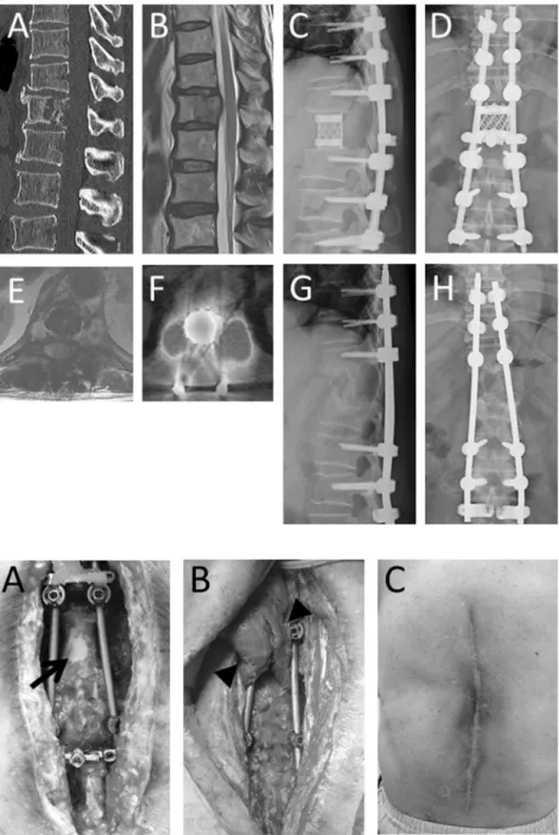
It is impossible to heal such a large dead space with unsuitable Fig. 1.Pre- and postoperative images. Preoperative multi-planner reconstruction sagittal image of computed tomo-graphy revealed osteolytic lesion at 12th thoracic vertebra (A). Magnetic resonance image (MRI) revealed tumor pro-truding to spinal canal (B). Total en bloc spondylectomy via single posterior approach was performed (C, D). Tumor was gloss totally resected. One year after primary surgery, deep surgical site infection occurred (MRI: E, positron emission tomography: F). Therefore we removed the infected cage followed by autologous iliac bone grafting (G, H).
Fig. 2.Photographs of wound.
After open wound therapy and irrigation, granulation for-mation was observed except for dura mater and exposed implants (A, arrow). Intraoperative photograph of muscle
flap using latissimus dorsi musclefilling the massive dead space besides the resected vertebra (B, arrowheads). Appearance of healed wound one year afterflap surgery (C).
M. Koda et al. Interdisciplinary Neurosurgery 11 (2018) 8–10
situation for wound healing described above by conservative therapy alone. Filling of the large dead space by well vascularized tissue is mandatory to control deep SSI after TES[5–8]. Therefore we performed
musculocutaneous flap using latissimus dorsi muscle. As a result, the large dead space could be successfully filled and the infection was successfully controlled in the present case.
4. Conclusion
In conclusion, musculo-cutaneousflap is one of treatment options to fill the dead space and to control deep surgical site infection after TES.
References
[1] P.C. Hsieh, K.W. Li, D.M. Sciubba, I. Suk, J.P. Wolinsky, Z.L. Gokaslan, Posterior-only approach for total en bloc spondylectomy for malignant primary spinal neoplasms: anatomic considerations and operative nuances, Neurosurgery 65 (2009) 173–181.
[2] Z.R. Cohen, D.R. Fourney, R.A. Marco, L.D. Rhines, Z.L. Gokaslan, Total cervical spondylectomy for primary osteogenic sarcoma. Case report and description of op-erative technique, J. Neurosurg. 97 (2002) 386–392.
[3] H. Hayashi, H. Murakami, S. Demura, S. Kato, K. Yoshioka, K. Shinmura, et al., Surgical site infection after total en bloc spondylectomy: Risk factors and the pre-ventive new technology, Spine J. 15 (2015) 132–137.
[4] M. Matsumoto, K. Watanabe, T. Tsuji, K. Ishii, M. Nakamura, K. Chiba, et al., Late instrumentation failure after total en bloc spondylectomy, J Neurosurg Spine 15 (2011) 320–327.
[5] L.O. Chieng, Z. Hubbard, C.J. Salgado, A.D. Levi, H. Chim, Reconstruction of open wounds as a complication of spinal surgery withflaps: a systematic review, Neurosurg. Focus. 39 (2015) E17.
[6] A.B. Cho, L.M. Rodrigues, R.J. Nicolau, G.M. Rugiero, W.Y. Fukushima, C. Milani, Treatment of hardware exposure after severe infections in spine surgery with pedi-cled muscularflaps, Clinics 63 (2008) 277–280.
[7] P.B. Garvey, M.W. Clemens, L.D. Rhines, J.M. Sacks, Vertical rectus abdominis musculocutaneousflow-throughflap to a freefibulaflap for total sacrectomy re-construction, Microsurgery 33 (2013) 32–38.
[8] L. de Weerd, T.K. Solberg, S. Weum, Closure of complex posterior midline defects after spinal surgery with sensate midline-based perforatorflaps and the long-term results, Spine 40 (2015) E1233–1238.
M. Koda et al. Interdisciplinary Neurosurgery 11 (2018) 8–10