Study of transcranial direct current
stimulation toward clinical application
0oyama, Soichiro
)octor of Philosophy
)epartment of Physiological Sciences
School of 1ife Science
SO0EN)AI (The Graduate University for
Advanced Studies)
Study of transcranial direct current stimulation toward clinical application
Koyama,Soichiro
SOKENDAI (The Graduate University for Advanced Studies)
School of Life Science
Department of Physiological Sciences
Table of contents
1 Summary ... 1
2 Introduction ... 4
3 Study 1: Enhancement of motor skill consolidation... 9
Introduction ... 9
Methods ... 11
Results ... 16
Discussion ... 17
4 Study 2: Modulation of pain-induced cortical response ... 21
Introduction ... 21
Methods ... 25
Results ... 33
Discussion ... 36
5 Conclusion ... 43
6 Acknowledgement ... 44
7 References ... 45
8 Tables ... 67
9 Figures ... 70
1 Summary
The main purpose of rehabilitation medicine is to enhance acquisition and/or reacquisition of
motor skills and reduce excessive pain sensations after various central nerve injuries.
Transcranial direct current stimulation (tDCS), a neuroscience-based approach, is a novel
rehabilitation tool for non-invasively modulating cortical excitability. Although the neural
mechanisms are not yet completely clear, tDCS not only alters the spontaneous firing rate of
neurons in the stimulated cerebral cortex by altering the resting membrane potential, but also
helps to produce transient neuroplastic changes by altering synaptic function. In addition to
inducing these neurophysiological changes, tDCS can influence motor learning, motor memory
consolidation, and sensory sensation, as well as suppress pain sensations, in healthy subjects
and patients with central nerve injury. Thus, tDCS could potentially enhance the therapeutic
effect of conventional rehabilitative approaches. In order to consolidate a novel rehabilitation
approach, further studies should test novel tDCS protocols with the goal of optimizing clinical
applications of tDCS. The two major objectives of this project were to examine the effects of
tDCS on motor skill acquisition and pain sensation, from the standpoint of clinical applications.
To achieve these objectives, I conducted a behavioral study and a neurophysiological study.
In the first study, I sought to elucidate the effect of tDCS on motor skill acquisition. Motor
performance is improved with repetitive practice (i.e., online process), and is subsequently
2
stabilized or improved without additional (i.e. consolidation or off-line process). The purpose of
rehabilitation is not only to improve motor skills by practice; it is also important that the
practiced motor skills can be maintained for a long period of time. To explore these concepts, 28
healthy subjects (age = 25.2 ± 2.7 years) participated in an experiment with a single-blind,
sham-controlled, between-group design. Fourteen subjects practiced a ballistic movement with
their left thumb during dual-hemisphere tDCS. Subjects received 1 mA anodal tDCS over the
contralateral primary motor cortex and 1 mA cathodal tDCS over the ipsilateral primary motor
cortex for 25 min during the training session. The remaining 14 subjects underwent identical
training sessions, except that dual-hemisphere tDCS was applied for only the first 15 s (sham
group). All subjects performed the task again at 1 h and 24 h later. Primary measurements
examined improvement in peak acceleration of ballistic thumb movement at 1 h and 24 h after
stimulation. The improvement in peak acceleration was significantly larger in the tDCS group
(144.2 ± 15.1%) than in the sham group (98.7 ± 9.1%) (p < 0.05) at 24 h, but not 1 h, after
stimulation. The results of the first study indicated that dual-hemisphere tDCS over primary
motor cortex enhanced acquisition of ballistic thumb movements in healthy adults.
The second study was aimed at elucidating the effect of tDCS on brain activation following
noxious stimulation, with the goal of evaluating the possible benefits of tDCS on moderate pain.
Although previous studies reported that transcranial magnetic stimulation over the opercular
3
somatosensory region, which is among the most common cortical areas to be activated
bilaterally by noxious pain stimuli, can modulate pain sensation, the effects of tDCS over this
region require clarification. To objectively quantify the effects of tDCS on noxious stimuli, I
utilized magnetoencephalography. Twelve healthy male subjects (age = 28.2 ± 2.6 years)
participated in a study with a single-blind, sham-controlled, cross-over trial design. The three
tDCS conditions investigated included left cathodal/right anodal tDCS, left anodal/right
cathodal tDCS (2 mA, 12 min each), and sham tDCS (2 mA, 15 sec). The center of each of two
stimulation electrodes was placed over one of the two bilateral opercular somatosensory regions.
Somatosensory-evoked magnetic fields following noxious intra-epidermal electrical stimulation
to the left index finger were recorded pre- and post-tDCS. The two anodal ("real") interventions
significantly decreased the activity of the opercular somatosensory region associated with
somatosensory-evoked magnetic fields following noxious intra-epidermal electrical stimulation
(p < 0.05), whereas sham tDCS did not (p > 0.05). The results of the second study indicated that
the opercular somatosensory region is a potential tDCS target area for pain mitigation.
Together, these findings suggest that tDCS might enhance the therapeutic effect of
conventional rehabilitative approaches in patients with motor dysfunction and pain.
4 Introduction
Rehabilitation is defined as the combined and coordinated use of medical, social, educational,
and vocational measures to retrain a person to the highest possible level of functional ability
(WHO Expert Committee on Medical Rehabilitation, 1969). The main targets of rehabilitation
medicine are to enhance acquisition and/or reacquisition of motor skills and reduce excessive
pain sensations. To improve impaired motor skills and ameliorate abnormal pain sensations,
various rehabilitation approaches have been used, e.g., constraint-induced movement therapy
(Taub et al., 1993, 2013), robot-based rehabilitation (Hughes et al., 2015), neuromuscular
electrical stimulation (Schuhfried et al., 2012; Vafadar et al., 2015), motor imagery (Giraux and
Sirigu, 2003), brain–machine computer interface (Bamdad et al., 2015), tactile discrimination
tasks (Moseley et al., 2008), and acceptance and commitment therapy (Wetherell et al., 2011).
However, recovery of these impairments after central nerve injury typically remains incomplete
despite the implementation of an appropriate rehabilitation program (Kwakkel et al., 2003; Go
et al., 2014).
Transcranial direct current stimulation (tDCS), a neuroscience-based rehabilitation method, has
recently been used to non-invasively modulate cortical excitability in humans. Compared to
transcranial magnetic stimulation (TMS), another non-invasive brain stimulation technique,
tDCS is safer and easier to use (Poreisz et al., 2007). tDCS is applied using a battery-powered
5
direct current generator connected to two relatively large rubber electrodes covered with
saline-soaked sponges (area, 20–35cm2) placed over the scalp. The current strength delivered
varies between 1 and 2 mA. During tDCS, weak direct current from the two electrodes
penetrates the skull to enter the brain. The penetrating direct currents modulates the cortical
excitability and spontaneous firing rate of neural activity (Bindman et al. 1964). The direction of
tDCS-induced cortical excitability changes depends on stimulation polarity. In general, the
cortical excitability of the primary motor cortex (M1) is increased by anodal tDCS over M1 and
decreased by cathodal tDCS (Nitsche and Paulus, 2000, 2001).The primary neural mechanism
underlying the effects of tDCS appears to be dependent on changes in membrane potential.
Pharmacological studies have shown that a calcium channel blocker (flunarizine) and a sodium
channel blocker (carbamazepine) abolished the modulatory effect on cortical excitability during
tDCS (Nitsche et al. 2003a). Following tDCS, motor cortical excitability increases for up to 90
minutes after the end of stimulation (Nitsche and Paulus, 2001). Pharmacological studies aimed
at elucidation of these after-effects revealed that N-methyl-D-aspartate (NMDA) receptor
antagonist (dextromethorphan) suppresses the post-stimulation increase in excitability (Nitsche
et al. 2003a; Liebetanz et al. 2002), indicating that the after-effects of tDCS are driven by
activation of the NMDA receptors in post-synaptic neurons. Moreover, paired-pulse TMS
studies revealed that the after-effects of tDCS result in a reduction of short latency intracortical
6
inhibition and an increase in intracortical facilitation, suggesting a decrease in
gamma-aminobutyric acid (GABA)-mediated interneuronal activity after the end of tDCS
stimulation (Nitsche et al., 2005). Thus, tDCS not only alters spontaneous firing rates of neurons
in stimulated cerebral cortex by altering the resting membrane potential, but also helps to
produce transient neuroplastic changes by altering synaptic function. The results of behavioral
experiments suggest that tDCS can influence motor learning (Boggio et al., 2006; Vines et al.,
2008), motor memory consolidation (Reis et al., 2009, 2015; Kang and Paik, 2011), and sensory
sensation (Fujimoto et al., 2014; Nakagawa et al., 2015), as well as reduce pain sensations
(Antal et al., 2008; Csifcsak et al., 2009; Reidler et al., 2012). Thus, previous
neurophysiological and behavioral studies of tDCS have raised the possibility that this method
represents a potential tool for enhancing the therapeutic effect of conventional rehabilitative
approaches.
My first primary aim was to test the effect of tDCS on the acquisition of motor skills, which
involves two main processes, practice and consolidation. Motor performance is improved by
repetitive practice (i.e., online process), and is subsequently stabilized and/or improved after the
end of practice without further activity (i.e., consolidation or offline process) (Robertson et al.,
2004, 2009). The purpose of rehabilitation is not only to improve motor skills by practice; it is
also important that the practiced motor skills be maintained at a high level for a long period of
7
time. tDCS over M1 enhances consolidation of various motor performance tasks, such as
visuomotor adaptation (Galea et al., 2011), serial reaction time (Kang and Paik, 2011; Kantak et
al., 2012), and sequential visual isometric pinch (Reis et al., 2009, 2015). However, it remains
unknown whether tDCS over M1 enhances consolidation of ballistic movement skills, which are
fundamental components of fine motor control (Hallett and Marsden, 1979). Therefore, the first
study tested the hypothesis that tDCS over M1 enhances consolidation of newly learned ballistic
movements in healthy adults.
My second primary aim was to test the effect of tDCS on pain sensation, i.e., the occurrence of
unpleasant somatic sensations. Previous brain imaging studies revealed that noxious stimuli can
activate a variety of brain regions, including the opercular somatosensory region (OP)
consisting of the secondary somatosensory cortex (S2) and insular cortex, primary
somatosensory cortex (S1), posterior parietal cortex, motor cortex, and limbic areas (Talbot et
al., 1991; Casey et al., 1994; Coghill et al., 1994, 1999; Kakigi et al., 1995b; Kanda et al., 2000;
Bingel et al., 2002; Bornhövd et al., 2002; Forss et al., 2005; Qiu et al., 2006; Baumgärtner et al.,
2010; Frot et al., 2013). Of these, the OP is among the cortical areas most commonly bilaterally
activated by noxious pain stimuli (Huttunen et al., 1986; Kakigi et al., 1995a; Ploner et al.,
1999; Kanda et al., 2000; Inui et al., 2003a, 2003b; Nakata et al., 2008). Although TMS over the
OP can modulate pain sensation, the detailed effects of tDCS over the OP require clarification.
8
Therefore, the present second study tested whether and how tDCS over the OP influences
cortical responses to a noxious stimulus and evoked pain sensation. To objectively quantify the
effect of tDCS on noxious stimuli, I utilized magnetoencephalography (MEG).
In this project, in order to obtain basic findings in healthy adults with the goal of developing
clinical applications, I undertook these two studies to test the effect of tDCS on consolidation of
newly learned motor skills and sensory evoked magnetic fields following noxious
intra-epidermal electrical stimulation (IES). To consolidate a novel rehabilitation approach, it is
necessary to perform basic research on the effect of tDCS on motor skill acquisition and pain
sensations. In the future, studies that test novel tDCS protocols might identify better approaches
for clinical application of tDCS.
9
Study 1: Enhancement of motor skill consolidation
Introduction
Acquisition of motor skills plays a fundamental role in daily life. Motor skill learning is the
process by which movements are executed more accurately and rapidly as a result of motor
training. In general, the effect of motor training occurs not only during training but also
afterward, a phenomenon termed consolidation (Muellbacher et al., 2002; Robertson et al.,
2004; Krakauer and Shadmehr, 2006; Robertson, 2009). Consolidation can result in increased
resistance to interference (memory stabilization), or even in improved motor performance after
training is completed (memory enhancement). These two types of consolidation play important
roles in the acquisition of motor skills (Robertson et al., 2004, 2009).
tDCS is a noninvasive technique that modulates cortical excitability via electrodes in humans
(Nitsche and Paulus, 2000). Anodal stimulation increases excitability of M1. Previous studies
have reported that various types of motor skill performance are improved in healthy adults and
in stroke patients when M1 is subjected to anodal tDCS (Nitsche et al., 2003b; Antal et al.,
2004; Boggio et al., 2006; Vines et al., 2006, 2008; Tanaka and Watanabe, 2009; Tanaka et al.,
2009, 2011; Hummel et al., 2010). In addition, tDCS over M1 enhances consolidation of various
motor performance tasks, including visuomotor adaptation (Galea et al., 2011), serial reaction
time (Kantak et al., 2012), and sequential visual isometric pinch (Reis et al., 2009, 2015).
10
Ballistic movements are elementary motor behaviors. For optimal performance of ballistic
movements, subjects must direct maximal drive to primary agonist muscles while minimizing
drive to antagonistic muscles (Hallett and Marsden, 1979; Muellbacher et al., 2001). The
electromyographic pattern of a ballistic movement is characterized by two bursts of phasic
agonist muscle activity and one burst of phasic antagonist muscle activity. The coordination of
reciprocal muscle activation in ballistic movement is a fundamental component of fine motor
control (Hallett and Marsden, 1979). Consolidation of ballistic movement skills involves M1
(Muellbacher et al., 2002), but it remains unknown whether tDCS over M1 can enhance
consolidation of ballistic movement skills.
The specific aim of this study was to investigate whether tDCS over M1 using a
dual-hemisphere protocol enhances consolidation of ballistic movements in healthy adults.
Dual-hemisphere tDCS, which excites one hemisphere and inhibits the other, is a powerful
strategy for improving behavioral performance (Vines et al., 2008; Williams et al., 2010; Karok
and Witney, 2013; Kasahara et al., 2013; Fujimoto et al., 2014). The mechanisms underlying
improved performance observed with dual-hemisphere tDCS may involve the combined effect
of increased excitability in one hemisphere and decreased excitability in the other, likely
mediated via interhemispheric connections (Vines et al., 2008; Tanaka et al., 2011; Karok and
Witney, 2013). Interhemispheric inhibition has long been thought of as a “rivalry” between the
11
two hemispheres, with motor function in the cortex of one hemisphere promoted by inhibitory
TMS of the contralateral cortex (Takeuchi et al., 2005).
Therefore, I postulated that decreased excitability of M1 in the left hemisphere via cathodal
tDCS would further increase M1 excitability in the right hemisphere, where consolidation of
ballistic thumb movements occurs (Muellbacher et al., 2001, 2002). This phenomenon is
mediated by interhemispheric inhibition (Takeuchi et al., 2005; Vines et al., 2008; Karok and
Witney, 2013), which further enhances consolidation of ballistic movements. In this study, I
tested the hypothesis that consolidation of a ballistic movement can be enhanced by
dual-hemisphere tDCS over M1 relative to sham stimulation.
Methods
Subjects
Twenty-eight healthy subjects (10 females and 18 males; mean age ± SD = 25.2 ± 2.7 years)
participated in the study. The subjects were neurologically healthy and had no family history of
epilepsy. The Human Research Ethics Committee at the National Institute for Physiological
Sciences approved all experimental procedures. All subjects gave informed consent before
participating in the experiment.
12 Experimental procedure
This study employed a single-blind, sham-controlled, between-group experimental design to
compare the effects of tDCS over M1 vs. sham stimulation on performance of a ballistic thumb
movement. M1 was chosen as the target based on evidence that consolidation of newly learned
ballistic movement involves this region (Muellbacher et al., 2002; Baraduc et al., 2004). To
measure consolidation of ballistic thumb movements, all subjects performed the same task at 1 h
and 24 h after completing the initial training.
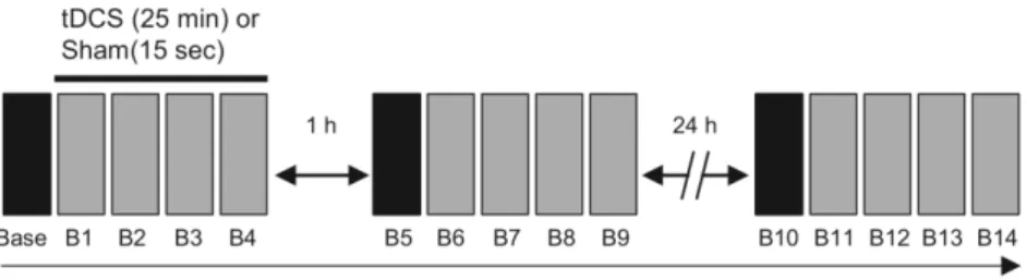
The experimental procedure is shown in Figure 1. First, all subjects underwent 20 trials of
ballistic thumb movement to gain familiarity with the task. Next, the subjects performed 60
trials to measure their baseline performance before the application of tDCS. After the baseline
measurements, the subjects were randomly assigned to two groups (tDCS or sham), and all
subjects performed four blocks (B1–B4) of the task while undergoing tDCS or sham stimulation.
Each block contained 60 trials, and subjects performed a total of five blocks during training
(total = 300 trials). Trials were paced at 0.5 Hz. To avoid fatigue, a 2-min break was included
between each block. In the tDCS group (14 subjects), stimulation of the anodal electrode over
right M1, and the cathodal electrode over left M1, was applied for 25 min during the training. In
the sham stimulation group (the remaining 14 subjects), tDCS electrodes were placed in the
13
same position as the tDCS group, but stimulation was delivered for only the first 15 s. The
subjects did not know whether they belonged to the tDCS or sham stimulation group.
At 1 h and 24 h after the initial tDCS or sham stimulation session, all subjects performed five
additional blocks (B5–B9 and B10–B14) of the same task to examine the effects of the
interventions on consolidation of the trained ballistic movements.
Motor task
Peak acceleration of thumb movement was used to measure ballistic thumb movement
performance (Muellbacher et al., 2001, 2002). The subjects were seated in front of a computer
screen. The subject’s left arm was flexed 70–80° at the elbow and slightly abducted the shoulder.
The forearm was held in a neutral position (between pronation and supination) with the thumb
free to move, while the fingers and forearm were fixed in place with a customized
upper-extremity orthotic. An accelerometer was then attached to the left thumb pad. The peak
acceleration of each ballistic thumb movement was recorded using an accelerometer with
integral electronics (model 25A; Endevco, CA, USA). The signal was amplified by a
battery-powered low-noise signal conditioner (model 4416B Isotron Signal Conditioner;
Endevco). Acceleration signals were amplified (10×), digitized at 2,000 Hz using an
analog–digital converter, and recorded on a computer for offline analysis. A customized
14
LabVIEW program was created to trigger movement onset (via an auditory signal), provide
visual feedback, and record the motor performance data.
All subjects were asked to flex the thumb as rapidly as possible following the auditory signal.
Acceleration signals were measured for 1.5 s after the auditory signal. At 1.5 s after the
accelerometer value was obtained, the subjects were provided with visual feedback regarding
peak acceleration of the ballistic thumb movement via a color signal displayed on the computer
screen. When subjects performed faster than the median of the previous five acceleration values,
a blue rectangle was presented on the computer screen. By contrast, when subjects performed
slower than the median of the previous five acceleration values, a red rectangle was presented.
tDCS
A DC-Stimulator Plus (NeuroConn, Ilmenau, Germany) was used to deliver direct current
through two sponge surface electrodes (surface area: 5 × 5 cm2) soaked with sodium chloride.
The anodal electrode was placed over M1 in the right hemisphere, whereas the cathodal
electrode was placed over M1 in the left hemisphere. The intensity of stimulation was 1 mA.
The fade-in/fade-out time was 15 s in both groups. In a preliminary experiment (n = 6), I
compared the size of the motor-evoked potential (MEP) in the flexor pollicis brevis before and
immediately after 25 min of 1 mA anodal tDCS over right M1 and cathodal tDCS over left M1
15
(for methodological details of the MEP experiment, see Nitche and Paulus, 2000) (Nitsche and
Paulus, 2000). Subsequently, the mean MEP amplitude of the right M1 significantly increased
after tDCS (mean ± SE; 158.7 ± 22.0%, p < 0.05). Thus, this tDCS protocol facilitated cortical
excitability of the right M1. For each participant, the location of M1 was identified using an
individual T1 anatomical image and a frameless stereotaxic navigation system (Brainsight2;
Rogue Research, Montreal, Canada).
Data analysis
Peak acceleration of ballistic thumb movement was analyzed as an indicator of motor
performance. First, the median value of peak accelerations in each block was calculated. The
median peak acceleration value of each block (60 trials) was normalized to the baseline
measurement (e.g., B1/baseline and B2/baseline); thus, the baseline performance value was
given a value of 1.0. Improvements in ballistic movement at 1 h after training were calculated
by dividing the value for the first block of training beginning 1 h after initial training (B5) by
the value of the last block of initial training (B4) and multiplying the result by 100 (e.g., B5/B4
× 100). Similarly, improvements in ballistic movement at 24 h after training were calculated by
dividing the value of the first block of training beginning 24 h after the initial training (B10) by
the value of the last block of training at 1 h after initial training (B9) (for example, B10/B9 ×
16
100). Because the data were not normally distributed, the Wilcoxon rank-sum test was used to
compare the rate of improvement for subjects in the tDCS group with the rate in the sham
group.
In addition, a measure of overall skill acquisition was calculated (as the mean percentage
change) by dividing the value of the last block of 24 h training (B14) by that of the baseline
measurement and multiplying the resulting value by 100 (B14/baseline × 100). The Wilcoxon
rank-sum test was used to compare the overall skill acquisition value of the tDCS group with
that of the sham group. p < 0.05 was considered statistically significant. Statistical analyses
were performed using SPSS 21.0 software (SPSS, Chicago, IL, USA).
Results
The application of tDCS was safely completed in all subjects with no adverse effects. For the
baseline measurement of ballistic movement, the Wilcoxon rank-sum test revealed no
significant difference between subjects in the tDCS and sham groups (p = 0.16). The mean peak
acceleration in the baseline blocks prior to normalization was 3.74 ± 0.51 g (mean ± SE) for the
tDCS group and 5.03 ± 0.72 g for the sham group. The normalized median accelerations in each
block are shown in Figure 2. Performance of the ballistic movement gradually improved during
the intervention in both the tDCS and sham groups (both groups; correlation coefficient r > 0.97,
17
p < 0.01) according to a regression analysis that calculated the correlation between the number
of training movements and peak acceleration (Muellbacher et al., 2002).
Improved performance of ballistic movement at 1 and 24 h after application of tDCS in the
tDCS or sham groups is shown in Figure 3. The improvement in motor performance observed at
1 h after training in both the tDCS and sham groups was not statistically significant (p = 0.69;
Figure 3A). By contrast, the improvement in motor performance at 24 h after training was
significantly greater in the tDCS group (mean ± SE; 144.2 ± 15.1%) than in the sham group
(98.7 ± 9.1%, p < 0.05; Figure 3B). These data indicate that motor training combined with tDCS
enhances consolidation of ballistic movement at 24 h, but not 1 h, after training. The overall
learning of ballistic movement skill in the tDCS and sham stimulation groups is shown in Figure
3C. Learning of this skill in the tDCS group (266.8 ± 48.4%) was significantly superior to that
in the sham group (159.4 ± 17.8%, p < 0.05; Figure 3C).
Discussion
Previous studies have reported that anodal tDCS over M1 enhances acquisition of various
finger motor skills in healthy adults, including the visuomotor adaptation task (Galea et al.,
2011), serial reaction time task (Kantak et al., 2012), and sequential visual isometric pinch task
(Reis et al., 2009, 2015).
18
Using a single-blind, sham-controlled design, this study examined the effect of
dual-hemisphere tDCS over bilateral M1 on consolidation of a ballistic movement. The results
demonstrated that bilateral M1 tDCS also facilitated acquisition of a newly learned ballistic
thumb movement, significantly improving peak acceleration of thumb movement relative to the
sham group at 24 h after training. These data suggest that bilateral M1 tDCS enhances
consolidation of newly learned ballistic thumb movements in healthy adults.
The results also demonstrated that tDCS facilitated performance of ballistic thumb
movements at 24 h, but not at 1 h, after tDCS ended. There are two plausible explanations for
this time-dependent effect. First, given that sleep is reportedly necessary for consolidation of
some types of motor skills (Fischer et al., 2002; Walker et al., 2002, 2003; Walker and Stickgold,
2004), it is possible that tDCS enhances sleep-dependent consolidation (Kantak et al., 2012).
The consolidation of motor skill acquisition during sleep appears to rely on covert reactivation
of brain areas involved in motor skill acquisition (Maquet et al., 2000). Anodal tDCS over M1
facilitates improvement of a serial reaction time task 24 h after tDCS ended (Kantak et al.,
2012). Thus, M1 tDCS may enhance sleep-dependent consolidation. Alternatively, it is also
possible that tDCS enhances consolidation independent of sleep (Reis et al., 2015). A previous
study reported that tDCS affected sleep-independent consolidation of a sequential visual
isometric pinch-force task (Reis et al., 2015). Thus, the tDCS protocol in the present study may
19
have enhanced this time-dependent consolidation of ballistic finger movement. However,
resolution of this issue will require further experiments that include sleep as an independent
variable.
In this study, I found that a dual-hemisphere tDCS protocol facilitated consolidation of a
ballistic finger movement, consistent with the results of a previous study showing that
dual-hemisphere tDCS over M1 enhanced consolidation of a sequential finger movement task
(Kang and Paik, 2011). In our dual-hemisphere tDCS protocol, the anodal tDCS may have
increased excitability of M1 in the right hemisphere, where the consolidation of ballistic thumb
movements occurs (Muellbacher et al., 2001, 2002). In addition, decreased excitability in the
left hemisphere M1 by cathodal tDCS might have further increased excitability in the right
hemisphere M1 by reducing interhemispheric inhibition (Vines et al., 2008; Tanaka and
Watanabe, 2009; Williams et al., 2010; Karok and Witney, 2013). I speculate that the combined
effect of increasing M1 excitability in the right hemisphere by anodal tDCS and decreasing M1
excitability in the left hemisphere by cathodal tDCS may underlie the observed behavioral gain.
Because I used only dual-hemisphere tDCS in this study, I cannot rule out the possibility that
single-hemisphere tDCS over M1 might have been sufficient to improve consolidation. In a
preliminary experiment with six healthy subjects, I investigated the effect of single-hemisphere
tDCS (anodal electrode over the right M1 and cathodal electrode over the contralateral orbit) on
20
consolidation of the same ballistic movement task. However, I did not observe any significant
improvement performance relative to sham stimulation. Therefore, it is reasonable to
preliminarily conclude that anodal tDCS over the M1 alone is insufficient to induce the
behavioral improvement observed in this study. Future studies should clarify this issue by
investigating single-hemisphere stimulation–induced effects on behavior.
There were some limitations to this study. First, a single-blind design was used; future studies
should employ a double-blind design in order to avoid the observer effect. Second, I
investigated the effect of tDCS only on performance of a trained task. Future studies should
examine a generalization of the effects of tDCS on performance of untrained tasks. Third, I
stimulated only one brain region. The lack of other control regions to be stimulated may limit
the strength of our results when the relatively low spatial resolution of tDCS is taken into
account. Finally, I investigated only behavioral changes induced by tDCS. Future studies should
examine the neurophysiological changes associated with the behavioral gain observed in this
study. Nevertheless, loss of thumb movement remains a problematic impairment after stroke
(Fritz et al., 2005; Lang and Beebe, 2007). Therefore, our findings may be useful in guiding the
rehabilitation of patients with upper limb dysfunctions following subcortical strokes.
21
Study 2: Modulation of pain-induced cortical response
Introduction
Pain, which is the occurrence of unpleasant somatic sensations, is defined as an emotional and
bodily experience associated with actual or probable tissue damage, or is described in terms of
such damage (Merskey and Bogduk, 1994). The discomfort accompanying pain results in
drastic reduction in activities and quality of daily life, as well as alterations of mental state
including negative emotionality, maladaptive stress responses, and depression (Baliki and
Apkarian, 2015). Therefore, it is critically important to manage pain sensation in human
patients.
Pain is generated in the brain. Brain imaging studies using positron emission tomography
(Talbot et al., 1991; Casey et al., 1994; Coghill et al., 1994, 1999), functional magnetic
resonance imaging (Bingel et al., 2002; Bornhövd et al., 2002; Qiu et al., 2006; Baumgärtner et
al., 2010), MEG (Kakigi et al., 1995b; Kanda et al., 2000; Forss et al., 2005; Frot et al., 2013),
and intracranial recording (Baumgärtner et al., 2011; Frot et al., 2013) demonstrated that
noxious stimuli can activate a variety of brain regions, including the OP consisting of the S2 and
insular cortex, S1, posterior parietal cortex, motor cortex, and limbic areas. Of these, the OP is
among the cortical areas most commonly activated by noxious pain stimuli.
Previous MEG studies consistently reported OP activation in both brain hemispheres following
22
laser stimulation (Kakigi et al., 1995a; Ploner et al., 1999; Kanda et al., 2000; Nakata et al.,
2008), IES (Inui et al., 2003a, 2003b), stimulation of the nasal mucosa with carbon dioxide gas
(Huttunen et al., 1986; Hari et al., 1997), and painful electrical stimulation of the tooth pulp
(Hari et al., 1983). These findings suggest that the OP plays an indispensable role in perceiving
pain. This view is supported by electrical stimulation mapping data obtained during brain
surgery showing that the OP is central to pain sensation (Mazzola et al., 2012). Moreover,
patients with OP lesions exhibit impaired pain sensations (Greenspan et al., 1999).
The sensation of pain is modulated by non-invasive brain stimulation applied to the OP. For
instance, repetitive TMS over the OP results in reduction of chronic visceral pain (Fregni et al.,
2005, 2011) and an increase in pain threshold (Valmunen et al., 2009). Likewise, single TMS
over this region impairs discrimination sensitivity to the intensity of pain stimuli (Lockwood et
al., 2013). Therefore, non-invasive brain stimulation over the OP could serve as an important
tool to manage pain.
tDCS is a non-invasive brain stimulation technique that provides prolonged shifts in cortical
excitability. Notably, tDCS also has a beneficial effect on pain reduction in healthy adults and
symptomatic pain patients. The excitability of M1 is transiently increased by anodal tDCS and
decreased by cathodal tDCS (Nitsche and Paulus, 2000; Tanaka and Watanabe, 2009; Tanaka et
al., 2009, 2011). tDCS has certain advantages over TMS. For example, the tDCS device is
23
portable, inexpensive, easy to use, and safe in the clinical setting (Poreisz et al., 2007; Tanaka
and Watanabe, 2009).
Anodal tDCS over the M1 disrupts pain sensation in healthy adults (Antal et al., 2008;
Csifcsak et al., 2009; Reidler et al., 2012) and patients with fibromyalgia (Fregni et al., 2006b;
Riberto et al., 2011), traumatic spinal cord injury (Fregni et al., 2006a), multiple sclerosis (Mori
et al., 2010), or chronic pelvic pain (Fenton et al., 2009). However, the effects of tDCS
administered over the OP on neurophysiological aspects and pain sensation remain unclear.
Applied tDCS current can cross into the brain through the highly conductive cerebrospinal fluid
(CSF) network (Datta et al., 2009; Antal et al., 2014; Opitz et al., 2015). Therefore, I
hypothesize that tDCS over the OP will effectively regulate cortical responses to a noxious
stimulus, as well as the magnitude of subjective pain sensation.
Several investigators have proposed a dual-hemisphere tDCS protocol as a powerful strategy
for controlling brain excitability and various neurological functions (Vines et al., 2008;
Kasahara et al., 2013; Fujimoto et al., 2014; Koyama et al., 2015; Nakagawa et al., 2015). Due
to its greater impact on interhemispheric projections, simultaneous application of tDCS over
both hemispheres is more effective than single-hemisphere tDCS for modulation of motor
performance, sensory perception, and cognitive performance (Vines et al., 2008; Kasahara et al.,
2013; Fujimoto et al., 2014; Koyama et al., 2015). Thus, dual-hemisphere tDCS potentiates the
24
effects of anodal (or cathodal) stimulation of one hemisphere through additional modulation of
interhemispheric interactions via cathodal (or anodal) stimulation of the contralateral
hemisphere. The bilateral OPs are thought to be linked either directly by transcallosal
connections or indirectly by thalamic and S1 circuitries (Krubitzer and Kaas, 1990; Krubitzer et
al., 1998; Disbrow et al., 2001; Blankenburg et al., 2008; Fregni et al., 2011). Moreover, as with
the bilateral M1s and bilateral S1s, inhibitory connections exist between the bilateral OPs in the
rat (Zhang and Oppenheimer, 2000). In humans, the bilateral OPs are tightly functionally
connected during painful stimulation (Peltz et al., 2011). Thus, dual-hemisphere tDCS is
expected to result in a clearer effect on the cortical responses to a noxious stimulus.
To objectively quantify the effect of tDCS on the noxious stimuli, I utilized MEG. Previous
MEG studies demonstrated the high positive correlation between the magnitude of subjective
pain sensation and activity in bilateral OPs following noxious stimulation (Timmermann et al.,
2001). To activate nociceptors selectively, I used noxious IES. This method relies on the fact
that nociceptive fiber terminals are located mainly in the epidermis, whereas other fibers end
deep in the dermis (Inui et al., 2002). The aim of this study was to employ MEG to gain insights
into the neurophysiological and analgesic effects of tDCS over the OP on cortical responses to a
noxious stimulus. I propose that moderation of such cortical responses and reduction of the
magnitude of subjective pain sensation will establish the OP as a novel tDCS target area for pain
25 relief.
Methods
Study design
A single-blind, sham-controlled, cross-over trial design was utilized to test the effects of tDCS
over the OP on OP activity following noxious IES, as determined using MEG. The magnitude of
subjective pain sensation was also investigated. Subjects underwent three tDCS conditions with
different stimulation protocols: 1) anodal tDCS applied over the left OP and cathodal tDCS
applied over the right OP (LA/RC tDCS), 2) cathodal tDCS applied over the left OP and anodal
tDCS applied over the right OP (LC/RA tDCS); and 3) sham tDCS. To avoid carry-over effects
of the various tDCS conditions, each session was separated by at least 1 week. The order of the
conditions was counterbalanced across subjects based on a Latin square design. Primary
outcome measures included post-IES activity in the OP in the hemisphere contralateral to the
stimulated side (cOP) and the hemisphere ipsilateral to the stimulated side (iOP), and the visual
analog scale (VAS) score for the assessment of subjective pain sensation. Secondary outcome
measures included S1 activity following innocuous medial nerve electrical stimulation, and
responses to a questionnaire designed to evaluate the subjective states of the study participants
(attention, fatigue, pain, sleepiness, and discomfort) during tDCS intervention.
26 Subjects and exclusion criteria
Twelve healthy male subjects (mean age ± SD = 28.2 ± 2.6 years, all right-handed) participated
in the study. Subjects were free from neurological diseases, psychiatric disorders, chronic pain
disorders, and a family history of epilepsy. Exclusion criteria included acute severe pain within
the previous 4 weeks, intake of analgesics within the previous 24 h, and implanted electrical
devices. All experimental procedures were approved by the Human Research Ethics Committee
of the National Institute for Physiological Sciences, and were in accordance with the
Declaration of Helsinki. All subjects provided informed consent prior to participation in the
study.
Experimental procedures
At the beginning of the study, the stimulus intensity was set at a level sufficient to evoke a pain
sensation with a VAS score of 5 in each subject. This intensity level was maintained throughout
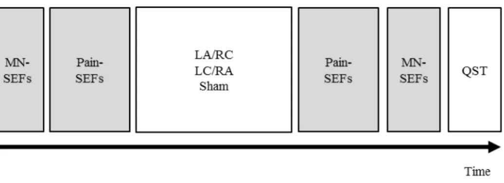
the experimental procedures. The timeline of MEG measurements and tDCS interventions in
each session consisted of five sequences (Figure 4). First, 1) S1 activity and 2) OP activity
baseline measurements were recorded. Next, the subjects received 3) a tDCS intervention
outside the MEG room. Immediately after tDCS intervention, 4) OP activity and 5) S1 activity
27
were again measured using the same protocol as that employed for baseline measurements.
Following all interventions, subjective states (attention, fatigue, pain, sleepiness, and
discomfort) of each participant during tDCS were assessed using a questionnaire and a
four-point scale (e.g., attention: 1 = no distraction of attention, 4 = highest distraction of
attention) (Poreisz et al., 2007).
tDCS protocol
The DC Stimulator Plus (NeuroConn, Ilmenau, Germany) was used to deliver a direct current
over the OP through two sponge surface electrodes (surface area = 5 × 5 cm2) soaked with
sodium chloride. These experiments were performed according to a dual-hemisphere tDCS
protocol in which the center of each of the two stimulation electrodes was placed over one of
the two bilateral OPs. Stimulation points were determined via anatomical brain images obtained
using a Magnetom Verio 3 Tesla magnetic resonance imaging system (Siemens, Ltd., Erlangen,
Bavaria, Germany) and a Brainsight2 frameless stereotaxic navigation system (Rogue Research
Inc., Montreal, Canada). The stimulus point of the OP was defined as the cortical area adjacent
to the junction of the rostral end of the post-central gyrus and the sylvian fissure (Kanda et al.,
2003; Fregni et al., 2011; Lockwood et al., 2013). In the anodal (“real”) tDCS conditions
(LA/RC and LC/RA), the current was ramped up over the first 15 sec to a maximum of 2 mA,
28
held constant at 2 mA for 690 sec, and then ramped down over the last 15 sec (total time of
current application = 12 min). For sham stimulation, the same procedure was used, but the
constant current was delivered for only 15 sec. This procedure enabled the blinding of study
participants to the experimental conditions.
MEG recording
OP and S1 activities were measured using a whole-head-type Vector View 306-channel MEG
system (Elekta Neuromag, Helsinki, Finland) comprising 102 identical triple-sensor elements.
Each sensor element contained two orthogonal planar gradiometers and one magnetometer
coupled to a multi-superconducting quantum interference device. Two hundred and four
planar-type gradiometers were employed in the present study. The signals were recorded with a
bandpass filter of 0.1–200 Hz and digitized at a sampling rate of 1000 Hz. The analysis was
conducted from 100 ms before the onset of pain stimulation to 500 ms afterward. The
pre-stimulus period was used as the direct current baseline. Epochs of somatosensory-evoked
magnetic fields following noxious IES (Pain-SEFs) and innocuous medial nerve electrical
stimulation (MN-SEFs) were averaged at least 60 and 200 times, respectively. Epochs with
MEG signals of > 2.7 pt/cm were rejected from the averaging.
29 Noxious electrical stimulation for Pain-SEFs
An IES electrode (Inui et al., 2002, 2006) and a portable peripheral nerve stimulator
(PNS-7000, Nihon, Koden, Tokyo, Japan) were used to produce the pain stimulus. The electrode
consisted of an outer ring with a diameter of 1.3 mm, and an inner needle protruding 0.02 mm
from the outer ring. Parameters of pain stimulation were as follows: The inner needle served as
the cathode, and the outer ring served as the anode; the electrical pulse corresponded to a
triangular wave with a rise and fall time of 0.5 ms; and the pulse train corresponded to four
pulses with an inter-stimulus interval of 5 ms to increase the magnitude of subjective pain
sensation (Mouraux et al., 2014). Participants received seven cycles of pain stimulation to the
dorsum of the left index finger, restricted to the first metacarpal bone. Each cycle consisted of
ten trials of pain stimulation, with an inter-trial interval of 10 sec. To avoid fatigue during the
recording of Pain-SEFs, the interval between cycles was set at 30 sec.
Innocuous electrical stimulation for MN-SEFs
The left medial nerve was stimulated percutaneously at a frequency of 1 Hz using a
conventional felt-tip bipolar electrode. The electrode was placed over the medial nerve at the
left wrist, and the optimal stimulus point was identified by a visible twitching movement of the
thumb. The ground electrode was placed around the wrist. The stimulus pulse corresponded to a
30
square monophasic waveform with a plus width of 0.3 ms. The stimulation intensity was
maintained just above the motor threshold, defined as the minimum intensity required to
produce a visible twitch of the thumb flexion muscle. During the recording of MN-SEFs,
participants watched a silent movie to maintain awareness.
Subjective pain measurement
Magnitude of subjective pain intensity was evaluated using the VAS, which is widely used in
tDCS studies of pain (Antal et al., 2008; Terney et al., 2008; Csifcsak et al., 2009; Hansen et al.,
2011) and has high validity and reproducibility (Bolton and Wilkinson, 1998). Participants were
asked to rate the magnitude of their subjective pain intensity during the MEG recording. After
each pain stimulation, a yellow-colored horizontal bar moved from the left (VAS = 0; no pain)
to the right (VAS = 10; worst imaginable pain) on a screen in front of the participants. The
participants manipulated a push-type button with their right hand, and stopped the movement of
the horizontal line at the optimal location for the perceived pain sensation. Presentation software
(Neurobehavioral Systems, Inc., San Francisco, CA, USA) was used to display the VAS scale
and to record the VAS data.
Data analysis
31
Because the position of the head relative to that of the sensors was not identical before and
after tDCS application or among the tDCS conditions, the source strength of the evoked
response was used to assess tDCS effects. A multiple dipole analysis was carried out to detect
temporally overlapping, equivalent current dipoles using the Brain Electric Source Analysis
(BESA) software package (NeuroScan, McLean, VA, USA). The averaged waveform was
filtered offline with a bandpass of 0.5–100 Hz.
A multiple dipole model was obtained for each session as described previously (Inui et al.,
2003a), with a focus on IES-evoked activity in the cOP and iOP. Two dipole sources (one in
each bilateral OP) were first determined. If necessary, one or more sources in each OP were
determined to explain the residual MEG data. However, the contribution of these sources to the
overall recorded fields was small, and consequently these source responses were not included in
the analysis. Dipole location and orientation were averaged before and after tDCS application
and among the tDCS conditions, and the averaged model was applied to all included study data,
as described previously (Otsuru et al., 2012; Kodaira et al., 2013). The obtained source strength
waveforms were used to evaluate OP activity. Because the duration of the initial component of
the Pain-SEFs was ~100–300 ms, the peak latency was measured within 200 ms after the
initiation of the pain stimulus. The onset of the source strength waveform was defined as the
minimum value at 50 ms before the peak (Figure 5). Peak-to-peak amplitude was calculated as
32 the magnitude of OP activity.
The equivalent current dipole for the MN-SEFs was estimated at 20–30 ms following the onset
of the stimulus, and the obtained source strength waveform was used to measure peak
amplitudes via the same procedure used for the Pain-SEFs. Peak latencies for the N20, P35, and
P60 MN-SEF latency components were measured as previously described (Nakagawa et al.,
2014; Sugawara et al., 2015), and peak amplitudes were measured from baseline. To confirm the
location of the obtained dipoles, the data were superimposed on individual magnetic resonance
images using the head position indicator system. Dipole location was transformed into Talairach
coordinates using Brain Voyager QX 1.4 (Maastricht, The Netherlands) and the BESA software.
Statistical analysis
Three cortical activities and the VAS score were subjected to a two-way repeated measures
analysis of variance (ANOVA) with three tDCS conditions (LA/RC, LC/RA, and sham tDCS)
and two time points (pre- and post-tDCS intervention) as within-subject factors. Post hoc
analyses consisted of paired t-tests with Bonferroni correction. Due to the non-parametric nature
of the distribution, questionnaire scores were analyzed using the Kruskal-Wallis test. SPSS
software (version 21, SPSS, Chicago, IL, USA) was used for statistical analyses. Statistical
significance was set at p < 0.05. Quantifiable data are given as means ± SD.
33 Results
All subjects completed the three experimental tDCS conditions (Figure 4) with no notable
adverse effects. Two subjects were excluded from the analysis because Pain-SEFs could not be
clearly recorded from them. Accordingly, the data included in the final analysis were obtained
from the ten remaining participants (mean age ± SD = 28.4 ± 2.7 years).
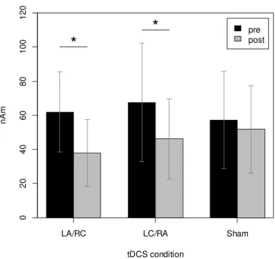
IES-evoked cOP activity
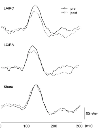
Figure 5 presents the superimposed waveforms recorded from 204 gradiometers following IES
(A), the source strength waveform pre- and post-tDCS (B), and the dipole source location
overlaid on the magnetic resonance images of a representative subject (Subject 1) (C). Figure 6
shows the source strength waveforms for Subject 1 under the three tDCS conditions. Results of
two-way (tDCS condition × time) repeated-measures ANOVA revealed significant two-way
interactions between the tDCS conditions and time (F2,18 = 9.425, p < 0.05, and partial η2 =
0.51), and a significant main effect of time (F1,9 = 28.70, p < 0.05, and partial η2 = 0.76). By
contrast, the main effect of tDCS intervention was not significant (F2,18 = 0.74, p = 0.49, and
partial η2 = 0.08). Post hoc analysis with Bonferroni correction revealed that the amplitude of
IES-evoked cOP activity was significantly lower than baseline after LA/RC and LC/RA tDCS (p
34
< 0.05), but not after sham tDCS (p > 0.05) (Figure 7).
Table 1 shows the mean dipole source location in standardized Talairach coordinates, and
Table 2 shows the peak amplitudes of cOP and iOP activity. The peak latency of the source
activities in this study (120–130 ms) was slightly shorter than that reported in previous studies
employing IES (e.g., see Inui et al., 2003a), probably because the pain stimulation I used (VAS
score = ~5) was stronger than that used in previous studies (VAS score = ~2).
IES-evoked iOP activity
Two-way (tDCS condition × time) repeated-measures ANOVA revealed significant two-way
interactions among the tDCS conditions and time (F2,18 = 4.76, p < 0.05, and partial η2 = 0.35),
and a significant main effect of time (F1,9= 10.92, p < 0.05, and partial η2 = 0.55). By contrast,
the main effect of tDCS was not significant (F2,18 = 0.86, p = 0.44, and partial η2 = 0.08). Post
hoc testing with Bonferroni correction again showed that the amplitude of IES-evoked iOP
activity was significantly lower than baseline after LA/RC and LC/RA tDCS (p < 0.05), but not
after sham tDCS (p < 0.05) (Figure 8).
Median nerve-evoked S1 activity in the hemisphere contralateral to the stimulated side
In all subjects, I estimated the dipole location for MN-SEFs as being in and around the
35
postcentral gyrus. The source strength waveform as a function of time exhibited several peaks
with different polarities at ~20, ~35, and ~60 ms. Therefore, I measured the peak amplitude for
these three latency components, N20, P35, and N60. Two-way (tDCS condition × time)
repeated-measures ANOVA revealed neither significant two-way interactions among the tDCS
conditions and time (N20: F2,18 = 1.68, p = 0.22, and partial η2 = 0.16; P35: F2,18 = 0.96, p = 0.40,
and partial η2 = 0.10; and P60: F2,18 = 0.11, p = 0.90, and partial η2 = 0.01) nor a significant
main effect of time (N20: F1,9= 0.05, p = 0.82, and partial η2 = 0.006; P35: F1,9 = 0.98, p = 0.35,
and partial η2 = 0.10; and P60: F1,9 = 0.80, p = 0.40, and partial η2 = 0.08) or tDCS condition
(N20: F2,18 = 0.40, p = 0.67, and partial η2 = 0.43; P35: F2,18= 3.10, p = 0.07, and partial η2 =
0.25; and P60: F2,18= 3.32, p = 0.06, and partial η2 = 0.27) (Figure 9). These results indicate that
S1 excitability is not modulated by tDCS intervention.
Magnitude of subjective pain sensation
Two-way (tDCS condition × time) repeated-measures ANOVA revealed neither significant
two-way interactions among the tDCS conditions and time (F2,18= 0.78, p = 0.47, and partial η2
= 0.08) nor a significant main effect of time (F1,9= 0.81, p = 0.39, and partial η2 = 0.08) or tDCS
condition (F2,18= 0.13, p = 0.88, and partial η2 = 0.014) (Figure 10). Therefore, the present study
failed to demonstrate significant differences among the three tDCS conditions (LA/RC, RA/LC,
36
and sham) on the magnitude of subjective pain intensity.
Questionnaire results
The subjective state of the subjects during tDCS intervention could potentially impact their
performance. To address this possibility, the study participants completed questionnaires
post-tDCS to rate their levels of attention, fatigue, pain, sleepiness, and discomfort. However,
no intervention-evoked alterations of subjective state were noted that might have affected the
overall results of the investigation (Table 3).
Discussion
This study used a single-blind, sham-controlled, cross-over trial design to evaluate the effects
of dual-hemisphere tDCS over the OP (2 mA, 12 min) on OP and S1 activity, as well as the
magnitude of subjective pain sensation. The results provide the first evidence that
dual-hemisphere tDCS can decrease IES-evoked OP activity in a polarity-independent manner
in healthy adults. By contrast, subjective pain sensation and median nerve-evoked S1 activity
were similar before and after tDCS intervention. The questionnaire results indicated that
attention, fatigue, pain, sleepiness, and discomfort were also similar between tDCS conditions
(LA/RC, LC/RA, and sham). Therefore, our findings did not stem from differences in subjective
37
state during tDCS. Because I used a cross-over trial design and employed only male participants,
the contributions of individual differences and gender effects to the obtained data were also
excluded.
Dual-hemisphere tDCS is a powerful strategy for modulating brain function (Vines et al.,
2008; Tanaka et al., 2011; Kasahara et al., 2013; Fujimoto et al., 2014; Koyama et al., 2015;
Nakagawa et al., 2015). Dual-hemisphere tDCS more effectively impacts motor and cognitive
performance and sensory perception than single-hemisphere tDCS (Vines et al., 2008; Kasahara
et al., 2013; Fujimoto et al., 2014; Koyama et al., 2015). The bilateral OPs are thought to be
connected either directly by transcallosal connections or indirectly by thalamic and S1 circuitry
(Krubitzer and Kaas, 1990; Krubitzer et al., 1998; Disbrow et al., 2001; Blankenburg et al.,
2008). In rats, OP activation is inhibited by electrical stimulation applied to the cOP (Zhang and
Oppenheimer, 2000). Therefore, I propose that the inhibitory effects of cathodal tDCS on one
hemisphere might be further augmented by simultaneous enhancement of interhemispheric
inhibitory inputs by administration of anodal tDCS to the other hemisphere.
Although I clearly documented polarity-independent actions of dual-hemisphere tDCS over the
OP, I observed no polarity-dependent effects on IES-evoked OP activity. The
polarity-independent effects of tDCS have also been documented in several other studies (Antal
et al., 2007; Ferrucci et al., 2008, 2012; Orban de Xivry et al., 2011; Shah et al., 2013). Given
38
that cathodal stimulation is generally inhibitory, whereas anodal stimulation is excitatory, it is
unclear why dual-hemisphere tDCS over the OP should elicit polarity-independent effects.
There are two possible explanations for this result. First, OP excitability might be decreased by
both anodal and cathodal stimulation. In support of this hypothesis, repetitive TMS studies
revealed that both facilitatory (high-frequency) (Valmunen et al., 2009; Lindholm et al., 2015)
and inhibitory (low-frequency) (Fregni et al., 2005) stimulation over the OP impairs pain
perception in healthy subjects, as well as in patients experiencing pain. Hence, my application
of anodal tDCS over the bilateral OP might have inhibited instead of facilitated OP excitability.
In the future, studies using monopolar stimulation should be performed to elucidate the
influence of tDCS with respect to polarity differences.
Second, the function of the connections between the two OPs must be considered. Earlier work
on Pain-SEFs reported that peak latency was shorter for the cOP than for the iOP by ~5–15 ms,
consistent with the results reported here (Kanda et al., 2000; Ploner et al., 2000; Nakata et al.,
2008). The latency difference between the hemispheres has been interpreted to reflect the time
required to transmit signals via the corpus callosum. This implies that when OP activity
following IES in the contralateral hemisphere is suppressed by cathodal stimulation, ipsilateral
activation by the callosal transmission is also reduced as a consequence. In this case, however,
the iOP receives anodal stimulation, presumably increasing excitation in the region. Therefore,
39
the final output in both hemispheres depends on the balance between excitatory and inhibitory
influences.
As noted above, inhibitory connections are present between the bilateral OPs in the rat (Zhang
and Oppenheimer, 2000). In humans, the OPs are tightly functionally connected during painful
stimulation (Peltz et al., 2011). Accordingly, our findings suggest that the inhibitory effects in
the hemisphere receiving cathodal tDCS outweighed the facilitatory effects in the opposite
hemisphere receiving anodal tDCS. Further research, in particular studies using
single-hemisphere tDCS restricted to the right or the left OP, is required in order to investigate
this possibility.
Despite the inhibitory effects of direct current stimulation on IES-evoked cortical responses,
the results of this study revealed only modest effects of all tDCS conditions on the magnitude of
subjective pain sensation. In some earlier studies, tDCS over the M1 exerted analgesic actions
on experimentally induced pain (Antal et al., 2008; Csifcsak et al., 2009; Reidler et al., 2012),
whereas other studies reported no such effects in healthy adults (Hansen et al., 2011; Jürgens et
al., 2012; Ihle et al., 2014). This discrepancy leads me to speculate that subjective pain sensation
in evoked responses is more complex than mere pain-related somatosensory processing (Ihle et
al., 2014). Moreover, these previous studies suggested that differences between
neurophysiological effects (e.g., pain-related evoked potentials following painful transcutaneous
40
electrical stimulation vs. hemodynamic responses following heat-pain stimulation) and the
subjective magnitude of pain sensation were due to differences in tDCS parameters (Hansen et
al., 2011; Ihle et al., 2014). Therefore, additional insights into optimal tDCS parameters are
essential for the establishment of the most efficacious tDCS-based approach to pain relief.
I observed no change in median nerve-evoked S1 activity before and after tDCS. Previous
work showed that anodal tDCS over the S1 facilitates the P22/N30, P25/N33, and N33/P40
latency components of MN-SEFs (Matsunaga et al., 2004). Furthermore, the source strengths
for the P35 and P60 components increases after tDCS over M1, and the strength for P60
increases after tDCS over S1 (Sugawara et al., 2015). Here, the P35 and P60 amplitudes
remained unaltered before and after tDCS. Therefore, the ability of dual-hemisphere tDCS over
the OP to modulate IES-evoked OP activity cannot be explained by changes in S1 excitability,
but instead appears to result from a variance in current density between the OP and S1.
Although the tDCS current is transferred widely to multiple brain areas through the CSF, current
density is highest at the position of the electrode. Because the effectiveness of tDCS on the
excitability of the stimulated cortex depends on current density (Wagner et al., 2007; Nitsche et
al., 2008; Bastani and Jaberzadeh, 2012, 2013), decreased IES-evoked cortical responses might
be attributed to modulation of OP excitability, but not S1 excitability.
This study had certain limitations. First, the small number of subjects (n = 12 original
41
participants, with two excluded from the final analysis) undoubtedly restricts the strength of the
conclusions. Second, I did not separate temporally overlapping OP sources, which might be
predicted to affect the present results given that multiple sources in the OP e.g., the S2 (Bingel
et al., 2004; Baumgärtner et al., 2010), anterior insula (Henderson et al., 2007; Baumgärtner et
al., 2010), and posterior insula (Brooks et al., 2005; Henderson et al., 2007; Mazzola et al.,
2009; Baumgärtner et al., 2010) all participate in the processing of noxious information. Third, I
focused on IES-evoked OP activity, because the OP is one of the cortical areas most commonly
influenced by noxious stimuli-evoked activation. Although previous MEG studies observed S1
activity following noxious stimuli (Ploner et al., 1999; Kanda et al., 2000; Inui et al., 2003b), I
did not observe any obvious IES-evoked S1 activity. Therefore, a contribution of pain-specific
S1 activity, if any, to the inhibitory effects of tDCS on the OP cannot be completely excluded.
Fourth, our study protocol included only dual-hemisphere tDCS, and consequently I could not
establish whether single-hemisphere tDCS over the OP is also effective for the suppression. Last,
pain research in healthy subjects using experimentally induced pain is widespread because the
procedures are readily standardized, and pain sensation is generally not influenced by
psychological comorbidities (Staahl and Drewes, 2004; Cavallone et al., 2013). Nonetheless,
data obtained by such means might not be directly transferable to the treatment of chronic pain
(Reddy et al., 2012). Indeed, patients with chronic pain reportedly exhibit functional (Flor et al.,
42
1995; Karl et al., 2001) and structural (Schmidt-Wilcke et al., 2007) changes to the central
nervous system. Ideally, future investigations should compare the efficacy of tDCS over the OP
in healthy subjects and pain patients.
43 Conclusion
The first study used a single-blind, sham-controlled design to test the effect of dual-hemisphere
tDCS over M1 on consolidation of ballistic movement skills in healthy adults. The results
showed that this treatment enhances the consolidation of a newly learned ballistic thumb
movements skill. The second study employed a single-blind, cross-over, sham-controlled trial
design to investigate whether dual-hemisphere tDCS over bilateral OPs can modulate OP
activity in healthy adults. The main finding of the study was that OP activity was decreased by
this treatment in a polarity-independent manner.
Together, these two basic findings obtained using healthy adults suggests that tDCS represents
a potentially useful tool for novel treatment approaches aimed at enhancing newly learned
motor skills or ameliorating abnormal pain sensations.
44
Acknowledgement
I would like to recognize the many people who have offered support and encouragement
throughout my PhD studies. I thank my supervisor, Prof. Sadato, for his continued support of
my PhD studies. In addition, I think Dr. Tanaka (Hamamatsu university school of medicine), Dr.
Nakagawa (Hiroshima University), Dr. Inui (NIPs), and Dr. Tanabe (Fujita Health University)
for their continued experimental and technical support of my PhD studies. I would also like to
thank the team members at the Department of Cerebral Research, Division of Cerebral
Integration in the NIPs. Studying at our laboratory has been truly enjoyable, and I am always
inspired by the other lab members.
I thank my parents and a younger sister for supporting me spiritually during the writing of my
PhD thesis and throughout my life in general. Finally, I would like to gratefully and sincerely
thank my wife. She is always there to cheer me up, and I greatly appreciate her kind words of
support.
I would like to acknowledge funding support from the SOKENDAI and the Sasakawa
Scientific Research Grant from The Japan Science Society.