17
Yonago Acta medica 2008;51:17–20
Abbreviation: CT, computed tomo-gram, -graphic, -graphy
An Autopsy Case of Acute Pulmonary Embolism after Lung
Cancer Surgery
Hiroshige Nakamura, Kenichiro Miyoshi, Yoshin Adachi, Shinji Fujioka, Ken Miwa, Yuji Taniguchi, Yukisato Kitamura* and Kazuhiko Hayashi*
Division of General Thoracic Surgery, Tottori University Hospital, Yonago 683-8504 and *Division of Molecular Pathology, Department of Microbiology and Pathology, School of Medicine, Tottori University Faculty of Medicine, Yonago, 683-8503 Japan
The patient was a 77-year-old female who had difficulty breathing at 48 h after undergo-ing a right upper lobectomy for lung cancer and experienced sudden cardiopulmonary arrest. Emergency resuscitation was performed, and she was therefore put under arti-ficial respiration, but she died 15 h after resuscitation. A pathological autopsy was per-formed upon obtaining inper-formed consent from the family. An examination of the lungs in which the right upper lobectomy was performed detected multiple embolisms block-ing the bilateral main pulmonary arteries to the periphery, and the cause of death was established as acute pulmonary embolism. The rate of occurrence of fatal pulmonary embolism after surgery is assumed to be 0.08%, but the rate of occurrence after tho-racic surgery shows a high rate of 0.34%. It is important to implement early detection and the early treatment of pulmonary embolism that occurred by misfortune while also reviewing all risk assessments and preventive measures established under the medical guidelines in order to prevent such cases from becoming fatal.
Key words: autopsy; lung cancer operation; pulmonary embolism
In Japan, the rate of occurrence of acute pulmo-nary embolism has been increasing gradually in recent years as our daily lifestyle becomes more Westernized. We experienced a patient who suf-fered a fatal acute pulmonary embolism after sur-gery for lung cancer, and we performed an autopsy. We herein present our results with a brief discus-sion regarding the prevention and treatment of post-operative pulmonary embolism.
Patient Report
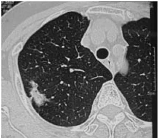
The patient was a 77-year-old female in whom an abnormal shadow was detected by chest com-puted tomography (CT) during medical examina-tion, resulting in hospitalization in October 2007
for the purpose of surgery. She was 157-cm tall, weighed 62 kg and had a body mass index of 24.9. Her medical history included surgery for thyroid cancer 10 years earlier. She had no past history of smoking habit or diabetes mellitus and was a only regular user of antihypertensive and thyroid hormones (Thyroxine). Her blood pressure was 130/85 mmHg and there were no abnormal find-ings in the thyroid function, blood coagulative test and cardiopulmonary examination. So, we and the anesthesiologists judged this patient low risk for pulmonary embolism and did not perform the ultrasound examination on the veins in both lower limbs and intrapelvic area. A 3-cm tumor on the right upper lobe was detected in the chest CT scan (Fig. 1), and the patient was diagnosed
18
H. Nakamura et al.
with pulmonary adenocarcinoma. She therefore underwent a thoracoscopic right upper lobectomy and a mediastinal lymph node dissection. The operation took 240 min to complete with 170 g of bleeding and an intraoperative massage using intermittent pneumatic compression applied to her lower limbs as her progress continued to improve. Postoperative pathological findings showed a well-differentiated adenocarcinoma without lymph node metastases, and we concluded the IA disease of tumor-node-metastasis staging. The patient’s postoperative vital signs were stable, and she started taking meals in a seated position at 18 h after surgery. She had difficulty breathing at 48 h after surgery when she was moved to a general ward, but her condition recovered immediately after oxygen was administered. After dinner, a nurse discovered that the patient was experiencing respiratory arrest, so cardiopulmonary resuscita-tion was immediately applied, and the patient was moved to the Intensive Care Unit, where she was put on artificial respiration. Echocardiography findings indicated a suspected collapse of the left ventricle and embolisms within the pulmonary arteries, so it was assumed to be a case of pul-monary embolism, and therefore the intravenous administration of 48,000 units of urokinase and 5,000 units of heparin was carried out. However,
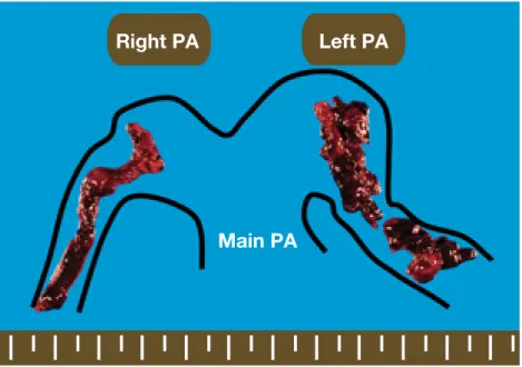
we observed pupil dilation and a loss of light re-flex, and the patient was pronounced dead 15 h after resuscitation. A pathological autopsy was performed after obtaining the informed consent from the family. Upon examining her lungs af-ter the right upper lobectomy, we detected many embolisms blocking the bilateral main pulmonary arteries to the periphery (Figs. 2A and B). The re-sected embolisms were fresh red embolisms that were 55 7 mm in size on the right side and 60
Fig. 2. Autopsy findings show many embolisms (arrows) blocking bilateral main pulmonary arteries to the periphery. A: Right lung. B: Left lung.
Fig. 1. In the preoperative chest computed tomogram,
a 3-cm tumor with irregular margin on the right upper lobe was detected. We suspected lung adenocarinoma.
19
An autopsy case of pulmonary embolism
13 mm on the left side in length. An autopsy for the brain and both lower limbs was not performed by the refusal from her family. No significant abnormalities were observed in the other organs, including intracardiac thrombus and vein throm-buses in the intrapelvic area. The cause of death was determined to be acute pulmonary embolism.
Discussion
It is said that 600,000 people per year experience pulmonary embolism in Europe and the United States, and that 16% to 25% of these cases result in death (Benotti and Dalen,1984). In Japan, there has been an increasing tendency of occur-rences of pulmonary embolism in recent years as the general life style becomes more Westernized: 80% to 90% of the causes are attributed to embo-lisms in the deep veins of the lower limbs (Saito et al., 2007). Embolism occurs easily through a congestion of blood flow and an increase in clotting function due to bed rest, particularly after surgery. The risk increases with surgery for cancer, which is prone to induce coagulation abnormalities: reported rates were 3.85% for
pancreatic cancers, 3.22% for esophageal cancers, 2.94% for lung cancers, 1.64% for rectal cancers and 1.28% for stomach cancers, and a correlation with surgical stress has also been observed (Sakon, 2005). According to one large-scale multicenter survey, the rate of occurrence of fatal postopera-tive pulmonary embolism, such as that seen in this patient, was 0.08%, with a high rate of 0.34% for thoracic surgeries and a low rate of 0.04% for abdominal surgeries (Sakon et al., 2004). Preven-tion is important for postoperative pulmonary em-bolism. The recommended actions described in the medical guidelines include the assessment of risk factors, which are age, body type (body mass index), diabetes, underlying diseases such as low-er-limb varicosity, a history of embolism and sur-gical stress before surgery: preventive measures corresponding to the risk levels should be taken during and after surgery (Geerts et al., 2004). In operations for lung cancer, intermittent pneumatic compression or the administration of heparin are recommended. Perioperative intermittent pneu-matic compression was performed in this patient as well. There is no way to guarantee prevention, but the addition of elastic stockings can further enhance the efficacy of intermittent compression
Fig. 3. The resected were fresh red embolisms 55 7 mm in size on the right
20
H. Nakamura et al.
erative pulmonary embolism occurs even in the less invasive thoracoscopic surgery. This was a valuable case that further reinforced the need for all medical workers to have an increased aware-ness regarding this disease, and we believe that it is important to ensure the prevention of any reoc-currences by making use of the lessons learned from this case.
References
1 Arcasoy SM, Kreit JW. Thtombolytic therapy of pul-monary embolism: a comprehensive review of cur-rent evidence. Chest1999;115:1695–1707.
2 Benotti JR, Dalen JE. The natural history of pulmo-nary embolism. Clin Chest Med 5 1984;5:403–410. 3 Clagett G, Reisch J. Prevention of venous
throm-boembolism in general surgical patients. Results of meta-analysis. Ann Surg 1988;208:227–240.
4 Geerts WH, Pineo GF, Heit JA, Bergqvist D, Las-sen MR, Colwell CW, et al. Prevention of venous thromboembolism: the Seventh ACCP Conference on Antithrombotic and Thrombolytic Therapy. Chest 2004;126:338–400.
5 Konstantinides S, Geibel A, Heusel G, Heinrich F, Kasper W. Heparin plus alteplase compared with heparin alone in patients with submasive pulmonary embolism. N Engl J Med 2002;347:1143–1150. 6 Ota M, Nakamura M, Yamada N, Yazu T, Ishikura K,
Hiraoka N, et al. Prognostic significance of early di-agnosis in acute pulmonary thromboembolism with circulatory failure. Heart Vessels 2002;17:7–11. 7 Saito G, Takasago K, Yoshida K, Nishimura H.
Acute pulmonary embolism following lung resec-tion. Kyoubu Geka 2007;60:946–949 (in Japanese with English abstract).
8 Sakon M. Postoperative pulmonary thromboem-bolism and its prevention from the perspective of the establishment of Japanese guidelines. Nippon Gekagakkai Zasshi 2005;106:237–240 (in Japanese with English abstract).
9 Sakon M, Kakkar AK, Ikeda M, Sekimoto M, Naka-mori S, Yano M, et al. Current status of pulmonary embolism in general surgery in Japan. Surg Today 2004;34:805–810.
Received December 21, 2007; accepted January 9, 2008 Corresponding author: Hiroshige Nakamura, MD
and the additional administration of heparin is the most effective preventive method known at pres-ent. On the other hand, it is a well-known fact that the administration of heparin increases the risk of perioperative and postoperative bleeding (Clagett and Reisch, 1988), thus indicating the dif-ficulty of preventing pulmonary embolism in any and all cases. Based on our experience with this patient, we have verified the need to assess risks in detail before surgery according to the medical guidelines, as well as the need to ensure the use of elastic stockings and intermittent pneumatic compression until postoperative ambulation, and to administer heparin to patients with a history of deep venous thrombus, and we have compiled these guidelines into a manual.
In diagnosing pulmonary embolism, key signs include sudden difficulty in breathing and a rapid decrease in saturation of percutaneous O2
that occurs with the first body movement. So, the common occurrence period of pulmonary embolism was usually considered to be at the 2nd or 3rd day after the surgery (Sakon, 2005). As medical workers, we are required to pay especial attentions to the patient in this dangerous period and accurately predict a diagnosis from such slight symptoms (Saito et al., 2007). By taking a contrast chest CT, embolisms can be detected by the filling in of defects within the pulmonary ar-teries, and a diagnosis can thus be determined. It has been reported that 32% of cases of acute pul-monary embolism result in death of the patient, as was the case with this patient, and of these cases, 43% died within 1 h of onset (Ota et al., 2002). Clearly, it is better for treatment to be adminis-tered earlier: that is, success or failure depends on how early thrombolytic therapy and anticoagu-lation therapy are initiated (Arcasoy and Kreit, 1999; Konstantinides et al., 2002). Because this patient complained of difficulty in breathing when she was being moved to the general ward at 48 h after the surgery, and although it was only tempo-rary, it is predicted that acute pulmonary embo-lism had already occurred by that time.