Title
High-Temperature Wide Thermal Hysteresis of an Iron(II)
Dinuclear Double Helicate( 本文(Fulltext) )
Author(s)
HORA, Shiori; HAGIWARA, Hiroaki
Citation
[Inorganics] vol.[5] no.[3] p.[49]
Issue Date
2017
Rights
©2017 by the authors. Licensee MDPI, Basel, Switzerland. This
article is an open access article distributed under the terms and
conditions of the Creative Commons Attribution (CC BY) license
(http://creativecommons.org/licenses/by/4.0/).
Version
出版社版 (publisher version) postprint
URL
http://hdl.handle.net/20.500.12099/74339
inorganics
Article
High-Temperature Wide Thermal Hysteresis of
an Iron(II) Dinuclear Double Helicate
Shiori Hora1and Hiroaki Hagiwara2,* ID
1 Graduate School of Education, Gifu University, Yanagido 1-1, Gifu 501-1193, Japan;
2 Department of Chemistry, Faculty of Education, Gifu University, Yanagido 1-1, Gifu 501-1193, Japan
* Correspondence: [email protected]; Tel.: +81-58-293-2253 Received: 28 June 2017; Accepted: 25 July 2017; Published: 28 July 2017
Abstract: Two new dinuclear iron(II) complexes (1·PF6 and 1·AsF6) of the general formula [FeII2(L2C3)2](X)4·nH2O·mMeCN (X = PF6, n = m = 1.5 for 1·PF6and X = AsF6, n = 3, m = 1 for 1·AsF6) have been prepared and structurally characterized, where L2C3is a bis-1,2,3-triazolimine type Schiff-base ligand, 1,10-[propane-1,3-diylbis(1H-1,2,3-triazole-1,4-diyl)]bis{N-[2-(pyridin-2-yl)ethyl]methanimine}. Single crystal X-ray structure analyses revealed that 1·PF6 and 1·AsF6 are isostructural. The complex-cation [FeII2(L2C3)2]4+of both has the same dinuclear double helicate architecture,
in which each iron(II) center has an N6 octahedral coordination environment. Neighboring helicates are connected by intermolecular π–π interactions to give a chiral one-dimensional (1D) structure, and cationic 1D chains with the opposite chirality exist in the crystal lattice to give a heterochiral crystal. Magnetic and differential scanning calorimetry (DSC) studies were performed only for 1·AsF6, since the thermal stability in a high-temperature spin crossover (SCO) region of 1·PF6is poorer than that of 1·AsF6. 1·AsF6shows an unsymmetrical hysteretic SCO between the low-spin–low-spin (LS–LS) and high-spin–high-spin (HS–HS) states at above room temperature. The critical temperatures of warming (Tc↑) and cooling (Tc↓) modes in the abrupt spin transition area are 485 and 401 K, respectively,
indicating the occurrence of 84 K-wide thermal hysteresis in the first thermal cycle.
Keywords:double helicate; hysteresis; spin crossover; iron; 1,2,3-triazolimine; π–π interaction
1. Introduction
The interconversion between a high-spin (HS) and a low-spin (LS) state, the so-called “spin crossover” (SCO), is one of the most attractive phenomena in the field of molecular bistability [1–3]. This phenomenon is observed in 3d4–3d7 transition metal complexes with various coordination geometries (e.g., octahedral, square-pyramidal, trigonal-bipyramidal, tetrahedral, etc.) [1,4–7], and is triggered by an external stimulus such as temperature, pressure, light irradiation, or a magnetic field [1–3]. While SCO originates from an individual metal center, a spin transition profile, for example, abruptness and multi-steps with or without hysteresis, is provided from the cooperative effect between SCO metal sites through intra- or intermolecular pathways [1–3]. There is a need for SCO materials which are capable of abrupt and complete HS–LS interconversion at around room temperature (RT) with wide thermal hysteresis width (∆T) for potential applications in molecular memories, switches, and display devices. As such, various cooperative SCO compounds have been produced with supramolecular assembly via intermolecular hydrogen bonding [8–10] and π–π stacking [11–13], or coordination polymeric architecture by using bridging ligands [14–18]. Multinuclear clusters have also been widely studied in the hope of developing multistable compounds with more accessible spin states towards denser information storage, or hybrid SCO materials with multifunctional properties (e.g., SCO with charge transfer, magnetic coupling, luminescence, etc.) arising from the combination of the SCO metal center and other metal centers [2,19–24]. In particular, dinuclear species have received
Inorganics 2017, 5, 49 2 of 15
much attention since they are a simple model for revealing intra- and intermolecular interactions for enhancing cooperativity [25–34]. From a more practical point of view, the importance of studying the reproducible nature and scan rate dependence of the hysteresis loop and high-temperature SCO with sufficient thermal stability has recently been pointed out in order to define the limiting characteristics of SCO materials [33,35–39].
Recently, we have focused on SCO molecules with 1-R-1H-1,2,3-triazole-containing Schiff-base (1-R-1H-1,2,3-triazolimine; R = Me and Ph) ligands since the ligand system can easily modify its structure with the choice of a substituent R of a precursor, 1-R-1H-1,2,3-triazole-4-carbaldehyde, and another amino precursor by simple click reaction [40–42], Schiff-base reaction [43] and replacement of R using the method reported by L’abbé and coworkers [44,45]. The iron(II) complexes with multidentate 1-R-1H-1,2,3-triazolimine ligand show a variety of SCO properties such as gradual, abrupt, and two-step transition with (or without) hysteresis in a wide temperature range (around RT, below RT and above RT over ca. 100◦C) [34,46,47]. In one of these studies, we reported the SCO iron(II) dinuclear double helicate for the first time [34]. This compound, with the formula [FeII2(L2C2)2](PF6)4·5H2O·MeCN, has two bis-tridentate type ligand strands (L2C2) in which two
1,2,3-triazolimine moieties of the ligand strand are bridged by an ethylene chain (Scheme1), and shows an anomalous two-step SCO with 11 K-wide hysteresis in a second step centered at 432 K, which is the highest hysteretic transition temperature in the dinuclear SCO system reported so far [26–29,33,48].
From the fascinating structure and function of the [FeII2(L2C2)2]4+ system, our own interest
in the system has increasingly concerned the effect of the bridging alkyl chain length. We report here the synthesis, thermal stability, and structure of the corresponding propylene-bridged iron(II) dinuclear double helicate complexes [FeII2(L2C3)2](PF6)4·1.5H2O·1.5MeCN (1·PF6) and [FeII2(L2C3)2](AsF6)4·3H2O·MeCN (1·AsF6), where L2C3= 1,10 -[propane-1,3-diylbis(1H-1,2,3-triazole-1,4-diyl)]bis{N-[2-(pyridin-2-yl)ethyl]methanimine} (Scheme1), and the high-temperature hysteretic SCO of 1·AsF6.
Inorganics 2017, 5, 49 2 of 15
[2,19–24].In particular, dinuclear species have received much attention since they are a simple model for revealing intra- and intermolecular interactions for enhancing cooperativity [25–34]. From a more practical point of view, the importance of studying the reproducible nature and scan rate dependence of the hysteresis loop and high-temperature SCO with sufficient thermal stability has recently been pointed out in order to define the limiting characteristics of SCO materials [33,35–39].
Recently, we have focused on SCO molecules with 1-R-1H-1,2,3-triazole-containing Schiff-base (1-R-1H-1,2,3-triazolimine; R = Me and Ph) ligands since the ligand system can easily modify its structure with the choice of a substituent R of a precursor, 1-R-1H-1,2,3-triazole-4-carbaldehyde, and another amino precursor by simple click reaction [40–42], Schiff-base reaction [43] and replacement of R using the method reported by L’abbé and coworkers [44,45]. The iron(II) complexes with multidentate 1-R-1H-1,2,3-triazolimine ligand show a variety of SCO properties such as gradual, abrupt, and two-step transition with (or without) hysteresis in a wide temperature range (around RT, below RT and above RT over ca. 100 °C) [34,46,47]. In one of these studies, we reported the SCO iron(II) dinuclear double helicate for the first time [34]. This compound, with the formula [FeII2(L2C2)2](PF6)4·5H2O·MeCN, has two bis-tridentate type ligand strands (L2C2) in which two
1,2,3-triazolimine moieties of the ligand strand are bridged by an ethylene chain (Scheme 1), and shows an anomalous two-step SCO with 11 K-wide hysteresis in a second step centered at 432 K, which is the highest hysteretic transition temperature in the dinuclear SCO system reported so far [26–29,33,48].
From the fascinating structure and function of the [FeII2(L2C2)2]4+ system, our own interest in the
system has increasingly concerned the effect of the bridging alkyl chain length. We report here the synthesis, thermal stability, and structure of the corresponding propylene-bridged iron(II) dinuclear double helicate complexes [FeII2(L2C3)2](PF6)4·1.5H2O·1.5MeCN (1·PF6) and
[FeII2(L2C3)2](AsF6)4·3H2O·MeCN (1·AsF6), where L2C3 =
1,1′-[propane-1,3-diylbis(1H-1,2,3-triazole-1,4-diyl)]bis{N-[2-(pyridin-2-yl)ethyl]methanimine} (Scheme 1), and the high-temperature hysteretic SCO of 1·AsF6.
Scheme 1. Synthesis of the dinuclear double helicate complex-cation [FeII2(L2R)2]4+ (R = C2 = ethylene [34], R = C3 = propylene (this work)).
2. Results and Discussion
2.1. Synthesis and Characterization of [FeII2(L2C3)2](PF6)4·1.5H2O·1.5MeCN (1·PF6) and [FeII2(L2C3)2](AsF6)4·3H2O·MeCN (1·AsF6)
The ligand L2C3 was prepared stoichiometric by the 1:2 condensation reaction of
1,1′-(propane-1,3-diyl)bis(1H-1,2,3-triazole-4-carbaldehyde) (4) and 2-(2-aminoethyl)pyridine. 4 was synthesized starting from 1-phenyl-1H-1,2,3-triazole-4-carbaldehyde and 1,3-diaminopropane (Scheme 2) based on the previously reported procedure of a related ethylene-bridged compound [34]. The dinuclear 1·PF6 and 1·AsF6 were prepared by mixing the ligand L2C3, FeIICl2·4H2O, and KX (X = PF6 and AsF6
for 1·PF6 and 1·AsF6, respectively)with a 1:1:2 molar ratio in MeOH/H2O mixed solution, which was
then recrystallized from the MeCN/MeOH mixed solution. All the synthetic procedures were performed in air and both complexes were obtained as dark-red block crystals. The infrared spectra of both showed a characteristic band at ca. 1609 cm−1, corresponding to the C=N stretching vibration
of the Schiff-base ligand [34,49]. In addition, characteristic bands at ca. 844 and 701 cm−1 were
observed, corresponding to the counter anions, PF6− and AsF6− for 1·PF6 and 1·AsF6, respectively [34,49,50]. The formula of both complexes was confirmed by elemental analyses and thermogravimetric analyses. Thermogravimetry and differential thermal analysis (TG/DTA) curves
Scheme 1.Synthesis of the dinuclear double helicate complex-cation [FeII2(L2R)2]4+(R = C2 = ethylene [34],
R = C3 = propylene (this work)). 2. Results and Discussion
2.1. Synthesis and Characterization of [FeII2(L2C3)2](PF6)4·1.5H2O·1.5MeCN (1·PF6) and [FeII
2(L2C3)2](AsF6)4·3H2O·MeCN (1·AsF6)
The ligand L2C3was prepared stoichiometric by the 1:2 condensation reaction of 1,10 -(propane-1,3-diyl)bis(1H-1,2,3-triazole-4-carbaldehyde) (4) and 2-(2-aminoethyl)pyridine. 4 was synthesized starting from 1-phenyl-1H-1,2,3-triazole-4-carbaldehyde and 1,3-diaminopropane (Scheme2) based on the previously reported procedure of a related ethylene-bridged compound [34]. The dinuclear 1·PF6and 1·AsF6were prepared by mixing the ligand L2C3, FeIICl2·4H2O, and KX (X = PF6and AsF6
for 1·PF6and 1·AsF6, respectively) with a 1:1:2 molar ratio in MeOH/H2O mixed solution, which
was then recrystallized from the MeCN/MeOH mixed solution. All the synthetic procedures were performed in air and both complexes were obtained as dark-red block crystals. The infrared spectra of both showed a characteristic band at ca. 1609 cm−1, corresponding to the C=N stretching vibration of the Schiff-base ligand [34,49]. In addition, characteristic bands at ca. 844 and 701 cm−1were observed,
Inorganics 2017, 5, 49 3 of 15
corresponding to the counter anions, PF6−and AsF6−for 1·PF6and 1·AsF6, respectively [34,49,50]. The formula of both complexes was confirmed by elemental analyses and thermogravimetric analyses. Thermogravimetry and differential thermal analysis (TG/DTA) curves of both are shown in Figure1. As shown in Figure1a, when the powdered sample of 1·PF6was heated from 30◦C (303 K) at a rate of 10◦C·min−1, the sample weight decreased gradually and a weight loss of 4.3% at 224◦C (497 K) was observed, which corresponds to the calculated weight percentages of 0.5H2O and 1.5MeCN molecules
per [FeII2(L2C3)2](PF6)4·1.5H2O·1.5MeCN (1·PF6) (4.2%). Above this temperature, the weight loss became abrupt, and then the weight loss of the remaining one H2O molecule (1.1%) was observed at
242◦C (515 K). In this region, a sharp thermal anomaly in DTA appeared (Tmax= 232◦C (505 K)). Finally,
above 242◦C (515 K), the weight loss became more and more abrupt, indicating the decomposition of 1·PF6. In the same way, as shown in Figure1b, when the powdered sample of 1·AsF6was heated from 30◦C (303 K) at a rate of 10◦C·min−1, the sample weight decreased gradually and the total weight loss was 5.0% at 215◦C (488 K) in agreement with the calculated weight percentage of 3H2O and
one MeCN molecule per [FeII2(L2C3)2](AsF6)4·3H2O·MeCN (1·AsF6) (5.1%). On further increasing the temperature from 215◦C (488 K), the desolvated sample of 1·AsF6was stable up to ca. 263◦C (536 K), and began to decompose above this temperature. Although a sharp thermal anomaly in DTA was also observed at Tmax= 212◦C (485 K) in 1·AsF6, this peak was far from the decomposition temperature (ca. 263◦C), which is obviously different from that of 1·PF6. To access the reason for exhibiting a sharp thermal anomaly in DTA around 485–505 K, thermochromic properties of both compounds were also investigated (Figure2). Although the orange-brown color of powdered samples of 1·PF6becomes slightly lighter from 300 K to ca. 483 K, possibly due to the partial SCO, further elevation of the temperature to 493 K induces rapid black coloration, suggesting the decomposition of the compound (Figure2a). This thermochromic tendency and TG/DTA results suggest that the decomposition temperature overlaps the spin transition temperature in 1·PF6. On the other hand, powdered samples of 1·AsF6showed a clearly distinguishable thermochromism from orange-brown at 300 K to orange-yellow at ca. 498 K, suggesting the occurrence of SCO (Figure2b). Then, further increasing the temperature to 503 K induces slight yellowish coloration, and ca. 5 min. later, a black coloration related to decomposition proceeds. This color change of 1·AsF6in a step-by-step-manner is comparable to the clear distinction between sharp thermal anomaly at Tmax= 485 K and decomposition
temperature observed in TG/DTA curves. Consequently, we decided to investigate SCO properties only for 1·AsF6which is more stable than 1·PF6in a high-temperature SCO region.
Inorganics 2017, 5, 49 3 of 15
of both are shown in Figure 1. As shown in Figure 1a, when the powdered sample of 1·PF6 was heated
from 30 °C (303 K) at a rate of 10 °C·min−1, the sample weight decreased gradually and a weight loss of 4.3% at 224 °C (497 K) was observed, which corresponds to the calculated weight percentages of 0.5H2O and 1.5MeCN molecules per [FeII2(L2C3)2](PF6)4·1.5H2O·1.5MeCN (1·PF6) (4.2%). Above this
temperature, the weight loss became abrupt, and then the weight loss of the remaining one H2O molecule (1.1%) was observed at 242 °C (515 K). In this region, a sharp thermal anomaly in DTA appeared (Tmax = 232 °C (505 K)). Finally, above 242 °C (515 K), the weight loss became more and more abrupt, indicating the decomposition of 1·PF6. In the same way, as shown in Figure 1b, when the
powdered sample of 1·AsF6 was heated from 30 °C (303 K) at a rate of 10 °C·min−1, the sample weight
decreased gradually and the total weight loss was 5.0% at 215 °C (488 K) in agreement with the calculated weight percentage of 3H2O and one MeCN molecule per [FeII2(L2C3)2](AsF6)4·3H2O·MeCN (1·AsF6) (5.1%). On further increasing the temperature from 215 °C (488 K), the desolvated sample of
1·AsF6 was stable up to ca. 263 °C (536 K), and began to decompose above this temperature. Although
a sharp thermal anomaly in DTA was also observed at Tmax = 212 °C (485 K) in 1·AsF6, this peak was
far from the decomposition temperature (ca. 263 °C), which is obviously different from that of 1·PF6.
To access the reason for exhibiting a sharp thermal anomaly in DTA around 485–505 K, thermochromic properties of both compounds were also investigated (Figure 2). Although the orange-brown color of powdered samples of 1·PF6 becomes slightly lighter from 300 K to ca. 483 K,
possibly due to the partial SCO, further elevation of the temperature to 493 K induces rapid black coloration, suggesting the decomposition of the compound (Figure 2a). This thermochromic tendency and TG/DTA results suggest that the decomposition temperature overlaps the spin transition temperature in 1·PF6. On the other hand, powdered samples of 1·AsF6 showed a clearly
distinguishable thermochromism from orange-brown at 300 K to orange-yellow at ca. 498 K, suggesting the occurrence of SCO (Figure 2b). Then, further increasing the temperature to 503 K induces slight yellowish coloration, and ca. 5 min. later, a black coloration related to decomposition proceeds. This color change of 1·AsF6 in a step-by-step-manner is comparable to the clear distinction
between sharp thermal anomaly at Tmax = 485 K and decomposition temperature observed in TG/DTA curves. Consequently, we decided to investigate SCO properties only for 1·AsF6 which is more stable
than 1·PF6 in a high-temperature SCO region.
Scheme 2. Synthetic scheme of the ligand L2C2. Reagents and conditions: (1) 1,3-diaminopropane,
MeOH, 1 h, 60 °C; 78%; (2) 1-PrOH, 16 h, 80 °C; 78%; (3) HCl (aq), 2 h, room temperature (RT); 86%; (4) 2-(2-aminoethyl)pyridine (2 equiv), MeOH, 1 h, RT.
Scheme 2.Synthetic scheme of the ligand L2C2. Reagents and conditions: (1) 1,3-diaminopropane, MeOH, 1 h, 60◦C; 78%; (2) 1-PrOH, 16 h, 80◦C; 78%; (3) HCl (aq), 2 h, room temperature (RT); 86%; (4) 2-(2-aminoethyl)pyridine (2 equiv), MeOH, 1 h, RT.
Inorganics 2017, 5, 49 4 of 15
Inorganics 2017, 5, 49 4 of 15
Figure 1. Thermogravimetry and differential thermal analysis (TG/DTA) curves of 1·PF6 (a) and
1·AsF6 (b).
Figure 2. Temperature-dependent optical microscope images of grinding samples of 1·PF6 (a) and
1·AsF6 (b) deposited on a heating stage. Unless otherwise noted, after increasing the sample
temperature at each target point, the sample was held for 5 min, and then an image was taken at each temperature.
2.2. Crystal Structure of 1·PF6 and 1·AsF6
The crystal structure of 1·PF6 and 1·AsF6 was determined at 120 K (low-spin–low-spin (LS–LS) state). As shown in Table 1, both complexes crystallize in the same monoclinic space group type C2/c (number 15) with Z = 4 and are isostructural. The structure consists of a [FeII2(L2C3)2]4+ complex-cation,
two disordered monovalent counterions (PF6− and AsF6− for 1·PF6 and 1·AsF6, respectively), and H2O
and MeCN molecules as lattice solvents. In the crystal lattice, both solvent molecules are in the one solvent site in a severely disordered manner in both compounds. Thus, these regions, including solvent molecules, are treated with the PLATON SQUEEZE program [51] (see Experimental Section 3.3 including the details about analyses of disordered counter anions and solvent molecules).
Figure 3 shows the molecular structure of the complex-cation [FeII2(L2C3)2]4+ of 1·AsF6 at 120 K.
As shown in Table 2, the structural feature of the complex-cation of 1·PF6 is similar to that of 1·AsF6. Thus, we only discuss the structure of 1·AsF6 below. The complex-cation has a dinuclear double-helical architecture, in which each ligand L2C3 bound as a bis-tridentate ligand to two different iron
centers, and conversely, the FeII centers are N6-coordinated pseudo-octahedral coordination
environments bound to two different ligand strands. The intra-helical metal–metal distance is 10.894(3) Å, which is much longer than that of the related ethylene-bridged helicate [FeII2(L2C2)2]4+
(7.855(3) Å) [34] due to the elongation of the alkyl chain from two to three C atoms. Each metal site is chiral, with either a Δ or a Λ configuration, and the complex-cation depicted in Figure 3 has a homochiral Λ–Λ pair. In the crystal, complex-cations with the Δ–Δ and Λ–Λ pairs coexist to form a racemic crystal since the complex crystallizes in a centrosymmetric space group, C2/c. The Fe–N coordination bond distances (1.954(3)–2.007(3) and 1.945(3)–2.007(3) Å for Fe1–N and Fe2–N, respectively) indicate that both centers are in the LS FeII state. While the average Fe–N bond distance
and octahedral volume of LS–LS [FeII2(L2C3)2]4+ are similar to those of LS–LS [FeII2(L2C2)2]4+, the Θ [52]
Figure 1. Thermogravimetry and differential thermal analysis (TG/DTA) curves of 1·PF6 (a) and
1·AsF6(b).
Inorganics 2017, 5, 49 4 of 15
Figure 1. Thermogravimetry and differential thermal analysis (TG/DTA) curves of 1·PF6 (a) and
1·AsF6 (b).
Figure 2. Temperature-dependent optical microscope images of grinding samples of 1·PF6 (a) and
1·AsF6 (b) deposited on a heating stage. Unless otherwise noted, after increasing the sample
temperature at each target point, the sample was held for 5 min, and then an image was taken at each temperature.
2.2. Crystal Structure of 1·PF6 and 1·AsF6
The crystal structure of 1·PF6 and 1·AsF6 was determined at 120 K (low-spin–low-spin (LS–LS)
state). As shown in Table 1, both complexes crystallize in the same monoclinic space group type C2/c (number 15) with Z = 4 and are isostructural. The structure consists of a [FeII2(L2C3)2]4+ complex-cation, two disordered monovalent counterions (PF6− and AsF6− for 1·PF6 and 1·AsF6, respectively), and H2O and MeCN molecules as lattice solvents. In the crystal lattice, both solvent molecules are in the one solvent site in a severely disordered manner in both compounds. Thus, these regions, including solvent molecules, are treated with the PLATON SQUEEZE program [51] (see Experimental Section 3.3 including the details about analyses of disordered counter anions and solvent molecules).
Figure 3 shows the molecular structure of the complex-cation [FeII2(L2C3)2]4+ of 1·AsF6 at 120 K. As shown in Table 2, the structural feature of the complex-cation of 1·PF6 is similar to that of 1·AsF6.
Thus, we only discuss the structure of 1·AsF6 below. The complex-cation has a dinuclear
double-helical architecture, in which each ligand L2C3 bound as a bis-tridentate ligand to two different iron centers, and conversely, the FeII centers are N6-coordinated pseudo-octahedral coordination environments bound to two different ligand strands. The intra-helical metal–metal distance is 10.894(3) Å, which is much longer than that of the related ethylene-bridged helicate [FeII2(L2C2)2]4+ (7.855(3) Å) [34] due to the elongation of the alkyl chain from two to three C atoms. Each metal site is chiral, with either a Δ or a Λ configuration, and the complex-cation depicted in Figure 3 has a homochiral Λ–Λ pair. In the crystal, complex-cations with the Δ–Δ and Λ–Λ pairs coexist to form a racemic crystal since the complex crystallizes in a centrosymmetric space group, C2/c. The Fe–N coordination bond distances (1.954(3)–2.007(3) and 1.945(3)–2.007(3) Å for Fe1–N and Fe2–N, respectively) indicate that both centers are in the LS FeII state. While the average Fe–N bond distance and octahedral volume of LS–LS [FeII2(L2C3)2]4+ are similar to those of LS–LS [FeII2(L2C2)2]4+, the Θ [52]
Figure 2. Temperature-dependent optical microscope images of grinding samples of 1·PF6(a) and
1·AsF6 (b) deposited on a heating stage. Unless otherwise noted, after increasing the sample temperature at each target point, the sample was held for 5 min, and then an image was taken at each temperature.
2.2. Crystal Structure of 1·PF6and 1·AsF6
The crystal structure of 1·PF6and 1·AsF6was determined at 120 K (low-spin–low-spin (LS–LS) state). As shown in Table1, both complexes crystallize in the same monoclinic space group type C2/c (number 15) with Z = 4 and are isostructural. The structure consists of a [FeII2(L2C3)2]4+complex-cation,
two disordered monovalent counterions (PF6−and AsF6−for 1·PF6and 1·AsF6, respectively), and H2O and MeCN molecules as lattice solvents. In the crystal lattice, both solvent molecules are in the
one solvent site in a severely disordered manner in both compounds. Thus, these regions, including solvent molecules, are treated with the PLATON SQUEEZE program [51] (see Experimental Section3.3
including the details about analyses of disordered counter anions and solvent molecules).
Figure3shows the molecular structure of the complex-cation [FeII2(L2C3)2]4+of 1·AsF6at 120 K. As shown in Table2, the structural feature of the complex-cation of 1·PF6is similar to that of 1·AsF6. Thus, we only discuss the structure of 1·AsF6below. The complex-cation has a dinuclear double-helical architecture, in which each ligand L2C3bound as a bis-tridentate ligand to two different iron centers,
and conversely, the FeIIcenters are N6-coordinated pseudo-octahedral coordination environments bound to two different ligand strands. The intra-helical metal–metal distance is 10.894(3) Å, which is much longer than that of the related ethylene-bridged helicate [FeII2(L2C2)2]4+(7.855(3) Å) [34]
due to the elongation of the alkyl chain from two to three C atoms. Each metal site is chiral, with either a∆ or a Λ configuration, and the complex-cation depicted in Figure3has a homochiralΛ–Λ pair. In the crystal, complex-cations with the∆–∆ and Λ–Λ pairs coexist to form a racemic crystal since the complex crystallizes in a centrosymmetric space group, C2/c. The Fe–N coordination bond distances (1.954(3)–2.007(3) and 1.945(3)–2.007(3) Å for Fe1–N and Fe2–N, respectively) indicate that both centers are in the LS FeIIstate. While the average Fe–N bond distance and octahedral volume of
Inorganics 2017, 5, 49 5 of 15
LS–LS [FeII2(L2C3)2]4+are similar to those of LS–LS [FeII2(L2C2)2]4+, theΘ [52] and continuous shape
measures (CShMs) relative to the regular octahedron with the center as the reference shape [53] of LS–LS [FeII2(L2C3)2]4+are lower than those of LS–LS [FeII2(L2C2)2]4+, indicating that the FeIIcenters of
LS–LS [FeII2(L2C3)2]4+have a more regular octahedral geometry than those of LS–LS [FeII2(L2C2)2]4+
with the one minor exception of theΣ [54] of LS–LS [FeII2(L2C2)2]4+, which is slightly lower than that
of LS–LS [FeII
2(L2C3)2]4+(average Fe–N bond distance,Σ, Θ, CShMs and octahedral volume of the
LS–LS [FeII2(L2C2)2]4+for [FeII2(L2C2)2](PF6)4·5H2O·MeCN [34] are 1.971 Å, 42.5◦, 133.7◦, 0.696 and
10.096 Å3, respectively).
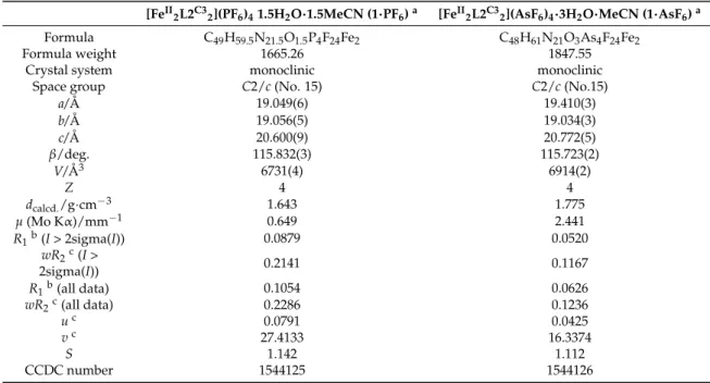
Table 1.X-ray crystallographic data for 1·PF6and 1·AsF6at 120 K.
[FeII2L2C32](PF6)41.5H2O·1.5MeCN (1·PF6)a [FeII2L2C32](AsF6)4·3H2O·MeCN (1·AsF6)a
Formula C49H59.5N21.5O1.5P4F24Fe2 C48H61N21O3As4F24Fe2
Formula weight 1665.26 1847.55
Crystal system monoclinic monoclinic
Space group C2/c (No. 15) C2/c (No.15)
a/Å 19.049(6) 19.410(3) b/Å 19.056(5) 19.034(3) c/Å 20.600(9) 20.772(5) β/deg. 115.832(3) 115.723(2) V/Å3 6731(4) 6914(2) Z 4 4 dcalcd./g·cm−3 1.643 1.775 µ(Mo Kα)/mm−1 0.649 2.441 R1b(I > 2sigma(I)) 0.0879 0.0520 wR2c(I > 2sigma(I)) 0.2141 0.1167 R1b(all data) 0.1054 0.0626 wR2c(all data) 0.2286 0.1236 uc 0.0791 0.0425 vc 27.4133 16.3374 S 1.142 1.112 CCDC number 1544125 1544126
aThe PLATON SQUEEZE program [51] was used to treat regions with highly disordered solvent molecules which could not be sensibly modeled in terms of atomic sites;bR
1=Σ||Fo|−|Fc||/Σ|Fo|;cwR2= [Σw(|Fo|2−|Fc|2)2/ Σw|Fo2|2]1/2, w = 1/[σ2(|Fo|2) + (uP)2+ vP] where P = (|Fo|2+ 2|Fc|2)/3.
The assembly structure of 1·AsF6 is shown in Figure4. As shown in Figure 4a, a cationic one-dimensional (1D) structure is formed along the b-axis by slightly inclined intermolecular π–π interactions between all pyridyl rings of neighboring complex-cations with Cg1···Cg2ii= 4.123(3) Å.
Inorganics 2017, 5, 49 5 of 15
and continuous shape measures (CShMs) relative to the regular octahedron with the center as the reference shape [53] of LS–LS [FeII2(L2C3)2]4+ are lower than those of LS–LS [FeII2(L2C2)2]4+, indicating
that the FeII centers of LS–LS [FeII2(L2C3)2]4+ have a more regular octahedral geometry than those of
LS–LS [FeII2(L2C2)2]4+ with the one minor exception of the Σ [54] of LS–LS [FeII2(L2C2)2]4+, which is
slightly lower than that of LS–LS [FeII2(L2C3)2]4+ (average Fe–N bond distance, Σ, Θ, CShMs and
octahedral volume of the LS–LS [FeII2(L2C2)2]4+ for [FeII2(L2C2)2](PF6)4·5H2O·MeCN [34] are 1.971 Å,
42.5°, 133.7°, 0.696 and 10.096 Å3, respectively).
Table 1. X-ray crystallographic data for 1·PF6 and 1·AsF6 at 120 K.
[FeII2L2C32](PF6)4 1.5H2O·1.5MeCN (1·PF6) a [FeII2L2C32](AsF6)4·3H2O·MeCN (1·AsF6) a
Formula C49H59.5N21.5O1.5P4F24Fe2 C48H61N21O3As4F24Fe2
Formula weight 1665.26 1847.55
Crystal system monoclinic monoclinic
Space group C2/c (No. 15) C2/c (No.15)
a/Å 19.049(6) 19.410(3) b/Å 19.056(5) 19.034(3) c/Å 20.600(9) 20.772(5) β/deg. 115.832(3) 115.723(2) V/Å3 6731(4) 6914(2) Z 4 4 dcalcd./g·cm−3 1.643 1.775 μ (Mo Kα)/mm−1 0.649 2.441 R1b (I > 2sigma(I)) 0.0879 0.0520 wR2c (I > 2sigma(I)) 0.2141 0.1167 R1b (all data) 0.1054 0.0626 wR2c (all data) 0.2286 0.1236 u c 0.0791 0.0425 v c 27.4133 16.3374 S 1.142 1.112 CCDC number 1544125 1544126
a The PLATON SQUEEZE program [51] was used to treat regions with highly disordered solvent molecules which could not be sensibly modeled in terms of atomic sites; b R1 = Σ||Fo| − |Fc||/Σ|Fo|; c wR2 = [Σw(|Fo|2 − |Fc|2)2/Σw|Fo2|2]1/2, w = 1/[σ2(|Fo|2) + (uP)2 + vP] where P = (|Fo|2 + 2|Fc|2)/3.
The assembly structure of 1·AsF6 is shown in Figure 4. As shown in Figure 4a, a cationic
one-dimensional (1D) structure is formed along the b-axis by slightly inclined intermolecular π–π interactions between all pyridyl rings of neighboring complex-cations with Cg1···Cg2 ii = 4.123(3) Å.
Figure 3. ORTEP drawing of the dinuclear double helicate complex-cation [FeII2(L2C3)2]4+ of 1·AsF6 at 120 K with the atom numbering scheme except for carbon atoms, where the thermal ellipsoids are drawn with a 50% probability level. H atoms have been omitted for clarity. Symmetry operation: (i) –x, y, 3/2 − z.
Figure 3.ORTEP drawing of the dinuclear double helicate complex-cation [FeII
2(L2C3)2]4+of 1·AsF6 at 120 K with the atom numbering scheme except for carbon atoms, where the thermal ellipsoids are drawn with a 50% probability level. H atoms have been omitted for clarity. Symmetry operation: (i) –x, y, 3/2−z.
Inorganics 2017, 5, 49 6 of 15
(Cg1 = centroid of the N1–C1–C2–C3–C4–C5 ring, Cg2 = centroid of the N6–C12–C13–C14–C15– C16 ring; symmetry operation: (ii) x,−1 + y, z; Table3). In the light of chiral assembly, homochiral complex-cations are linked together in the π–stacked 1D structure to give a homochiral 1D chain (∆–∆···∆–∆···∆–∆···orΛ–Λ···Λ–Λ···Λ–Λ···). Figure4b shows the stacking observed between adjacent cationic 1D chains viewed along the ac plane. One-dimensional chains of opposite chirality are alternately arrayed along the c-axis to give a heterochiral crystal. Each cationic 1D chain is separated by AsF6−ions and solvent molecules occupying the space between the 1D chains in a strongly disordered
manner even at 120 K. Although the assembly structure of 1·PF6is almost same as that of 1·AsF6, these 1D-based structures are remarkably different from the two-dimensional (2D) assembly of the related ethylene-bridged helicate [FeII
2(L2C2)2](PF6)4·5H2O·MeCN by intermolecular π–π and CH/π
interactions [34].
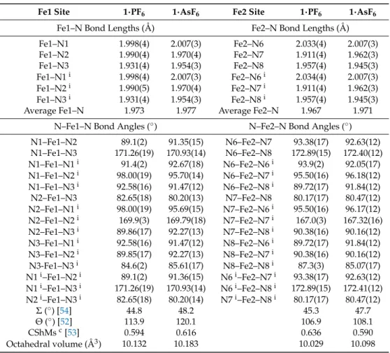
Table 2.Coordination bond lengths, angles, and structural parameters for 1·PF6and 1·AsF6at 120 K. CShMs: continuous shape measures.
Fe1 Site 1·PF6 1·AsF6 Fe2 Site 1·PF6 1·AsF6
Fe1–N Bond Lengths (Å) Fe2–N Bond Lengths (Å) Fe1–N1 1.998(4) 2.007(3) Fe2–N6 2.033(4) 2.007(3) Fe1–N2 1.990(4) 1.970(4) Fe2–N7 1.911(4) 1.962(3) Fe1–N3 1.931(4) 1.954(3) Fe2–N8 1.957(4) 1.945(3) Fe1–N1i 1.998(4) 2.007(3) Fe2–N6i 2.034(4) 2.007(3) Fe1–N2i 1.990(5) 1.970(4) Fe2–N7i 1.911(4) 1.962(3) Fe1–N3i 1.931(4) 1.954(3) Fe2–N8i 1.957(4) 1.945(3) Average Fe1–N 1.973 1.977 Average Fe2–N 1.967 1.971
N–Fe1–N Bond Angles (◦) N–Fe2–N Bond Angles (◦) N1–Fe1–N2 89.1(2) 91.35(15) N6–Fe2–N7 93.38(17) 92.63(12) N1–Fe1–N3 171.26(19) 170.93(14) N6–Fe2–N8 172.89(15) 172.40(12) N1–Fe1–N1i 91.4(2) 92.67(18) N6–Fe2–N6i 93.9(2) 92.05(17) N1–Fe1–N2i 98.00(19) 95.70(14) N6–Fe2–N7i 95.50(16) 96.18(12) N1–Fe1–N3i 92.58(16) 91.47(12) N6–Fe2–N8i 89.72(17) 91.84(12) N2–Fe1–N3 82.65(18) 80.20(13) N7–Fe2–N8 80.17(17) 80.47(12) N2–Fe1–N1i 98.00(19) 95.69(15) N7–Fe2–N6i 95.50(16) 96.17(12) N2–Fe1–N2i 169.9(3) 169.79(18) N7–Fe2–N7i 167.0(3) 167.32(16) N2–Fe1–N3i 89.86(17) 92.27(13) N7–Fe2–N8i 90.38(16) 90.16(12) N3–Fe1–N1i 92.58(16) 91.47(12) N8–Fe2–N6i 89.72(17) 91.84(12) N3–Fe1–N2i 89.85(17) 92.27(13) N8–Fe2–N7i 90.38(16) 90.16(12) N3-Fe1–N3i 84.6(2) 85.61(17) N8–Fe2–N8i 87.3(3) 85.07(17) N1i–Fe1–N2i 89.1(2) 91.36(15) N6i–Fe2–N7i 93.38(17) 92.63(12) N1i–Fe1–N3i 171.26(19) 170.93(14) N6i–Fe2–N8i 172.89(15) 172.41(12) N2i–Fe1–N3i 82.65(18) 80.20(14) N7i–Fe2–N8i 80.17(17) 80.47(12) Σ (◦) [54] 44.8 48.2 45.3 47.7 Θ (◦) [52] 113.9 120.1 106.9 108.1 CShMsc[53] 0.594 0.616 0.636 0.590 Octahedral volume (Å3) 10.132 10.183 10.029 10.098
Symmetry operation: (i)−x, y, 3/2 – z;cThe reference shape is the regular octahedron with center.
Table 3.Intermolecular contacts (Å) of π–π interaction for 1·PF6and 1·AsF6at 120 K.
1·PF6 1·AsF6 Cg1a···Cg2b,ii 4.139(4) 4.123(3) C3···C14ii 3.582(13) 3.610(9) C3···C15ii 3.403(12) 3.423(8) C4···C14ii 3.432(11) 3.421(7) C4···C15ii 3.711(11) 3.703(6)
aCg1 = centroid of the N1–C1–C2–C3–C4–C5 ring; bCg2 = centroid of the N6–C12–C13–C14–C15–C16 ring. Symmetry operation: (ii) x, 1 + y, z for 1·PF6and x,−1 + y, z for 1·AsF6.
Inorganics 2017, 5, 49 7 of 15
Inorganics 2017, 5, 49 7 of 15
Figure 4. (a) Intermolecular interactions of 1·AsF6 at 120 K. Adjacent complex-cations [FeII2(L2C3)2]4+
with the same chirality are connected by intermolecular π–π interactions (green dotted line) to form the homochiral 1D chain structure along the b-axis. Symmetry operations: (i) −x, y, 3/2 − z; (ii) x, −1 + y, z; (iii) −x, −1 + y, 3/2 − z; (b) Packing diagram of 1·AsF6 at 120 K viewed along the ac plane.
Δ–Δ-[FeII2(L2C3)2]4+ and Λ–Λ-[FeII2(L2C3)2]4+ enantiomers are represented by green and red colors,
respectively. Cationic 1D chains with the opposite chirality running along the b-axis exist in a crystal lattice to give a heterochiral crystal. Each cationic chain is separated by the space-occupying two disordered AsF6− ions (blue and orange; only the major component is indicated) and solvent
molecules such as 3H2O and one MeCN molecule (not indicated), which could not be sensibly
modeled due to the strong disorder and regions including these solvent molecules being treated with the PLATON SQUEEZE program. H atoms have been omitted for clarity.
Table 3. Intermolecular contacts (Å) of π–π interaction for 1·PF6 and 1·AsF6 at 120 K.
1·PF6 1·AsF6 Cg1 a···Cg2 b,ii 4.139(4) 4.123(3) C3···C14 ii 3.582(13) 3.610(9) C3···C15 ii 3.403(12) 3.423(8) C4···C14 ii 3.432(11) 3.421(7) C4···C15 ii 3.711(11) 3.703(6)
a Cg1 = centroid of the N1–C1–C2–C3–C4–C5 ring; b Cg2 = centroid of the N6–C12–C13–C14–C15–C16
ring. Symmetry operation: (ii) x, 1 + y, z for 1·PF6 and x, −1 + y, z for 1·AsF6.
2.3. Magnetic Property of 1·AsF6
The magnetic susceptibilities for polycrystalline samples of 1·AsF6 were measured upon heating
from 300 to 498 K and subsequent cooling to 300 K at a sweep rate of 0.5 K·min−1 under an applied magnetic field of 1 T using a superconducting quantum interference device (SQUID) magnetometer with a special heating setup. The χMT vs. T plots are shown in Figure 5, where χM is the molar magnetic susceptibility per Fe and T is the absolute temperature. As shown in Figure 5, 1·AsF6
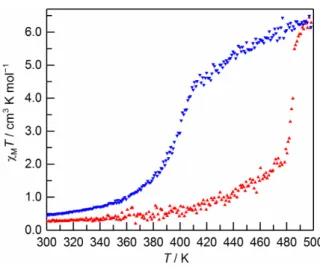
showed an unsymmetrical hysteretic SCO above room temperature. The initial χMT value is 0.3 cm3·K·mol−1 at 300 K, which is consistent with the theoretical value for a LS–LS FeII system. On raising the temperature from 300 K, the χMT value increases gradually to reach ca. 2.1 cm3·K·mol−1 at 478 K, and then increases abruptly to reach ca. 6.3 cm3·K·mol−1 at 498 K, which is consistent with the theoretical value for a high-spin–high-spin (HS–HS) FeII system (χMT = 6.0 cm3·K·mol−1). On lowering the temperature from 498 K, the χMT value decreases gradually, reaching ca. 4.3 cm3·K·mol−1 at 409 K, and then decreases abruptly to reach ca. 1.5·cm3·K mol−1 at 379 K. Then, the χMT value further decreases gradually to reach ca. 0.5 cm3·K·mol−1 at 300 K. The critical temperatures of the warming (Tc
↑
) and cooling (Tc↓
) modes in the abrupt spin transition area are 485 and 401 K, respectively, indicating the occurrence of ΔT = 84 K, which is the widest thermal hysteresis loop in the dinuclear system reported so far [26–29,33,34,48]. It is also noteworthy that the Tc↑
of 1·AsF6 is considerablyhigher than that of the related ethylene-bridged helicate [FeII2(L2C2)2](PF6)4·5H2O·MeCN (Tc
↑
= 437 K) [34] by about 48 K.Figure 4.(a) Intermolecular interactions of 1·AsF6at 120 K. Adjacent complex-cations [FeII2(L2C3)2]4+
with the same chirality are connected by intermolecular π–π interactions (green dotted line) to form the homochiral 1D chain structure along the b-axis. Symmetry operations: (i)−x, y, 3/2−z; (ii) x, −1 + y, z; (iii)−x,−1 + y, 3/2−z; (b) Packing diagram of 1·AsF6at 120 K viewed along the ac plane. ∆–∆-[FeII
2(L2C3)2]4+andΛ–Λ-[FeII2(L2C3)2]4+enantiomers are represented by green and red colors,
respectively. Cationic 1D chains with the opposite chirality running along the b-axis exist in a crystal lattice to give a heterochiral crystal. Each cationic chain is separated by the space-occupying two disordered AsF6−ions (blue and orange; only the major component is indicated) and solvent molecules
such as 3H2O and one MeCN molecule (not indicated), which could not be sensibly modeled due to
the strong disorder and regions including these solvent molecules being treated with the PLATON SQUEEZE program. H atoms have been omitted for clarity.
2.3. Magnetic Property of 1·AsF6
The magnetic susceptibilities for polycrystalline samples of 1·AsF6were measured upon heating from 300 to 498 K and subsequent cooling to 300 K at a sweep rate of 0.5 K·min−1under an applied magnetic field of 1 T using a superconducting quantum interference device (SQUID) magnetometer with a special heating setup. The χMT vs. T plots are shown in Figure5, where χMis the molar
magnetic susceptibility per Fe and T is the absolute temperature. As shown in Figure5, 1·AsF6showed an unsymmetrical hysteretic SCO above room temperature. The initial χMT value is 0.3 cm3·K·mol−1at
300 K, which is consistent with the theoretical value for a LS–LS FeIIsystem. On raising the temperature from 300 K, the χMT value increases gradually to reach ca. 2.1 cm3·K·mol−1at 478 K, and then increases
abruptly to reach ca. 6.3 cm3·K·mol−1at 498 K, which is consistent with the theoretical value for a
high-spin–high-spin (HS–HS) FeIIsystem (χMT = 6.0 cm3·K·mol−1). On lowering the temperature
from 498 K, the χMT value decreases gradually, reaching ca. 4.3 cm3·K·mol−1at 409 K, and then
decreases abruptly to reach ca. 1.5·cm3·K mol−1 at 379 K. Then, the χMT value further decreases
gradually to reach ca. 0.5 cm3·K·mol−1at 300 K. The critical temperatures of the warming (Tc↑) and
cooling (Tc↓) modes in the abrupt spin transition area are 485 and 401 K, respectively, indicating
the occurrence of∆T = 84 K, which is the widest thermal hysteresis loop in the dinuclear system reported so far [26–29,33,34,48]. It is also noteworthy that the Tc↑of 1·AsF6is considerably higher than that of the related ethylene-bridged helicate [FeII2(L2C2)2](PF6)4·5H2O·MeCN (Tc↑= 437 K) [34]
Inorganics 2017, 5, 49 8 of 15
Inorganics 2017, 5, 49 8 of 15
Figure 5. The magnetic behavior of 1·AsF6 in the form of the χMTvs. T plots. 1·AsF6 was warmed from
300 to 498 K (filled triangles; red), and then cooled from 498 to 300 K (filled inverted triangles; blue) at a sweep rate of 0.5 K·min−1.
2.4. Reproducibility of Hysteretic SCO of 1·AsF6
To reveal the desolvation effect and reproducibility of hysteretic SCO of 1·AsF6,
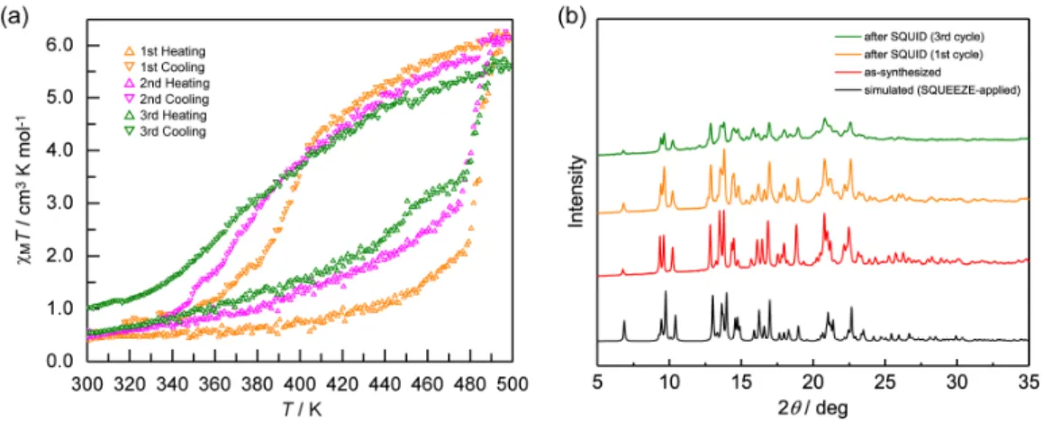
temperature-dependent magnetic susceptibilities were also measured over three consecutive cycles. As shown in Figure 6a, although 1·AsF6 shows a more gradual spin transition in the second cycle than in the first
cycle, probably due to the in situ solvent liberation in the first heating, an asymmetric wide hysteresis loop is then still observed. Further thermal cycling (third cycle) causes the spin transition pattern to have a much more gradual and partial SCO fashion, which does not occur in the related ethylene-bridged helicate [FeII2(L2C2)2](PF6)4·5H2O·MeCN [34]. Comparing powder X-ray diffraction (PXRD) patterns of as-synthesized 1·AsF6 and after first thermal cycle of SQUID measurement confirms that
there is no structural phase transition associated with the desolvation, because the peak patterns of the sample are not changed, although the peak positions increase slightly possibly due to the removal of solvent molecules and concomitant lattice contraction (Figure 6b). However, an intensity decrease and peak broadening are observed after three thermal cycles of SQUID measurement, presumably due to a loss of crystallinity and/or partial decomposition.
These properties were also confirmed by differential scanning calorimetry (DSC) experiments of four consecutive thermal cycles at a sweep rate of 10 K·min−1, in the temperature range of 222–502 K (Figure 7). As shown in Figure 7, from the first cycle in heating and following cooling modes, a thermal hysteresis of ca. 85 K was detected (Tmax↑ and Tmax↓ are 498 and 413 K, respectively) and the hysteresis width is consistent with the ΔT value observed by the magnetic measurement. As the thermal cycling is repeated, the shape of DSC peaks becomes broader and smaller, and the position of the peak shifts to a lower temperature in both the heating and cooling modes of the second cycle due to the solvent loss in the first heating (Tmax↑ and Tmax
↓
are 487 and 401 K, respectively, in the second cycle). However, further thermal cycling from the second cycle induces the peak shift to the opposite directions, in which the temperature variation in cooling modes is larger than that in heating modes (Tmax↑
and Tmax↓
are 490 and 392 K, respectively, in the third cycle, and 492 and 384 K, respectively, in the fourth cycle). In addition, peak broadening and reduction also continue in the third and fourth cycles. As a whole, thermal hysteresis of 1·AsF6 is retained for at least four cycles,although the spin transition becomes gradual and incomplete with shifting the transition temperature due to the solvent liberation in the first cycle and a loss of crystallinity and/or partial decomposition upon further thermal cycling.
Figure 5.The magnetic behavior of 1·AsF6in the form of the χMT vs. T plots. 1·AsF6was warmed from 300 to 498 K (filled triangles; red), and then cooled from 498 to 300 K (filled inverted triangles; blue) at a sweep rate of 0.5 K·min−1.
2.4. Reproducibility of Hysteretic SCO of 1·AsF6
To reveal the desolvation effect and reproducibility of hysteretic SCO of 1·AsF6, temperature-dependent magnetic susceptibilities were also measured over three consecutive cycles. As shown in Figure6a, although 1·AsF6shows a more gradual spin transition in the second cycle than in the first cycle, probably due to the in situ solvent liberation in the first heating, an asymmetric wide hysteresis loop is then still observed. Further thermal cycling (third cycle) causes the spin transition pattern to have a much more gradual and partial SCO fashion, which does not occur in the related ethylene-bridged helicate [FeII
2(L2C2)2](PF6)4·5H2O·MeCN [34]. Comparing powder X-ray diffraction
(PXRD) patterns of as-synthesized 1·AsF6and after first thermal cycle of SQUID measurement confirms that there is no structural phase transition associated with the desolvation, because the peak patterns of the sample are not changed, although the peak positions increase slightly possibly due to the removal of solvent molecules and concomitant lattice contraction (Figure6b). However, an intensity decrease and peak broadening are observed after three thermal cycles of SQUID measurement, presumably due to a loss of crystallinity and/or partial decomposition.
These properties were also confirmed by differential scanning calorimetry (DSC) experiments of four consecutive thermal cycles at a sweep rate of 10 K·min−1, in the temperature range of 222–502 K (Figure7). As shown in Figure7, from the first cycle in heating and following cooling modes, a thermal hysteresis of ca. 85 K was detected (Tmax↑and Tmax↓are 498 and 413 K, respectively) and the hysteresis
width is consistent with the∆T value observed by the magnetic measurement. As the thermal cycling is repeated, the shape of DSC peaks becomes broader and smaller, and the position of the peak shifts to a lower temperature in both the heating and cooling modes of the second cycle due to the solvent loss in the first heating (Tmax↑and Tmax↓are 487 and 401 K, respectively, in the second cycle). However,
further thermal cycling from the second cycle induces the peak shift to the opposite directions, in which the temperature variation in cooling modes is larger than that in heating modes (Tmax↑and Tmax↓are
490 and 392 K, respectively, in the third cycle, and 492 and 384 K, respectively, in the fourth cycle). In addition, peak broadening and reduction also continue in the third and fourth cycles. As a whole, thermal hysteresis of 1·AsF6is retained for at least four cycles, although the spin transition becomes gradual and incomplete with shifting the transition temperature due to the solvent liberation in the first cycle and a loss of crystallinity and/or partial decomposition upon further thermal cycling.
Inorganics 2017, 5, 49 9 of 15
Inorganics 2017, 5, 49 9 of 15
Figure 6. (a) Temperature dependence of the χMT product of 1·AsF6 in the heating (empty triangles)
and cooling (empty inverted triangles) modes over three successive thermal cycles at a sweep rate of 0.5 K·min−1. Orange: first cycle; magenta: second cycle; olive: third cycle; (b) Powder X-ray diffraction (PXRD) patterns of 1·AsF6 at RT in different states. Black: simulated from the SQUEEZE-applied single
crystal X-ray data at 120 K; red: as-synthesized 1·AsF6; orange: 1·AsF6 after the first thermal cycle of
superconducting quantum interference device (SQUID) measurement; olive: 1·AsF6 after a third
thermal cycle of SQUID measurement.
Figure 7. Differential scanning calorimetry (DSC) curves of 1·AsF6 in the heating mode (a) and cooling
mode (b) recorded over four successive thermal cycles at a sweep rate of 10 K·min−1, in the temperature range of 222–502 K. Orange: first cycle; magenta: second cycle; olive: third cycle; navy blue: fourth cycle.
3. Materials and Methods
3.1. Synthesis of FeII Complexes
3.1.1. General
All reagents and solvents were purchased from commercial sources and used for the syntheses without further purification. The 1-Phenyl-1H-1,2,3-triazole-4-carbaldehyde was prepared according to methods in the literature [47]. The synthetic procedure of 1,1′-(propane-1,3-diyl)bis(1H-1,2,3-triazole-4-carbaldehyde) (4) was constructed by modifying the reported procedures [34]. All the synthetic procedures were carried out in air.
3.1.2. Preparation of 1,1′-(propane-1,3-diyl)bis(1H-1,2,3-triazole-4-carbaldehyde) (4)
To a warm pale yellow solution of 1-phenyl-1H-1,2,3-triazole-4-carbaldehyde (3.464 g, 20 mmol) in MeOH (30 mL), 1,3-diaminopropane (0.741 g, 10 mmol) in MeOH (1 mL) was added. The resulting Figure 6.(a) Temperature dependence of the χMT product of 1·AsF6in the heating (empty triangles) and cooling (empty inverted triangles) modes over three successive thermal cycles at a sweep rate of 0.5 K·min−1. Orange: first cycle; magenta: second cycle; olive: third cycle; (b) Powder X-ray diffraction (PXRD) patterns of 1·AsF6at RT in different states. Black: simulated from the SQUEEZE-applied single crystal X-ray data at 120 K; red: as-synthesized 1·AsF6; orange: 1·AsF6after the first thermal cycle of superconducting quantum interference device (SQUID) measurement; olive: 1·AsF6after a third thermal cycle of SQUID measurement.
Inorganics 2017, 5, 49 9 of 15
Figure 6. (a) Temperature dependence of the χMT product of 1·AsF6 in the heating (empty triangles)
and cooling (empty inverted triangles) modes over three successive thermal cycles at a sweep rate of 0.5 K·min−1. Orange: first cycle; magenta: second cycle; olive: third cycle; (b) Powder X-ray diffraction (PXRD) patterns of 1·AsF6 at RT in different states. Black: simulated from the SQUEEZE-applied single
crystal X-ray data at 120 K; red: as-synthesized 1·AsF6; orange: 1·AsF6 after the first thermal cycle of
superconducting quantum interference device (SQUID) measurement; olive: 1·AsF6 after a third
thermal cycle of SQUID measurement.
Figure 7. Differential scanning calorimetry (DSC) curves of 1·AsF6 in the heating mode (a) and cooling
mode (b) recorded over four successive thermal cycles at a sweep rate of 10 K·min−1, in the temperature range of 222–502 K. Orange: first cycle; magenta: second cycle; olive: third cycle; navy blue: fourth cycle.
3. Materials and Methods
3.1. Synthesis of FeII Complexes
3.1.1. General
All reagents and solvents were purchased from commercial sources and used for the syntheses without further purification. The 1-Phenyl-1H-1,2,3-triazole-4-carbaldehyde was prepared according to methods in the literature [47]. The synthetic procedure of 1,1′-(propane-1,3-diyl)bis(1H-1,2,3-triazole-4-carbaldehyde) (4) was constructed by modifying the reported procedures [34]. All the synthetic procedures were carried out in air.
3.1.2. Preparation of 1,1′-(propane-1,3-diyl)bis(1H-1,2,3-triazole-4-carbaldehyde) (4)
To a warm pale yellow solution of 1-phenyl-1H-1,2,3-triazole-4-carbaldehyde (3.464 g, 20 mmol) in MeOH (30 mL), 1,3-diaminopropane (0.741 g, 10 mmol) in MeOH (1 mL) was added. The resulting Figure 7. Differential scanning calorimetry (DSC) curves of 1·AsF6 in the heating mode (a) and cooling mode (b) recorded over four successive thermal cycles at a sweep rate of 10 K·min−1, in the temperature range of 222–502 K. Orange: first cycle; magenta: second cycle; olive: third cycle; navy blue: fourth cycle.
3. Materials and Methods 3.1. Synthesis of FeIIComplexes
3.1.1. General
All reagents and solvents were purchased from commercial sources and used for the syntheses without further purification. The 1-Phenyl-1H-1,2,3-triazole-4-carbaldehyde was prepared according to methods in the literature [47]. The synthetic procedure of 1,10 -(propane-1,3-diyl)bis(1H-1,2,3-triazole-4-carbaldehyde) (4) was constructed by modifying the reported procedures [34]. All the synthetic procedures were carried out in air.
3.1.2. Preparation of 1,10-(propane-1,3-diyl)bis(1H-1,2,3-triazole-4-carbaldehyde) (4)
To a warm pale yellow solution of 1-phenyl-1H-1,2,3-triazole-4-carbaldehyde (3.464 g, 20 mmol) in MeOH (30 mL), 1,3-diaminopropane (0.741 g, 10 mmol) in MeOH (1 mL) was added. The resulting
Inorganics 2017, 5, 49 10 of 15
solution was stirred for 1 h at 60◦C, and was then cooled in a fridge. The precipitated white powders were collected by suction filtration, washed with Et2O (10 mL×3), and dried in vacuo. White solid of
N,N’-(propane-1,3-diyl)bis[1-(1-phenyl-1H-1,2,3-triazol-4-yl)methanimine] (2). Yield: 3.005 g (78%). Mp 127–128◦C (from MeOH), 1H NMR (600 MHz; DMSO-d6; TMS): δ 9.24 (s, 2H), 8.55 (s, 2H), 8.00–7.98 (m, 4H), 7.63–7.60 (m, 4H), 7.53–7.51 (m, 2H), 3.72 (t, J = 6.5 Hz, 4H), 2.05–2.00 (m, 2H). Anal. Calcd. for 2·0.2H2O = C21H20.4N8O0.2: C, 65.00; H, 5.30; N, 28.88%. Found: C, 65.05; H, 5.19; N, 29.01%.
A suspension of 2 (2.898 g, 7.5 mmol) in 1-PrOH (25 mL) was heated at 80◦C for 16 h, turning into a pale yellow solution, and the resulting solution was then cooled in a fridge. The precipitated white powders were collected by suction filtration, washed with Et2O (10 mL), and dried in vacuo. White
solid of 1,10-[propane-1,3-diylbis(1H-1,2,3-triazole-1,4-diyl)]bis(N-phenylmethanimine) (3). Yield: 2.260 g (78%). m.p. 192–193◦C (from 1-PrOH),1H NMR (600 MHz; DMSO-d6; TMS): δ 8.78 (s, 2H), 8.67 (s, 2H), 7.43–7.41 (m, 4H), 7.28–7.25 (m, 6H), 4.52 (t, J = 6.9 Hz, 4H), 2.59–2.55 (m, 2H). Anal. Calcd. for 3·0.35H2O = C21H20.7N8O0.35: C, 64.55; H, 5.34; N, 28.68%. Found: C, 64.15; H, 5.20; N, 29.06%.
Hydrochloric acid (36%) (1.671 g, 16.5 mmol) in H2O (28 mL) was added to white powders of
3(2.125 g, 5.5 mmol), and the mixture was stirred for 2 h at room temperature. After turning the mixture into a yellowish-white suspension, the residual white solids of 4 were collected by suction filtration, washed with H2O (10 mL) and then Et2O (10 mL), and dried in vacuo. Yield: 1.103 g (86%).
m.p. 124–125◦C (from H2O),1H NMR (600 MHz; DMSO-d6; TMS): δ 10.03 (s, 2H), 8.89 (s, 2H), 4.52 (t,
J = 6.9 Hz, 4H), 2.55–2.51 (m, 2H). Anal. Calcd. for C9H10N6O2: C, 46.15; H, 4.30; N, 35.88%. Found: C,
45.87; H, 4.19; N, 35.56%.
3.1.3. Synthesis of the Bis-Tridentate Ligand L2C3= 1,10 -[propane-1,3-diylbis(1H-1,2,3-triazole-1,4-diyl)]bis{N-[2-(pyridin-2-yl)ethyl]methanimine}
The ligand L2C3was prepared by mixing 4 and 2-(2-aminoethyl)pyridine with 1:2 molar ratio in MeOH. The ligand solution thus prepared was used for the synthesis of FeIIcomplexes without further purification and isolation.
3.1.4. Preparation of [FeII
2(L2C3)2](PF6)4·1.5H2O·1.5MeCN (1·PF6)
2-(2-Aminoethyl)pyridine (0.122 g, 1 mmol) in MeOH (1 mL) was added to a solution of 4 (0.118 g, 0.5 mmol) in MeOH (20 mL), and the resulting mixture was stirred at ambient temperature for 1 h. A pale yellow solution of the ligand (0.5 mmol) thus prepared was treated first with a solution of FeIICl2·4H2O (0.099 g, 0.5 mmol) in MeOH (1 mL), turning into a dark orange-red solution, and
then with KPF6(0.184 g, 1 mmol) in 4 mL of a mixed solution of MeOH and H2O (1/1 by volume).
The resulting mixture was stirred at ambient temperature for 1 h, during which time the precipitated orange-brown crude product was collected by suction filtration. The collected precipitate was dissolved in MeCN (4 mL) and then filtered. Dark-red block crystals were obtained by slow diffusion of MeOH (8 mL) into the filtrate (liquid–liquid diffusion) for a day. Yield: 0.154 g (37%). Anal. Calcd. for [FeII2(L2C3)2](PF6)4·1.5MeCN·1.5MeOH = C49H59.5N21.5O1.5P4F24Fe2: C, 35.34; H, 3.60; N, 18.08%.
Found: C, 35.48; H, 3.37; N, 18.06%. IR (KBr): νC=N1609 cm−1, νP–F(PF6−) 844 cm−1.
3.1.5. Preparation of [FeII2(L2C3)2](AsF6)4·3H2O·MeCN (1·AsF6)
2-(2-Aminoethyl)pyridine (0.122 g, 1 mmol) in MeOH (2 mL) was added to a solution of 4 (0.118 g, 0.5 mmol) in MeOH (20 mL), and the resulting mixture was stirred at ambient temperature for 1 h. A pale yellow solution of the ligand (0.5 mmol) thus prepared was treated first with a solution of FeIICl2·4H2O (0.099 g, 0.5 mmol) in MeOH (2 mL), turning into a dark orange-red solution, and
then with KAsF6(0.228 g, 1 mmol) in 4 mL of a mixed solution of MeOH and H2O (1/1 by volume).
The resulting mixture was stirred at ambient temperature for 30 min, during which time the precipitated red-orange crude product was collected by suction filtration. The collected precipitate was dissolved in MeCN (3 mL) and then filtered. Dark-red block crystals were obtained by slow diffusion of MeOH (7 mL) into the filtrate (liquid–liquid diffusion) for a day. Yield: 0.155 g (33%). Anal. Calcd. for
Inorganics 2017, 5, 49 11 of 15
[FeII2(L2C3)2](AsF6)4·3H2O·MeCN = C48H61N21O3As4F24Fe2: C, 31.21; H, 3.33; N, 15.92%. Found: C,
31.46; H, 3.04; N, 16.25%. IR (KBr): νC=N1609 cm−1, νAs–F(AsF6−) 701 cm−1.
3.2. Physical Measurements
Elemental C, H, and N analyses were performed on a J-Science Lab (Kyoto, Japan) MICRO CORDER JM-10. IR spectra were recorded at room temperature using a JASCO (Tokyo, Japan) FT/IR 460Plus spectrophotometer or a PerkinElmer (Waltham, MA, USA) Spectrum100 FT-IR spectrometer with the samples prepared as KBr disks. 1H NMR spectra were recorded on a JEOL (Tokyo, Japan) ECA-600 spectrometer. The melting point was measured through a Yanaco (Kyoto, Japan) MP-S3 micro melting point meter and was uncorrected. Thermogravimetric data were collected on a TG/DTA6300 (SII Nano Technology Inc., Chiba, Japan) instrument in the temperature range of 30–318◦C (303–591 K) for 1·PF6and 30–381◦C (303–654 K) for 1·AsF6at a rate of 10 K·min−1under a nitrogen atmosphere. DSC measurements for 1·AsF6were performed with a DSC6200 (SII Nano Technology Inc., Chiba, Japan) over the temperature range of 222–502 K, at a sweep rate of 10 K·min−1 under a nitrogen atmosphere using aluminum hermetic pans with an empty pan as reference. Magnetic susceptibilities of 1·AsF6were measured in the temperature range of 300–498 K at a sweep rate of 0.5 K·min−1under an applied magnetic field of 1 T using a Quantum Design (San Diego, CA, USA) MPMS-7 SQUID magnetometer with a special heating setup of a sample space oven option. The sample was wrapped in an aluminum foil and was then inserted into a quartz glass tube with a small amount of glass wool filler. Corrections for diamagnetism of the sample were made using Pascal’s constants [55] and a background correction for the sample holder was applied. PXRD patterns were recorded at room temperature on a portion of polycrystalline powders placed on a non-reflecting silicon plate, using a Rigaku (Tokyo, Japan) MiniFlex600 diffractometer with Cu Kα radiation (λ = 1.5418 Å) operated at 0.4 kW power (40 kV, 10 mA). Images of grinding samples were recorded under an optical microscope SMZ800N (Nikon, Tokyo, Japan). The sample temperature was controlled by using a Linkam (Tadworth, UK) THMS 600 heating and freezing stage.
3.3. Crystallographic Data Collection and Structure Analyses
X-ray diffraction data of 1·PF6and 1·AsF6were collected by a Rigaku (Tokyo, Japan) AFC7R Mercury CCD diffractometer using graphite monochromated Mo Kα radiation (λ = 0.71075 Å) operated at 5 kW power (50 kV, 100 mA). A single crystal was mounted on a MiTeGen (Ithaca, NY, USA) MicroMount (200 µm) with liquid paraffin, then rapidly frozen in a nitrogen-gas stream at 120 K to avoid the loss of crystal solvents and the diffraction data were collected at 120 K. The temperature of the crystal was maintained by means of a Rigaku cooling device to within an accuracy of±2 K. The data were corrected for Lorentz, polarization, and absorption effects. The structures were solved by the direct method [56] and refined on F2data using the full-matrix least-squares algorithm using SHELXL-2014 [57]. Non-hydrogen atoms were refined anisotropically. The structure of 1·PF6contains two disordered PF6−ions. Both PF6− ions were disordered over two positions and the occupancy
factors for the possible two positions of F atoms (F1A–F6A:F1B–F6B = 0.518(15):0.482(15) for P(1)F6−
and F7A–F12A:F7B–F12B = 0.635(16):0.365(16) for P(2)F6−) were refined using the tools available
from the SHELXL-2014 program package. In the refinement of these disordered PF6−ions, the SADI
command for all P–F bonds and F···F distances and the RIGU command for all P and F atoms were also applied. In addition, diffraction quality of the crystal of 1·PF6was not sufficient to model highly disordered solvent molecules. The PLATON SQUEEZE program (290617, A.L. Spek, Utrecht, NL-UT, The Netherlands) [51] was used to treat regions with disordered solvent molecules which could not be sensibly modelled in terms of atomic sites. Available void volume was 892 Å3. Then, 196 electrons
per unit cell were located and these were assigned to 1.5 H2O and 1.5 MeCN molecules per complex
(196/4 = 49 e per complex; 1.5 H2O (15) + 1.5 MeCN (33) = 48 electrons). In the same way, the structure
of 1·AsF6contains two disordered AsF6−ions and severely disordered solvent molecules. In 1·AsF6, both AsF6− ions were disordered over three positions and the occupancy factors for the possible
Inorganics 2017, 5, 49 12 of 15
three positions of F atoms (F1A–F6A:F1B–F6B:F1C–F6C = 0.434(3):0.291(3):0.275(3) for As(1)F6−and
F7A–F12A:F7B–F12B:F7C–F12C = 0.513(3):0.219(3):0.268(3) for As(2)F6−) were refined using the SUMP
command to keep the sum of three occupancy factors of each ion as 1.0. In the refinement of these disordered AsF6−ions, the SADI command for all As–F bonds and F···F distances, the RIGU command
for all As and F atoms, and the EADP command for F5A, F5B, F5C and F6A, F6B, F6C were also applied. The PLATON SQUEEZE program was also used to treat regions with disordered solvent molecules in the structure of 1·AsF6. Available void volume was 926 Å3, and 220 electrons per unit cell were located. These were assigned to 3 H2O and 1 MeCN molecules per complex (220/4 = 55 e
per complex; 3 H2O (30) + 1 MeCN (22) = 52 electrons). All H atoms were placed in geometrically
calculated positions, with the distances of C–H = 0.95 (aromatic) and 0.99 (CH2) Å, and refined as
riding atoms, with Uiso(H) = 1.2 Ueq(C). All calculations were performed by using the Yadokari-XG software package [58]. The CShMs of the FeIIcenters relative to the ideal octahedron were calculated by SHAPE 2.1 [53]. The octahedral volumes of the FeIIcenters were calculated by OLEX2 [59]. CCDC 1544125–1544126 contains the supplementary crystallographic data for this paper. These data can be obtained free of charge viahttp://www.ccdc.cam.ac.uk/conts/retrieving.htmlor from the CCDC (12 Union Road, Cambridge CB2 1EZ, UK; Fax: +44 1223 336033; E-mail: [email protected]).
4. Conclusions
In conclusion, the present work reveals the effects of the extension of the bridging alkyl chain length (from C2 to C3) on the structure and SCO properties of the iron(II) dinuclear double helicate [FeII2(L2R)2](X)4·solvent, with the concomitant effects of the coexistent counter anions and lattice
solvents. While propylene-bridged dinuclear complexes [FeII2(L2C3)2](PF6)4·1.5H2O·1.5MeCN (1·PF6) and [FeII2(L2C3)2](AsF6)4·3H2O·MeCN (1·AsF6) are isostructural, the thermal stability of 1·PF6is lower than that of 1·AsF6in a high-temperature SCO region. 1·AsF6shows an unsymmetrical hysteretic SCO between the LS–LS and HS–HS states at above room temperature with∆T = 84 K (Tc↑= 485 K
and Tc↓= 401 K in the abrupt spin transition area) in the first thermal cycle, which is the widest
hysteresis loop in the dinuclear system reported so far. In addition, the Tc↑of 1·AsF6is considerably higher than that of the related ethylene-bridged compound, by about 48 K. This high-temperature wide thermal hysteresis may be related to the 1D assembly structure composed of inter-helicate π–π interactions, which is obviously different from the 2D assembly of the related ethylene-bridged compound. Although the desolvation of 1·AsF6 does not directly affect the assembly structure, consecutive thermal cycles cause a loss of crystallinity and/or partial decomposition, and subsequent modification of the hysteretic SCO loop to a more gradual and partial fashion. As a consequence, the extension of the bridging alkyl chain length (from C2 to C3) and subsequent elongation of the dinuclear cation makes the 2D supramolecular assembly one-dimensional, and enhances the width of the hysteresis loop. Simultaneous raising of the Tc↑may also cause the overlapping between spin
transition temperature and decomposition temperature, which is related to the thermal durability upon SCO.
Supplementary Materials:The following are available online atwww.mdpi.com/2304-6740/5/3/49/s1. Cif and cif-checked files.
Acknowledgments: This work was partly funded by the Gifu University for the promotion of the research of young scientists. Part of this work was conducted at the Institute for Molecular Science, supported by the Nanotechnology Platform Program (Molecule and Material Synthesis) of the Ministry of Education, Culture, Sports, Science and Technology (MEXT), Japan. The authors would like to thank Professor O. Sakurada (Gifu University, Japan) for his assistance in collecting PXRD data.
Author Contributions: The experimental work was performed mainly by Shiori Hora with assistance from Hiroaki Hagiwara. Hiroaki Hagiwara supervised the experiments. Both authors analyzed the data and contributed to the preparation of the manuscript.