Acta Med. Nagasaki 43: 1-11
-Review Article -
High-Resolution CT Evaluation of Ground-Glass Opacity in Diffuse Lung Disease
Tetsuji YAMAGUCHI, Kazuto ASHIZAWA, Kenji NAGAOKI, Kuniaki HAYASHI
Department of Radiology, Nagasaki University School of Medicine
Ground-glass opacity of the lung is often demonstrated on high-resolution CT (HRCT) in various diffuse lung diseases.
Ground-glass opacity generally results from minimal thickening of the alveolar interstitium or partial filling in the alveolar spaces. We classify diffuse infiltrative lung diseases into two clinical categories, namely, noninfectious and infectious lung disease. In noninfectious lung disease, al- though ground-glass opacity can be observed as an isolated finding, it is commonly observed to be combined with other findings such as centrilobular micronodules, interlobular septal thickening, and cystic air-spaces. These associated findings may be important for the differential diagnosis.
Ground-glass opacity may also be seen as a consequence of increased capillary blood volume in redistribution of blood flow. This condition of hemodynamic origin is observed in
chronic obstructive pulmonary disease, airway disease, and vascular lung disease. Although ground-glass opacity is a nonspecific finding, it can suggest a specific diagnosis in certain clinical circumstances or indicate a potentially
treatable disease. Therefore, accurate recognition and differential diagnosis of ground-glass opacity are important.
ble, if the underlying disorder is treated early. Therefore, accurate diagnosis of underlying disorders in their early stages is important. Because ground-glass opacity per se is a nonspecific finding on HRCT, associated findings may be important for the differential diagnosis. This article deals with various causes of ground-glass opacity and provides a guide to the differential diagnosis of the diseases that can produce this finding. We will also describe some important pitfalls in evaluating ground-glass opacity on HRCT.
Definition of Ground-Glass Opacity
Ground-glass opacity can be defined by four main criteria : (1) areas of hazy and amorphous increased lung density ; (2) no obscuration of the underlying vascular markings and bronchial walls ; (3) identification on HRCT sections ; and (4) photographed with wide window settings [3].
Key Words : lung, high-resolution CT,
diffuse lung disease, ground-glass opacity
Introduction
'Ground -glass opacity' is defined as nonspecific in- creased density of the pulmonary parenchyma that is not associated with obscuration of the underlying pulmonary vessels on high-resolution CT (HRCT) [1-4]. This appear- ance is caused by a change in the relative proportions of air in the alveolar space and thickening of the alveolar interstitium that is out of the resolution capabilities of HRCT. In contrast to ground-glass opacity, increased lung density with obscuration of the underlying pulmonary vessels is referred to as `consolidation' [1].
Generally, ground-glass opacity is potentially reversi- Address Correspondence :
Dr. Tetsuji Yamaguchi, Department of Radiology, Nagasaki University School of Medicine, 1-7-1 Sakamoto, Nagasaki 852-8501, Japan
Causes of Ground-Glass Opacity
Lung density results from the relative proportions of the blood, air, extravascular water, and pulmonary tissue [3].
Generally, increased lung density histologically reflects
thickening of the alveolar walls and septal interstitium or
the filling of fluid, cells, or amorphous material in the
alveolar spaces. Increased lung density can be classified
into 'ground-glass opacity' and `consolidation'. Ground-
glass opacity reflects the presence of minimal thickening
of the alveolar interstitium or the presence of fluid, cells,
or amorphous material partially filling the alveolar spaces
(Fig. 1) [1-41. If there are more materials in the alveolar
spaces or more thickening of the interstitium and less air
in the alveolar space, the condition is increased lung
density of higher degree and referred to as `consolidation'
on HRCT. Therefore, ground-glass opacity and consolida-
tion are combined to varying degrees in a large number of
lung diseases. The HRCT findings in some diffuse lung
diseases are predominated by ground-glass opacity, and in
others by consolidation (Table 1). In this article, we
Normal alveoli A. Thickening of the alveolar interstitium
B. Partial filling
in the alveolar spaces
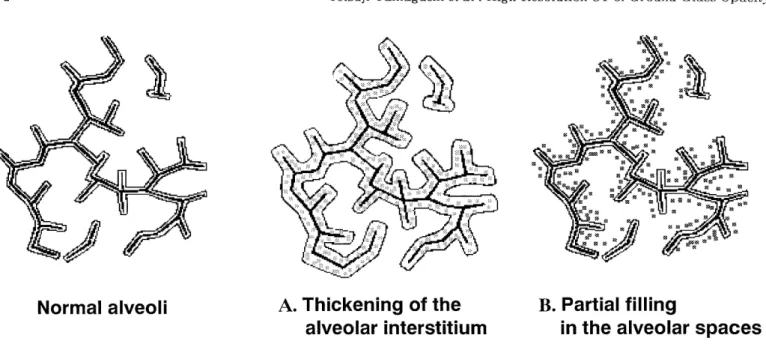
Fig. 1. Anatomic diagram of ground-glass opacity (modified from figure of reference 2)
A. Thickening of the alveolar interstitium occurs for cellular infiltration or fibrosis, and decreases the amount of airspaces.
B. Alveolar spaces can be partially filled with cellular debris, fluid, or amorphous material.
In the states of A or B, these events are not individually resolvable by HRCT. The change in proportions between air in the alveolar spaces and thickening of the alveolar interstitium leads to an increase of lung density on HRCT. It is expressed as a
"ground -glass opacity" .
Table 1. Classification by predominant findings of diffuse lung diseases with ground-glass opacity and/or consolida- tion
1. Ground-glass opacity dominant Hypersensitivity pneumonitis
Respiratory bronchiolitis (-interstitial lung disease) Alveolar proteinosis
Usual interstitial pneumonia (UIP) Desquamative interstitial pneumonia
Nonclassifable interstitial pneumonia
Lymphocytic interstitial pneumonia
Sarcoidosis
Atypical pneumonia
(e.g. Pneumocystis carinii pneumonia) Drug induced pneumonitis
Redistribution of pulmonary blood flow (e.g. chronic thromboembolic disease) 2. Consolidation dominant
Bronchiolitis obliterans with organizing pneumonia Eosinophilic pneumonia
Bacterial pneumonia
Radiation pneumonitis
3. Ground-glass opacity and consolidation
Acute interstitial pneumonia (Diffuse alveolar damage)
Accelerated deterioration of UIP (Diffuse alveolar damage)
Bronchioloalveolar carcinoma
Pulmonary edema
Pulmonary hemorrhage
Metastatic calcification
mainly discuss the diseases in which ground-glass opacity is more predominant than consolidation on HRCT. We divide diffuse infiltrative lung diseases into two clinical categories, namely, noninfectious and infectious lung disease.
Increased lung density may also result from an increase in pulmonary capillary blood volume (hyperperfusion). It causes diffuse or focal ground-glass opacity on HRCT.
Because the change of lung density due to increased pulmo- nary blood volume is commonly minimal, however, it may easily be overlooked on HRCT. Regional alterations in pulmonary blood volume are common hemodynamic factors shared by various clinical situations [3, 5-81.
Ground-glass opacity of hemodynamic origin is also discussed in this article.
Ground-Glass Opacity in Noninfectious Lung Disease
Ground-glass opacity has been known in various noninfectious lung diseases, including alveolar proteinosis, usual interstitial pneumonia (UIP), desquamative inter- stitial pneumonia (DIP), lymphocytic interstitial pneumo- nia (LIP), bronchiolitis obliterans with organizing pneumonia (BOOP), eosinophilic pneumonia, hypersensi- tivity pneumonitis (HP), respiratory bronchiolitis, sar- coidosis, radiation pneumonitis/fibrosis, metastatic calcification, and alveolar microlithiasis.
A large number of noninfectious lung diseases begin with
an accumulation of inflammatory cells in the alveolar septa and air-spaces and often culminate in severe fibrosis of the interstitium. Ground-glass opacity often reflects mild parenchymal alterations and may provide informa- tion about disease activity and prognosis.
The best known significance of ground-glass opacity is to reflect an active inflammatory process, representing the acute phase of lung injury referred to as alveolitis. In acute phase, the main pathologic features are observed to involve both the alveolar interstitium and the air-spaces.
Pathologic specimen shows thickening of the alveolar interstitium and incomplete filling of the alveolar spaces with inflammatory cells, cellular debris, and edema [3].
Ground-glass opacity on HRCT may sometimes reflect isolated inflammation of alveolar interstitium without air-space filling. In this phase, corticosteroid treatment is useful for a large number of these diseases.
Ground-glass opacity may also result from chronic changes that occur following acute lung injury in the absence of complete healing corresponding to lung fibrosis with or without lung distortion. The fibrotic reaction associated with residual alveolar infiltrates and debris leads to reduction of the amount of air in the alveolar spaces. Healing by fibrosis leads to formation of dense areas of collagen interspersed with parenchymal destruc- tion. The resultant cystic and destructive changes consti- tute the hallmarks of late-phase lung disease (pulmonary fibrosis). When cystic changes (honeycombing) are within
the resolution capabilities of HRCT, a classical `macro- scopic' honeycombing appears. When cystic changes are too small to be detected by HRCT, volume averaging of tiny cysts that characterize the microcystic (`micro- scopic') honeycombing may lead to ground-glass opacity.
Bronchi and bronchioles in the regions of lung distortion appear dilated and deformed secondary to the underlying destructive process, resulting in traction bronchiectasis or bronchiolectasis [9], another hallmark of pulmonary fibrosis.
Although ground-glass opacity can be observed as an isolated HRCT finding, it is more commonly observed to be combined with other HRCT findings. The most frequent associations are micronodular pattern and cystic air- spaces [3]. Micronodular pattern associated with ground- glass opacity usually reflects peribronchiolar or bronchiolar abnormalities and is demonstrated in centrilobular distribution. Cystic air-spaces associated with ground-glass opacity often reveal honeycombing (honeycomb cysts). Reticular opacities may also be combined with ground-glass opacity and they reflect fibrosis. Thus, we classify the ground-glass opacity seen in noninfectious lung diseases on the basis of the associated HRCT findings (Table 2).
1. Isolated Ground-Glass Opacity
The conditions showing isolated ground-glass opacities include mild inflammation or fibrosis of alveolar interstitium (increased cellularity of the alveolar interstitium) and partial filling of the alveolar spaces with cells or fluid.
Desquamative interstitial pneumonia (DIP) is charac- terized histologically by the presence of mild inflamma- tion and fibrosis of the alveolar septa with relative preservation of the alveolar structures, and the presence of cells (alveolar macrophages) within the alveolar spaces [10, 111. The areas of ground-glass opacity are predomi-
Table 2. Classification by combination with other findings in noninfectious lung disease 1. Isolated Ground-Glass Opacity
Desquamative interstitial pneumonia Nonclassifable interstitial pneumonia
Radiation pneumonitis (without fibrosis) Pulmonary hemorrhage
2. Ground-Glass Opacity Combined with Other Findings
1) Centrilobular micronodules with ground-glass opacity Respiratory bronchiolitis (- interstitial lung disease)
Hypersensitivity pneumonitis (acute or subacute stage)
2) Interlobular septal thickening within ground-glass opacity
Alveolar proteinosis
Pulmonary edema (interstitial edema) 3) Cystic air-spaces with ground-glass opacity
Usual interstitial pneumonia
Accelerated deterioration of UIP (Diffuse alveolar damage) Hypersensitivity pneumonitis (chronic stage)
Various end-stage lungs (e.g. sarcoidosis)
Pulmonary emphysema (redistribution of pulmonary blood flow)
nantly located in the posterior, basal, and peripheral regions on HRCT [111. Because interstitial fibrosis is mild or absent, the alteration in the structure of the lung parenchyma is commonly absent.
A recently identified new entity termed nonclassifiable interstitial pneumonia (NCIP) or nonspecific interstitial pneumonia has been reported in pathological literatures [12]. In comparison with UIP, fibrosis is uncommon in NCIP. On HRCT, the areas of ground-glass opacity and consolidation may be combined without lung distortion and honeycombing [13].
Radiation pneumonitis without fibrosis may be demon- strated as an isolated ground-glass opacity. It usually corresponds to radiation field and pathologically reflects to diffuse alveolar damage (DAD). Pulmonary hemor- rhage may also be depicted as an isolated ground-glass opacity on HRCT.
consolidation on HRCT reflects alveolar filling by cells (such as lymphocytes) or obstructive pneumonitis [171.
The nodular appearance correlates with the presence of alveolitis, interstitial infiltrates, small granulomas, and cellular bronchiolitis : histologic abnormalities are usu- ally most severe in a peribronchiolar distribution [181.
These peribronchiolar abnormalities cause obstructive change to the small airway, and the areas of airtrapping
2. Ground-Glass Opacity Combined with Other Findings 1) Centrilobular micronodules with ground-glass opacity
Centrilobular micronodules result from inflammation or fibrosis of the peribronchial interstitium and bronchial wall, and bronchiolar dilatation [2, 14, 15]. Differential
diagnosis of centrilobular micronodules includes endo- bronchial dissemination of infection and neoplasm such as pulmonary tuberculosis and bronchioloalveolar carcinoma.
Respiratory bronchiolitis is referred to as "smokers' bronchiolitis" [14-161. It mainly involves the respiratory bronchioles and is characterized by mild chronic inflamma- tion of the bronchioles associated with prominent accumu- lation of pigmented macrophages in the respiratory bronchioles and adjacent alveolar ducts and alveoli [14-
18]. The vast majority of cases produce no clinical symp- toms. Occasionally, however, it may be severe enough to produce clinical abnormalities, a condition referred to as respiratory bronchiolitis-interstitial lung disease [12, 15].
Ground-glass opacity associated with centrilobular ill- defined nodules are seen in respiratory bronchiolitis (Fig.
2) [14-18]. Only the upper lung zones may be involved, but the abnormality can be diffuse [18]. The prognosis of patients with respiratory bronchiolitis is good ; progres- sion to pulmonary fibrosis or respiratory failure has not been reported during follow-up periods of several years.
Acute or subacute stage of hypersensitivity pneumonitis (HP) can also be combined with centrilobular micro- nodular opacities (Fig. 3). Consolidation may be combined with ground-glass opacity. Ground-glass opacity and consolidation are often patchy in multilobular distribu- tion and they are sharply demarcated by relatively spared regions, making the so-called geographic pattern (Fig. 4).
This finding is typical in HP, but a large number of diffuse infiltrative lung diseases may also show similar findings.
In acute or subacute stage, ground-glass opacity or
Fig. 2. Suspected respiratory bronchiolitis. III -defined centrilobular nodules associated with diffuse ground-glass opacity are depicted in left upper lobe. These findings pre- dominantly distributed in bilateral upper lobes on HRCT.
Fig. 3. III -defined centrilobular nodules associated with
diffuse ground-glass opacity is depicted in a patient with
acute hypersensitivity pneumonitis. Relatively low attenua-
tion indicating spared lung region of lobular units is also
demonstrated (arrow).
predominates in BOOP, reflecting organizing pneumonia.
Fig. 4. Geographic pattern of ground-glass opacity in a patient with subacute hypersensitivity pneumonitis. Lobular
low attenuation is also demonstrated in left upper lobe (arrow).
may be demonstrated as lobular low attenuation on expiratory CT (Fig. 5) [21, 221. Chronic stage of HP is characterized by the presence of fibrosis [23, 24]. Since it is demonstrated as honeycombing or reticular opacities, it can not be distinguished from UIP on HRCT (Fig. 6).
Bronchiolitis obliterans organizing pneumonia (BOOP ) is sometimes associated with centrilobular nodules [141.
Centrilobular nodules mainly indicate cellular infiltration in the bronchiolar wall. Patchy consolidation commonly
2) Interlobular septal thickening within ground-glass
opacity
Interlobular septal thickening may correspond to the fibrosis in perilobular region in idiopathic pulmonary fibrosis (IPF) or chronic interstitial pneumonia (e.g. UIP) [25]. Lung distortion, volume loss, honeycombing, and traction bronchiectasis are generally associated in these disorders.
This pattern without structural alterations are often seen in pulmonary alveolar proteinosis and pulmonary edema. Pulmonary alveolar proteinosis is a disease charac- terized by filling of alveolar spaces with a PAS-positive proteinaceous material, rich in lipid. A geographic distri- bution of ground-glass opacity and/or consolidation is demonstrated on HRCT. Smooth thickening of interlobu- lar septa is superimposed and present only within areas of ground-glass opacity (Fig. 7) [26]. This finding on HRCT is well known as 'crazy-paving' appearance, reflecting interlobular septal edema and interstitial accumulation of the proteinaceous materials [27]. Crazy-paving appear- ance can be seen in other disorders, including bronchioloalveolar carcinoma [28], pulmonary hemor- rhage, and Pneumocystis carinii and cytomegalovirus infection.
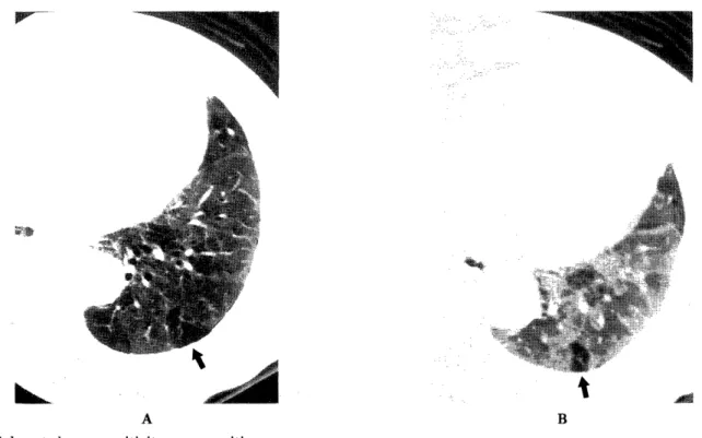
Fig. 5. Subacute hypersensitivity pneumonitis.
A. HRCT on deep inspiration : Faint ground-glass opacity is seen in left lower lobe. Relatively low attenuation area (LAA) of lobular units is demonstrated (arrow).
B. HRCT on deep expiration : Relatively LAA on inspiratory scan remains low attenuation (arrow). This finding suggests
focal air trapping due to obstructive change of small airway. Other lung fields heterogeneously increase lung density, which
may reflect parenchymal abnormalities such as alveolitis or normal lung parenchyma.
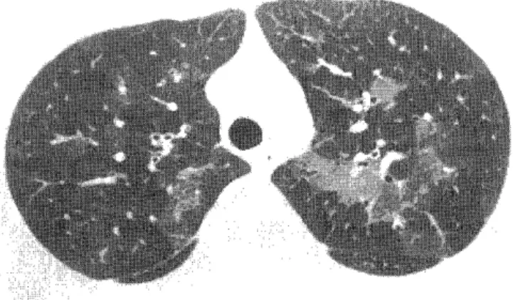
Fig. 6. Suspected chronic hypersensitivity pneumonitis.
HRCT shows diffuse ground-glass opacity associated with small cystic air-spaces (honeycombing) and traction bronchiectasis (arrows). Diffuse ground-glass opacity may reflect alveolitis or fibrosis, and honeycombing and traction bronchiectasis may indicate pulmonary fibrosis.
Fig. 8. Suspected idiopathic pulmonary fibrosis (pathologi- cally UIP).
HRCT shows ground-glass opacity associated with honey- combing in peripheral predominance.
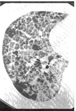
Fig. 7. Crazy-paving appearance in a patient with pulmo- nary alveolar proteinosis.
Linear opacities indicating interlobular and intralobular septal thickening is depicted within areas of ground-glass opacity.
Fig. 9. UIP associated with rheumatoid arthritis.
Diffuse ground-glass opacity and reticular opacities are seen
on HRCT. Diffuse ground-glass opacity may reflects fibrosis
or/and alveolitis. However, it should be considered as
fibrosis, because it is combined with traction bronchiectasis.
3) Cystic air-spaces with ground-glass opacity
This HRCT pattern is generally seen in chronic intersti- tial pneumonia or IPF (Fig. 8). Cystic air-spaces in these disorders are generally honeycomb cysts (honeycombing).
UIP is the most common type of chronic interstitial pneumonia or IPF. UIP can be seen in patients with other diseases, particularly collagen-vascular diseases (Fig. 9) such as rheumatoid arthritis and progressive systemic sclerosis, and certain drug reactions (e.g. from bleomy- cin).
The early stage of UIP is characterized histologically by alveolitis and increased cellularity of alveolar wall.
Ground-glass opacity may be demonstrated as only HRCT finding corresponding to these pathological changes. But this process progresses to fibrosis. Histopathologically, one lung specimen in patients with UIP may have the changes of various stages, from normal lung parenchyma and alveolitis to fibrosis (so called `temporal heterogene- ity'). Therefore, ground-glass opacity is commonly associ- ated with honeycombing and reticular opacities. Reticular opacities on HRCT correspond to areas of irregular fibrosis with a predominance of perilobular region, honey- combing, and traction bronchiectasis. Lung changes frequently show a peripheral, subpleural, and basal pre- dominance. Areas of ground-glass opacity also usually predominate in the subpleural region.
Ground-glass opacity on HRCT usually indicates the presence of disease activity and potentially treatable disease [29, 30], but can also be seen in the presence of
fibrosis or honeycombing below the resolution of HRCT (Fig. 9). However, ground-glass opacity should be consid- ered to represent an active process (alveolitis) only when there are no findings of fibrosis on HRCT. Definitive findings of fibrosis on HRCT are traction bronchiectasis and honeycombing.
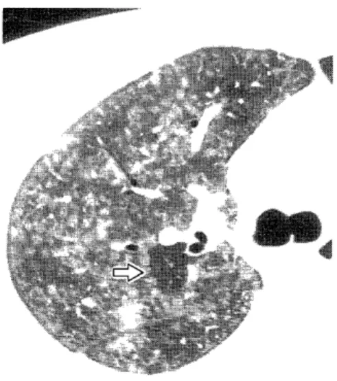
Most patients with UIP show slow progression, but accelerated deterioration (acute exacerbation) may also occur (Fig. 10). Pathologically, this condition represents diffuse alvelar damage (DAD) or active fibroblastic foci [31]. DAD is a pathologic diagnosis in most patients with the clinical diagnosis of adult respiratory distress syn- drome. Acute interstitial pneumonia (AIP) also shows the pathological findings of DAD [32]. Therefore, the findings of acute deterioration of UIP and AIP are essentially similar on HRCT. DAD is pathologically classified into three phases ; exudative (or acute) phase, proliferative (or organizing) phase, and f ibrotic phase [331. Characteristic findings on HRCT are diffuse and symmetrical ground- glass opacity and/or consolidation. They are sharply demarcated by relatively spared areas. Traction bro- nchiectasis is associated with ground-glass opacity and/or consolidation in proliferative or fibrotic phase of DAD [34]. However, honeycombing is a rare finding in patients with AIP. In patients with accelerated deterioration of UIP, honeycombing is commonly combined with ground- glass opacity. The patients with AIP and accelerated deterioration of UIP should be treated by high-dose corticosteroid therapy and are generally mortal without any therapies.
Fig. 10 A, B. Accelerated deterioration of UIP associated with rheumatoid arthritis. She had progressive dyspnea, dry cough, and fever. Chest radiograph revealed diffuse infiltrates. HRCT demonstrates sharply demarcated areas of ground-glass
opacity with segmental or panlobular distribution. Focal spared areas of lobular units are seen in right lower lobe (arrows).
Traction bronchiectasis and honeycombing are depicted within ground-glass opacity. Despite high-dose corticosteroid
therapy, she died due to respiratory failure after two weeks.
Ground-glass opacity combined with honeycombing is also seen in many end-stage lungs such as chronic HP (Fig.
6), sarcoidosis, Langerhans cell histiocytosis (eosinophilic granuloma), and asbestosis.
Ground-Glass Opacity in Infectious Lung Disease Ground-glass opacity in infectious lung disease is generally considered to represent mild to moderate alveo- lar filling [3]. When alveoli become completely airless, it leads to air-space consolidation. Ground-glass opacity is also seen in resolving stage of pneumonia.
Radiographic and pathologic findings vary by types of pneumonia, namely, bacterial pneumonia and atypical pneumonia. Because atypical pneumonia often shows bilateral diffuse shadows mimicking other acute or subacute infiltrative lung diseases, it must be considered as a possibility when diagnosing those diseases. Atypical pneumonia includes Mycoplasma pneumonia, Chlamydia psittaci or pneumoniae pneumonia, Pneumocystis carinii pneumonia (PCP), viral pneumonia (influenza virus, cytomegalovirus), and so on.
In Mycoplasma pneumonia, the target organ is regarded to be the ciliated cells of respiratory tract, and pathologic changes are usually confirmed to the areas of bronchiolar and peribronchilolar regions. Therefore, ground-glass opacity or consolidation tends to extend with lobular distribution and to be combined with thickening of the bronchovascular bandles, and centrilobular nodular opaci- ties [35].
Chlamydia psittaci and Chlamydia pneumoniae cause bronchitis and pneumonia in adults. In pulmonary infec- tion caused by chlamidia, typical pathologic changes were
Fig. 11. Chlamydia pneumoniae pneumonia.
Patchy ground-glass opacities with bronchicentric distribu- tion are demonstrated on HRCT.
Fig. 12. Pneumocystis carinii pneumonia associated with AIDS patient.
Diffuse ground-glass opacity with focal spared areas is seen in bilateral lungs.
reported to arise primarily from the respiratory bronchi- ole and then spread centrifugally to the adjacent alveoli.
The changes never arise from the distal portion of alveolar ducts, atria, and alveoli, resulting in pneumonia that is anatomically lobular [36]. CT findings of Chlamydia psittaci pneumonia are reported to be seen centrilobular
nodules and consolidation with lobular distribution [351.
These opacities may migrate and slowly regress [36, 371.
Chlamydia pneumoniae pneumonia (Fig. 11) has been described as a milder atypical pneumonia than Chlamydia psittaci pneumonia [38] .
Immunocompromised patients have an increased risk for developing PCP, especially patients with acquired immu- nodeficiency syndrome (AIDS), those with lymphopro- liferative disorders, and organ transplant recipients [39].
CT shows ground-glass opacities in most patients with PCP (Fig. 12) [40, 41]. Ground-glass opacity may form a geographic pattern involving secondary lobules while sparing others. Thickened interlobular septa within the areas of ground-glass opacity may be also identified in PCP [41]. Thin-walled cystic spaces are sometimes seen in AIDS patients [40]. Pneumocystis carinii infects the alveoli and results in lymphocytic infiltration and edema of the interstitium. Histopathologic features of PCP probably contribute to ground-glass opacity : the nature of fluid present within alveoli, which is foamy in appear- ance and is known to consist of surfactant, fibrin, and cellular debris ; and thickening of alveolar interstitium by edema and cellular infiltrates [42].
Immunocompromised patients also have an increased risk for developing cytomegalovirus (CMV) pneumonia.
CT findings of CMV pneumonia are reported that the most common pattern includes small nodules and consoli- dation. Ground-glass opacities are also identified in some patients, and histologically have early changes of DAD
1431.
Ground-Glass Opacity of Hemodynamic Origin
Blood in the pulmonary capillary bed is a major con- tributor to lung density. Alterations in regional blood flow affect lung density by increasing or decreasing blood volume in the capillary bed [5]. Therefore, adjacent areas of hyperperfused and hypoperfused lung have some degree of attenuation difference in most cases, referred to as
`mosaic oligemia' or `mosaic perfusion' [5
, 12]. The presence of increased vascular diameters through the abnormal lung density will help identify ground-glass opacity of hemodynamic origin [3].
1) Chronic obstructive pulmonary disease and airway disease
Regional blood flow in the affected areas of chronic obstructive pulmonary disease (COPD) and airway disease decreases due to vascular smooth muscle contraction and/
or structural changes in vessel wall [44]. If large areas of pulmonary vascular bed are narrowed or occluded, there must be a reciprocal increase in blood flow to the remain- ing vascular patent areas to maintain the cardiac output [5, 44]. Ground-glass opacity may reflects a regional increased blood flow. Destruction of alveolar wall occurs in pulmonary emphysema and obstructive change of bronchioles occurs in small airway diseases such as con- strictive bronchiolitis (obliterative bronchiolitis) (Fig.
13). Ground-glass opacity with prominent vascular struc- tures is often sharply demarcated with lobular borders in patients with constrictive bronchiolitis. Decreased lung density occurs as the result of the overinflation caused by emn)hvsema and air trannine'. Thus. the chane'e of lone
Fig. 13. Constrictive bronchiolitis in an allograft bone marrow trasplant recipient.
Sharply demarcated areas of ground-glass opacity with panlobular distribution are demonstrated on HRCT.
Presumably, decreased attenuated areas reflect air trapping due to obliterative change of bronchioles and areas of ground-glass opacity indicate regional increased blood flow due to redistribution (mosaic perfusion).
density may appear with higher contrast between nonaffected (hyperperfused) areas and affected (hypoper- fused and overinflated) areas on CT. The differentiation between affected and nonaffected areas may be revealed by expiratory scans, because increase of lung density and loss of volume do not usually occur in the affected areas and these changes occur in the spared areas on expiration [8].
2) Vascular lung disease
Regional alteration of pulmonary blood flow secondary to chronic thromboembolic diseases causes increased lung density. Ground-glass opacity with prominent vascular structures is often sharply demarcated with lobular borders [6]. A mosaic pattern of lung density is demon- strated with geographic areas of low density without evidence of emphysema or lung destruction. Low density areas probably reflect hypoperfused areas caused by thromboembolism. Therefore, these low density areas usually show increase of lung density as well as areas of ground-glass opacity on expiratory CT [8]. By another proposition, increased lung density may occur in occluded areas caused by the development of systemic collateral supply as antegrade systemic-to-pulmonary arterial shunts [45].
Similar CT findings may be also demonstrated in a patient with Takayasu arteritis caused by involvement of pulmonary artery [46]. In primary pulmonary hyperten- sion [3] and pulmonary venoocculusive disease [47], sharply demarcated areas of ground-glass opacity are also seen due to redistribution of blood flow.
3) Pulmonary edema of cardiac origin
Ground-glass opacity is seen in a patient with pulmo- nary edema. Pulmonary edema is the consequence of high pulmonary capillary and venous pressure due to failure of the left side of the heart. Fluid may locate in the alveolar spaces or may be restricted to the interstitium of the lung.
HRCT findings of pulmonary edema are thickening of interlobular septa and peribronchiolar interstitium, and featureless or sharply demarcated areas of ground-glass opacity [48]. These findings reflect pulmonary extra- vascular lung water either in the interstitium or alveolar spaces. There may also be dilated vessels through the abnormal lung density due to hemodynamic changes [3].
The sequence of fluid accumulation in lung as all or none,
with the pattern of reaction of each individual alveolus
being stereotyped as either air-filled and expanded or
fluid-filled and collapsed [491. The distribution of
parenchymal abnormalities is usually bilateral and sym-
metrical.
Fig. 14 A, B
A : window width/window level (500HU/-900HU) B : window width/window level (1500HU/-750HU)
Mimicking of ground-glass opacity on narrow window width and low window level. Thickening of vessels and bronchial walls are also seen in Fig. A. Multiple cystic air-spaces indicating centrilobular emphysema and small nodules are depicted.
Pitfalls in Ground-Glass Pattern Recognition 1) Technical pitfalls
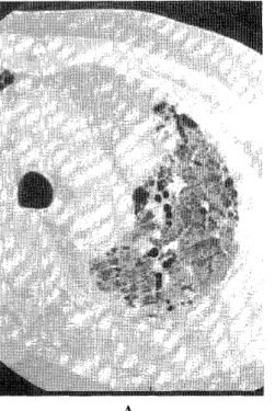
Varing window widths and levels greatly affect the interpretation of CT images. Reducing the window width or level may result in the mimicking of ground-glass opacity in pulmonary parenchyma (Fig. 14) [1, 50].
Thickening of vascular and bronchial walls are also seen.
Window widths of 1500-2000 HU and window levels of - 700 - 750 HU are recommended , when ground-glass opacity is evaluated.
2) Pitfalls by patient-related artifacts
Because atelectasis frequently develops in depending lung region, band-like areas of ground-glass opacity are seen in subpleural region of bilateral dorsal regions at supine position. Subpleural linear opacities may be also combined. Distinguishing reversible dependent atelectasis
from true lesion is easily made by the scan at prone position. Respiratory and cardiac motion may also pro- duce ground-glass opacity. Respiratory motion during acquisition of a scan usually causes patchy areas of ground-glass opacity throughout lung parenchyma.
Because low lung volume results in increased lung density, ground-glass opacity may be demonstrated on scans obtained without deep inspiration (Fig. 15). These areas of ground-glass opacity may be diffuse or patchy in distribu- tion [50] .
Fig. 15. Ground-glass opacity is demonstrated on scans obtained without deep inspiration. Ground-glass opacity is diffuse, but predominantly distributes in dependent portion of each lobes.