• 検索結果がありません。

Evaluation of an Experimental Animal Model for Estimating the Pathogenicity and the Efficacy of

N/A
N/A
Protected

Academic year: 2021

シェア "Evaluation of an Experimental Animal Model for Estimating the Pathogenicity and the Efficacy of "

Copied!
7
0
0

読み込み中.... (全文を見る)

全文

(1)

Acta Med. Nagasaki 30 : 68-74

Evaluation of an Experimental Animal Model for Estimating the Pathogenicity and the Efficacy of

Antibiotic Treatment in Bacterial Infection

Masaru NASU*, Jun GOTO, Yoichiro GOTO, Takayoshi TASHIRO and Takashi ITOGA

The Second Department of Internal Medicine, Medical College of Oita,

Oita, Japan

Received for publication, January 30, 1985

An experimental model of intracutaneous infection in the guinea pig, which was based on the study of MILES et al. (Br. J. Exp. Path. 38: 79-96, 1957) :and MASKELL (J.

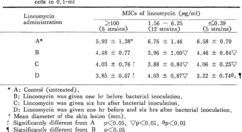
Med. Microbiol. 14: 131-140, 1981), was used to investigate the pathogenicity and the effect of chemotherapy with lincomycin, using 36 strains of the genus Bacteroides. The pathogenicity was estimated by intracutaneous injection of 0.1-ml quantities of bacterial suspension into guinea pigs and the value was expressed as the number of inoculated organisms required to induce a skin lesion 10 mm in diameter 24 hours after injection.

Bacteroides fragilis was, on average, three times as pathogenic as non-B. fragilis strains (p<0.1). Two intramuscular injections of lincomycin were more effective than a single one against the intracutaneous infection, and the lower was the MIC value, the greater was the effect (p<0.001). This experimental animal model was simple and convenient for estimating the pathogenicity and efficacy of antimicrobial agents. However, the skin lesions were often not clear if the bacteria with low virulence were inoculated and the induced infection is not natural.

Key words: Bacteroides,fragilis; Pathogenicity; Chemotherapy; Experimental infection

INTRODUCTION

Many kinds of experimental animal model in bacterial infection have been developed for estimating the pathogenicity and the effect of antimicrobial treatment. We attempted to evaluate the method of intracutaneous infection in the guinea pig based on the those

那 須   勝,後 藤  純,後 藤 陽 一 郎,田 代  隆 良,糸 賀   敬

Request for reprints should be addressed to Dr. Masaru Nasu, 2nd Department of Internal

Medicine, Medical College of Oita, 1-1506, Hazama-cho, Oita 879-56, Japan

(2)

of MILES") and MASKELLI5) using the genus Bacteroides. Bacteroides species are fre- quently isolated from human clinical specimens and play an important role in anaerobic infection. It is well known that Bacteroides fragilis among the species of the genus Bac- teroides is most frequently recognized as a causative pathogen in intraabdominal, posto- perative, female genital and respiratory infections, and in bacteremia6). Bacteroides spe- cies are the major components of the normal flora of the large intestine, and B. fragilis is a minor component9"17>, but it is more often isolated from clinical specimens and causes infections more frequently than non-B. fragilis strains. As a reason for the higher incidence of endogenous infections due to B. fragilis compared with non-B. fragilis species, several pathogenic factors have been proposed for B. fragilis3)4)1O>11)12)13)1e)21) In the present study, the pathogenicity of B. fragilis was compared with that of non- B. fragilis species by using intracutaneous infection in the guinea pig as an experimental model. The effect of the administration of an antibiotic, lincomycin, on infection with B.

fragilis was also investigated. This paper describes the results of these studies with some discussion.

MATERIALS AND METHODS

Bacteria. Twenty-five strains of B. fragilis and 11 of non-B. fragilis strains were used ; 23 were from clinical specimens such as pus, wound swabs, blood and ascitic fluid, one was B. fragilis G-2 provided by the Institute for Anaerobic Bacteriology, Gifu Uni- versity School of Medicine, Gifu, Japan ; and the remaining one was a reference strain, B. fragilis NCTC 9343. The non-B. fragilis species used consisted of 10 from clinical sp- ecimens (8 of B. distasonis and 2 of B. thetaiotaomicron) and one of B. uniformis G-

3 from Gifu University.

Animals. Female Hartley strain guinea pigs weighing 450-550g were used in this study.

Preparation of suspensions for animal inoculation. The bacterial strains were cultured on a GAM agar medium (NISSUI, JAPAN) for two days at 37°C in an anaerobe box incubator (FORMA, USA) in an atmosphere of 10% carbon dioxide, 7 % hydrogen and 83% nitrogen maintained at a humidity of 60%. The growth was scraped off with a pla- tinum spatula and suspended densely in sterile saline (NaC1 0.8% w/v in water) in the anaerobe box. The number of bacteria in the suspensions was determined by the dilution culture method or with a standard curve prepared by photodensitometry. The original su- spensions and 10-fold dilutions of both living bacteria and organisms which were killed by heating in a water bath at 80°C for 30 min were prepared for animal inoculation.

Intracutaneous inoculation in guinea pigs. The intracutaneous inoculation was car-

ried out according to the method of MILES et al. 16) . Guinea pigs were divided into

groups of three, and the hair of the dorsal skin and flanks was shaved off before the

injection. The bacterial suspension in 0.1-ml volumes was then randomly injected into

the skin of each guinea pig with a tuberculin needle from 16 to 20 sites at about 2.5-cm

(3)

intervals.

Evaluation of intracutaneous pathogenicity. Pathogenicity was evaluated according to MASKELL's method") as follows. At 24 hours after the inoculation, a barium sulphate paste was applied for about 5 min on the shaved skin area of the guinea pig for complete depilation and then the diameter of the skin lesion at each inoculated site was measured with slide calipers. The pathogenicity of each Bacteroides species was expressed in terms of the number of bacteria in 0.1-m1 capable of producing a lesion 10 mm in mean di- ameter. This value was termed the ED10, and for living and dead bacteria it was ex- pressed as ED10 (L) and ED10 (D) respectively ; these values were calculated from a graph on which were plotted the nlumber of bacteria in the original suspension and7the 10-fold dilutions injected and the diameter of the lesion produced by each strain. Pathogenicity was estimated on the assumption that the ED10 (L) value expresses the intracutaneous multiplication, persistance and invasiveness of the organisms, while the ED10 (D) value indicates the toxicity of the bacterial substances. Moreover, the ED10 (D)/ED10 (L) ratio (potency ratio) was thought to represent the intracutaneous multiplication or persistence of bacteria in the guinea pig. Thus, a low ED10 value means strong pathogenicity, and a potency ratio equal to or less than one (L1) indicates lack of multiplication, while a potency ratio greater than one (>1) means high multiplication and persistence of bacteria in guinea pig skin.

Antibiotic and the method of administration. Lincomycin (Upjohn) was employed in the current study. Lincomycin at 200 mg/kg was intramuscularly injected into each guinea pig in four groups, A-D, each group containing three guinea pigs. Group A was the control to which no antibiotic was given, group B was injected with lincomycin one hour before the B. fragilis inoculation, group C was given lincomycin six hours after the bacterial inoculation, and group D was treated with the antibiotic both one hour be- fore and six hours after the inoculation of B. fragilis.

Measurement of the antibacterial activity and the tissue levels of lincomycin. The minimum inhibitory concentrations (MICs) of lincomycin for B. fragilis were measured by an agar dilution technique using GAM agar medium. The strains were grown for 24 hours in GAM broth (NISSUI) and further diluted in broth to provide 104 colony forming units when delivered to the surface of the GAM agar medium with a multipoint i noculator (SAKUMA, Japan). Doubling dilutions of lincomycin were prepared in 10 mM phos- phate buffer (pH 7.0) to final concentrations of 100-0.05 ug/ml in GAM agar plate.

The plates were inocubated in an anaerobe box incubator at 37°C for 24 hours, and then the MIC was defined as the lowest concentration resulting in the total suppression of growth. The levels of lincomycin in the blood and in the skin lesions of three guinea pigs administered 200 mg/kg intramuscularly were measured by the agar well diffusion assay method').

RESULTS

Comparison of the pathogenicity of B. fragilis and non-B. fragilis strains.

(4)

In the preliminary study the diameter of the skin lesion was approximately directly proportional to the number of the organisms inoculated (r=0.75). The pathogenicity of 11 strains of B. fragilis was compared with that of 11 non-B. fragilis strains. The results obtained are shown in Table 1, in which the lesions caused by living bacteria are seen to be generally larger than those caused by the dead organisms. The mean values of ED 10 (L) and ED 10 (D) for B. fragilis were approximately two or three times smaller than those for non-B. fragilis strains ; these results may suggest stronger pathogenicity of B. fragilis than non-B. fragilis strains, but the differences were not statistically significant according to Student's t-test. The potency ratio of B. fragilis was greater than that of non-B. fragilis strains (p<0.1), suggesting that B. fragilis strains have a greater ability to persist or multiply in the guinea pig skin.

Tissue levels of lincomycin. Mean lincomycin levels in the serum and skin lesions of three guinea pigs are shown in Table 2. Peak levels of lincomycin in the serum and skin lesions reached 54 and 23.7 pg/ml, respectively, and generally exceeded the MICs used in the current study except for six resistant strains with MICs of X100 pg/ml.

The efficacy of lincomycin in intracutaneous infection in the guinea pig. Twenty- three strains of B. fragilis isolated from clinical specimens were used. The efficacy of lincomycin was expressed in terms of the diameter (mm) of the skin lesion 24 hours after the intracutaneous injection of 0.1-ml of a suspension with 109 living bacteria/ml.

The results obtained are shown in Table 3. The skin lesions in the lincomycin treated groups B, C and D were smaller than those in the untreated group A (p<0.01), and

Table 1. Comparison of ED10 (D)*, ED10 (L) and ED10 (D): ED10 (L) (potency ratio) of Bacteroides fragilis and non- B. fragilis strains

(x 10 cells) ED10 (D) (x 10 cells) ED10 (L) ED10 (D) : ED10 (L)

B. fragilis (11 strains) 5.06± 3.4+ 2.23 ± 2.4 3.89 ± 3.07 Non-Bl fraglis (11 strains) 10.3 ± 8.1 6.45 ± 7.5 2.35 ± 1.60

Statistical analysis NS NS p<0.1

* ED10 values are expressed as the number of organisms in 0 .1-m1 required to induce a mean lesion diamter of 10 mm in guinea pig skin; this value was

calculated for living and dead organisms and termed ED10 (L) and ED10 (D)

respectively. See Maskell (15) and Miles et al. (16).

} Mean ± SE

Table 2. Mean serum and skin lesion levels (tpg/ml) of lincomycin after intramuscular injection of 200 mg/kg in three guinea pigs

Time after injection (hr)

Sample

1/2 1 2 4 6

Serum 54 36 22.2 9.1 4.0

Skin lesion 13.2 23.7 17.3 7.7 4.1

(5)

the effect especially strong in the lesions caused by lincomycin sensitive strains in group D (p<0.001). The skin lesions due to highly resistant strains were also diminished significantly in groups C and D (p<0.05). There was no significant difference between groups B and C or C and D by Student's t-test.

Table 3. Relative efficacy of lincomycin administration (200 mg/kg) in intradermal infection of guinea pig caused by injection of 108 Bacteroides fragilis

cells in 0.1-ml

Lincomycin MICs of lincomycin (pg/ml)

administration >100 1.56 - 6.25 50.39

(6 strains) (12 strains) (5 strains)

A* 5.92 ± 1.38+ 6.76 ± 1.46 6.58 ± 0.70

B 4.48 ± 0.77 3.96 ± 1.OOV 4.46 ± 0.847

C 4.03 ± 0.76 1 3.88 ± 0.847 4.06 ± 0.257

D 3.85 ± 0.67 ( 4.03 ± 0.877 3.22 ± 0.740, 

* A : Control (untreated) .

B: Lincomycin was given one hr before bacterial inoculation.

C: Lincomycin was given six hrs after bacterial inoculation.

D: Lincomycin was given one hr before and six hrs after bacterial inoculation.

+ Mean diameter of the skin lesion (mm) .

Significantly different from A p(0.05, 7p<0.01, Op<0.01

¶ Significantly different from B p<0.05

DISCUSSION

The goal of this work was to estimate the pathogenicity of the Bacteroides species and to select the strains with strong pathogenicity for applying to further experimentally infected animal model, and also to evaluate the superiority or inferiority of the method of intracutaneous infection in guinea pig. The Bacteroides species are the major group of bacteria composing the resident bacterial flora in the intestinal tract, particularly in the large intestine. Non-B. fragilis strains such as B. vulgatus, B. distasonis, B. thetaiotao-

micron are more common than B. fragilis. However, B. fragilis is most frequently isolated from human specimens and clinical infections due to this organism are often encountered 9)19) As for the greater pathogenicity of B . fragilis strains, the capsular polysaccharide of B. fragilis has been considered to be a virulence factor by many investigators 10) 11)12)13)

14)18)2°) On the other hand , it has also been reported that the capsule is not only characteristic of B. fragilis, but that it is also present rather commonly in non-B. fragilis strains except for B. distasonis18). Superoxide dismutase production2l) and resistance to opsonin and to the bactericidal action of serum2)3)4)2°) have been reported as other possible pathogenic factors of B. fragilis. The results of the present study showed that B. fragilis was about three times, on the average, as pathogenic as the non-B. fragilis strains.

MASKELL15), who used the same experimental model, reported that B. fragilis is 17

times as pathogenic as non-B. fragilis strains. As a reason for these differences in

pathogenicity between B. fragilis and non fragilis strains, it was assumed that B. fragilis

(6)

would be more persistent in the guinea pig skin. B. fragilis was consistently recovered from skin lesions 24 hours after inoculation of the bacteria into the guinea pig skin, while non-B. fragilis strains were not recovered in many cases in the present study.

The results of investigation on antibiotic therapy showed that two administrations of lincomycin, given intramuscularly one hour before and six hours after intracutaneous inoculation of the bacteria, were therapeutically most effective (p<0.001). This finding seems to be in agreement with the results reported by EAGLE et al .1)6) in a series of ex- periments. Even if the intracutaneous level of lincomycin was lower than the in vitro MIC required to kill the inoculated bacterial strain, this antibiotic showed a significant therapeutic effect. This apparent potentiation of the effect of lincomycin was thought to be a protective reaction by the neutrophils, macrophages and complements which were produced in vivo in response to inoculation of B. fragilis. In the majority of the inoculated foci, there were macroscopically observable abscess formation, and histologically many polynucleocytes were seen to have infiltrated to the skin along with some macrophages, lymphocytes, eosinophils and so on. These findings were observed uniformly in all groups of animals inoculated with living or dead suspensions of B. fragilis and non-B. fragilis strains. An advantage of the present experimental model is that the pathogenicity of

many bacterial strains and the in vivo efficacy of the antibiotics can be tested with a small number of guinea pigs. On the other hand, the disadvantages are that the skin lesions are often not clear if small numbers of bacteria of low virulence are inoculated, physical reactions may possibly occur because of the inoculation of a large number (more than 107) of bacteria, and the induced infections are not natural.

ACKNOWLEDGEMENTS

We wish to thank Dr. J. P. MASKELL of the Department of Medical Microbiol- ogy, The London Hospital Medical College, University of London, for his kind advice in this study.

REFERENCES

1) BABB, J. L. , and CUMMINS, C. S. 1978. Encapsulation of Bacteroides species. Infect.

Immun. 19: 1088-1091.

2) BJORNSON, A. B., BJORNSON, H. S., ASHAF, M., and LANG, T. J. 1983.

Quantitative variability in requirements for opsonization of strains within the Bacteroides

fragilis group. J. Infect. Dis. 148: 668-675.

3) CASCIATO, D. A., ROSENBLATT, J. E., GOLDBERG, L. S., and BLUESTONE, R.

1975. In vitro interaction of Bacteroides fragilis with polymorphonuclear leucocytes

and serum factors. Infect. Immun. 11: 337-342.

4) CASCIATO, D. A., ROSENBLATT, J. E., BLUESTONE, R., GOLDBERG, L. S., and FINEGOLD,S. M. 1979. Susceptibility of isolates of Bacteroides to the bactericidal activity

of normal human serum. J. Infect. Dis. 140: 109-113.

5) EAGLE, H., FLEISCHMAN, R., and LEVY, M. 1953. "Continuous" vs "discontinuous"

therapy with penicillin. N. Engl. J. Med. 248: 481-488.

(7)

6) EAGLE, H., FLEISCHMAN, R., and MUSSLMAN, A. D. 1950. The bacterial action of penicillin in vivo. Ann. Intern. Med. 33: 544-571.

7) EDBERG, S. C., and SABATH, L. D. 1980. Determination of antibiotic levels in body fluids: techniques and significance. Bactericidal tests in endocarditis and other severe

infections. Antibiotics in laboratory medicine. Edited by V. Lorian. Baltimore: Williams

and Wilkins. pp206-264.

8) EINEGOLD, S. M. 1977. Anaerobic bacteria in human disease. New York, San Fran- cisco, London: Academic Press.

9) FINEGOLD, S. M., ATTEBERY, H. R., and SUTTER, V. L. 1974. Effect of diet on human fecal flora: comparison of Japanese and American diets. Am. J. Clin.

Nutr. 27: 1456-1469.

10) KASPER, D. L. 1976. The polysaccharide capsule of Bacterides fragilis subspecies fragilis: Immunochemical and morphologic definition. J. Infect. Dis. 133: 79-87.

11) KASPER, D. L., HAYES, M. E., REINAP, B. G., CRAFT, F. O. , ONDERDONK,

A. B., and POLK, B. G. 1977. Isolation and identification of encapsulated strains of Bacteroides fragilis, J. Infect. Dis. 136: 75-81.

12) KASPER, D. L., and ONDERDONK, A. B. 1982. Infection with Bacteroides fragilis:

Pathogenesis and immuno-prophylaxis in an animal model. Scand. J. Infect. Dis. Sup-

p1. 31: 28-33.

13) KASPER, D. L., ONDERDONK, A. B., POLK, B. G., and BARTLETT, J. G. 1979.

Surface antigens as virulence factors in infection with Bacteroides fragilis. Rev.Infect.

Dis. 1: 278-288.

14) LINDBERG, A. A., WEINTRAUB, A., KASPER, D. L., and LpNNGREN, J. 1982.

Virulence factors in infection with Bacteroides fragilis: Isolation and characterization

of capsular polysaccharide and lipopolysaccharide. Scand. J. Infect. Dis. Suppl. 35:

45-52.

15) MASKELL, J. P. 1981. The pathogenicity of Bacteroides fragilis and related species estimated by intracutaneous infection in the guinea pig. J. Med. Microbiol. 14: 131-

140.

16) MILES, A. A., MILES, E. M., and BURKE, J. 1957. The value and duration of defence reactions of the skin to the primary lodgement of bacteria. Br, J. Exp. Path.

38: 79-96.

17) MOORE, W. E. C., and HOLDMAN, L. V. 1974. Human fecal flora: The normal flora of 20 Japanese-Hawaians. Appl. Microbiol. 27: 961-979.

18) ONDERDONK, A. B., KASPER, D. L., CISNEROS, R. L., and BARTLETT, J. G. 1977.

The capsular polysaccharide of Bacteroides fragilis as a virulence factor: comparison

of the pathogenic potential of encapsulated and unencapsulated strains, J. Infect. Dis, 136: 82-89.

19) POLK, B. F., and KASPER, D. L. 1977. Bacteroides fragilis subspecies in clinica isolates. Ann. Intern. Med. 86: 569-571.

20) SIMON, G, L., KLEMPNER, M. S., KASPER, D. L., and GORBACH, S. L. 1982.

Alteration in opsonophagocytic killing by neutrophils of Bacteroiaes fragilis associated

with animal and laboratory passage: effect of capsular polysaccharide. J. Infect, Dis,

145: 72-77.

21) TALLY, F. P., GOLDIN, B, R., JACOBUS, N. V., and GORBACH, S. L. 1977.

Superoxide dismutase in anaerobic bacteria of clinical significance. Infect. Immun. 16:

20-25.

Table  1.  Comparison  of  ED10  (D)*,  ED10  (L)  and  ED10  (D):  ED10  (L)          (potency  ratio)  of Bacteroides  fragilis  and  non-  B
Table  3.  Relative  efficacy  of  lincomycin  administration  (200  mg/kg)  in  intradermal           infection  of  guinea  pig  caused  by  injection  of  108  Bacteroides  fragilis

参照

関連したドキュメント

Keywords: continuous time random walk, Brownian motion, collision time, skew Young tableaux, tandem queue.. AMS 2000 Subject Classification: Primary:

This paper develops a recursion formula for the conditional moments of the area under the absolute value of Brownian bridge given the local time at 0.. The method of power series

Answering a question of de la Harpe and Bridson in the Kourovka Notebook, we build the explicit embeddings of the additive group of rational numbers Q in a finitely generated group

Then it follows immediately from a suitable version of “Hensel’s Lemma” [cf., e.g., the argument of [4], Lemma 2.1] that S may be obtained, as the notation suggests, as the m A

Applications of msets in Logic Programming languages is found to over- come “computational inefficiency” inherent in otherwise situation, especially in solving a sweep of

Our method of proof can also be used to recover the rational homotopy of L K(2) S 0 as well as the chromatic splitting conjecture at primes p &gt; 3 [16]; we only need to use the

In this paper we focus on the relation existing between a (singular) projective hypersurface and the 0-th local cohomology of its jacobian ring.. Most of the results we will present

Shi, “The essential norm of a composition operator on the Bloch space in polydiscs,” Chinese Journal of Contemporary Mathematics, vol. Chen, “Weighted composition operators from Fp,