dynamics over long timescales
Author Viktoras Lisicovas Degree Conferral
Date
2020‑03‑31
Degree Doctor of Philosophy Degree Referral
Number
38005甲第46号 Copyright
Information
(C) 2020 The Author.
URL http://doi.org/10.15102/1394.00001212
Thesis submitted for the degree
Doctor of Philosophy
Improvements in optical techniques to investigate the behavior and neuronal network dynamics over long timescales
by
Viktoras Lisicovas
Supervisor: Keshav M. Dani
March, 2020
Declaration of Original and Sole Authorship
I, Viktoras Lisicovas, declare that this thesis entitled Improvements in optical techniques to investigate the behavior and neuronal network dynamics over long timescales and the data presented in it are original and my own work.
I confirm that:
● No part of this work has previously been submitted for a degree at this or any other university.
● References to the work of others have been clearly acknowledged.
Quotations from the work of others have been clearly indicated, and attributed to them.
● In cases where others have contributed to part of this work, such
contribution has been clearly acknowledged and distinguished from my own work.
● None of this work has been previously published elsewhere, with the exception of the following:
o Lisicovas, Viktoras, Bala Murali Krishna Mariserla, Chakradhar Sahoo, Reuben T. Harding, Michael KL Man, E. Laine Wong, Julien Madéo, and Keshav M. Dani. "Improving Signal and
Photobleaching Characteristics of Temporal Focusing Microscopy with the Increase in Pulse Repetition Rate." Methods and protocols 2, no. 3 (2019): 65. (Selected as the cover of the quarterly issue by the editors).
Date: 18th March, 2020 Signature:
experimental neuroscience from electrophysiological methods for observation and stimulation to all-optical solutions. One expects this trend to continue as future developments continue to deliver, and improve upon, the original promises of the technology: 1) minimally invasive actuation and recording of neurons, and 2) a drastic increase in targets that can be treated simultaneously. Moreover, as the high costs of the technology are reduced, one may expect its larger-scale adoption in the neuroscience community. In this thesis, I describe the development and implementation of two all- optical solutions for the analysis of behavior, neuronal signaling, and stimulation, which improve on previous state-of-the-art: (1) A minimally-invasive, high signal-to-noise two- photon microscopy setup capable of simultaneous, live-imaging of a large subset of sensory neurons post activation, and (2) a low-cost tracking solution to stimulate and record behavior. I begin this thesis with a review of recent advances in optical
neuroscience techniques for the study of neuronal networks with the focus on work done in Caenorhabditis elegans. Then, in chapter 2, I describe my implementation of a two-photon temporal focusing microscopy setup and show significant improvements through the use of a high power/ high pulse repetition rate excitation system, enabling live imaging with high resolution for extended periods of time. I model temperature increase during a
physiological imaging scenario for different repetition rates at fixed peak intensities and find range centered around 1 MHz to be optimal. Lastly, I describe the low-cost tracking setup with the ability to stimulate and record behavior over the course of hours. The setup is capable of two-color stimulation of optogenetic proteins over the area of the behavioral arena in combination with volatile chemicals. To showcase the utility of the system, I demonstrate behavioral analysis of integration of contradictory cues. In summary, I present a set of techniques for the interrogation of neural networks from animal behavior to
neuronal activity, over timescales of potentially hours and days. These techniques can be used to address a new dimension of scientific questions.
Acknowledgment
Putting this work from thought to paper has been a long and winding road. As luck would have it, I have been blessed to interact with a number of remarkable people along the way, who showed me the true meaning of solid science, professionalism, comradery, compassion, and perseverance. I would like to express my kindest thanks to the people, without whom this work could not have been:
My supervisor, Prof. Dr. Keshav M. Dani, whose laser-cut guidance in things big and small have kept me sane through the years. I am yet to meet anyone with more energy and enthusiasm for either science or people. Also for helping with research design, data interpretation, and writing.
My Academic Mentor, Prof. Dr. Tadashi Yamamoto, whose kind words, advice, and perspective always helped me see the bright side.
My Third Committee Member, Prof. Dr. Ichiro N. Maruyama, for providing support, resources and lab bench to assist with the development of the tracking system.
All of the present and past members of the Femtosecond Spectroscopy Unit and in particular: Prof. Dr. Bala Murali Krishna Mariserla, for technical assistance and showing me a masterclass in optical alignment; Dr. Julien Madéo for numerous discussions and rock-solid advice in data acquisition, interpretation and French cuisine; Dr. Michael Ka Lun Man for advice on data analysis, interpretation and an inspiring example of effortless
professionalism; Chakradhar Sahoo for help with optical alignment and valuable late-night discussions; and Reuben Harding for taking those hard first steps with me towards what became the microscopy setup described herein.
All of the present and past members of the Information Processing Biology Unit and in particular: Dr. Takashi Murayama, who painstakingly mentored me in finer points of C. elegans research and always found time for a lengthy midnight discussion; Dr. Eichiro Saita, who provided invaluable advice on implementation of odor stimulation; Dr. Yuto Momohara and Andrew Mugo for refreshing breaks; and (now Dr.) Tosif Ahamed, who is
positivity.
OIST Engineering Section for granting access, training, and help with microscopy and manufacturing, in particular: Dr. Kieran Deasy for always finding time in his busy schedule to help with microfabrication; and Takuya Miyazawa for assistance with fluorescence lifetime measurements.
Yumi Ito and Hitomi Ohtaki, Research Associates from Femtosecond Spectroscopy Unit and Information Processing Biology Unit for positive energy and helping out with all manner of paperwork.
Dr. William Schafer, Dr. Edward Boyden, Dr. Cornellia Bargmann, Dr.
Kevin M. Collins, Caenorhabditis Genetics Center, and Addgene for providing strains and plasmids used in this work and Nanoscribe for valuable advice on microfluidics manufacturing.
OIST Graduate University for generous funding and support through this journey.
My beautiful wife, Sakurako, family, and friends, who kept faith in spite of all the trials and tribulations.
Abbreviations
AMPA α-amino-3-hydroxy-5-methyl-4-isoxazolepropionic acid
ATR All-trans-Retinal
C. elegans Caenorhabditis elegans
C1V1-ET/ET A variant of chimeric fusion of channelrhodopsin-1 and Volvox VChR1
CGC Caenorhabditis Elegans Genetics Center
ChR2 Channelrhodopsin 2
CMOS Short for complementary metal-oxide-semiconductor CREB cAMP response element-binding protein
CRISPR Clustered regularly interspaced short palindromic repeats CrR ChrimsonR red-shifted channelrhodopsin variant
CTX chemotaxis
GCaMP Genetically encoded calcium indicator
GFP Green fluorescent protein
LED Light-emitting diode
NGM Nematode growth medium
NMDA N-methyl-D-aspartate
NOPA Non-colinear optical parametric amplifiers
NpHR Halorodopsin
OP50 E. coli uracil auxotroph
OPA Non-linear optical parametric amplifier
PC Principal component
PCR Polymerase chain reaction
PDMS Polydimethylsiloxane
TRPV1 The transient receptor potential cation channel subfamily V member 1
WF-TeFo Wide-field two-photon temporal focusing microscope
Nomenclature
µa Absorption coefficient
C Typical concentration of fluorophores
c Speed of light in vacuum
Fa
The fraction of absorbed photons that do not result in two- photon emission
h Planck constant
I Peak power density
k Specific thermal conductivity of brain tissue
d Propagation length (two Rayleigh lengths)
N1𝜆 Number of photons absorbed due to one-photon absorption of the medium
N2𝜆-Nem
Number of absorbed photons that do not result in two- photon emission
Na Number of absorbed photons
N𝜏 Total number of photons
PA Average laser power
qr Heat
S Coherence parameter
T Temperature
V The volume of the excitation cylinder
ε Heat source
υ Tangential velocity
Φ Quantum efficiency
ω Angular velocity
𝛿2𝜆 Two-photon cross-section at 960 nm
𝜆 The wavelength used for two-photon imaging
To my little Maya, who came into this world and beamed away all shadows.
Contents
Declaration of Original and Sole Authorship ... ii
Acknowledgment ... iv
Abbreviations ... vi
Nomenclature... vii
Contents ... ix
List of Figures ... xii
List of Tables ... xiv
Outline of the thesis ... xvi
1 Literature Review ... 1
1.1 Introduction ... 1
1.2 Overview of the nervous system of C. elegans ... 4
1.3 Olfaction and nociception in C. elegans ... 7
1.4 Long-term behavioral plasticity ... 9
1.5 Optogenetic toolkit... 13
1.6 Behavioral tracking of C. elegans ... 15
1.7 Optical microscopy for live imaging of neural activity ... 19
1.8 Two-photon temporal focusing microscopy ... 21
1.9 Problem statement and aims ... 24
2 Long-term neuronal activity recording with two-photon temporal microscopy with high pulse repetition rates ... 25
2.1 Introduction ... 26
2.2 Materials and methods ... 28
2.2.1 Configuration of the temporal focusing microscope ... 28
2.2.2 Preparation of the microfluidic devices ... 32
2.2.3 Preparation of imaging samples ... 32
2.3.1 Fluorescence signal intensity scales linearly with the repetition rate ... 35
2.3.2 Photobleaching accelerates faster with increased intensity than with repetition rate ... 37
2.3.3 Modeling of temperature constraints suggest an optimal repetition rate in the vicinity between 0.5 and 5 MHz ... 40
2.3.4 Design of a novel immobilization device for imaging with aversive cues ... 47
2.3.5 Application of increased repetition rates for live animal imaging ... 49
2.4 Discussion ... 52
3 Long-term tracking of population behavior in response to olfactory and multicolor optogenetic stimuli ... 54
3.1 Introduction ... 55
3.2 Materials and Methods ... 56
3.2.1 Animal cultivation and preparation of transgenic strains ... 56
3.2.2 Population assay conditions ... 60
3.3 Results ... 61
3.3.1 Design and implementation of the tracking system ... 61
3.3.2 Design of tracking software ... 66
3.3.3 Long-term tracking reveals eigenshapes on bacterial food ... 70
3.3.4 Pulsed odor stimulus of opposite valence induce locomotion features consistent with attraction and repulsion ... 73
3.3.5 Optical stimuli recapture changes in tangential velocity ... 76
3.3.6 Repeated co-stimulation of appetitive and aversive neurons leads to reversal inhibition ... 80
3.4 Discussion ... 82
4 Conclusion ... 84
Bibliography ... 86
List of Figures
Figure 1.1 Historical comparison of electrophysiological and optical techniques for
stimulation and recording. ... 3
Figure 1.2. Olfactory and nociceptive system of C. elegans. ... 8
Figure 1.3 Relevant long time scale behavioral plasticity paradigms. ... 10
Figure 1.4 An example toolkit for two-color stimulation of neurons and readout of neuronal activity. ... 15
Figure 1.5 Principles of automated tracking and behavioral complexity. ... 18
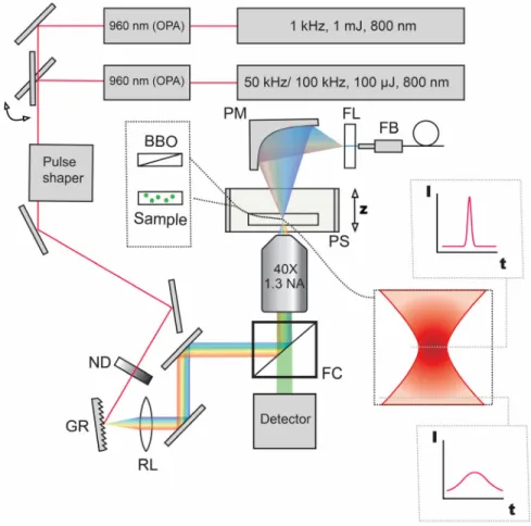
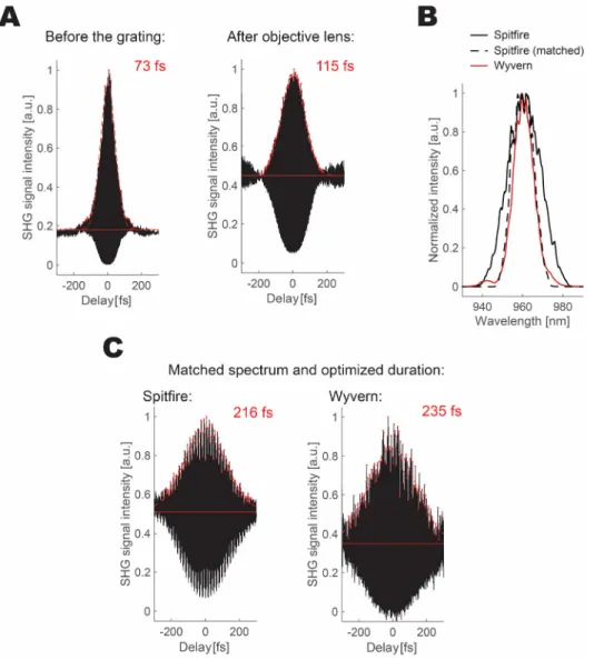
Figure 2.1 WF-TeFo setup diagram with switchable lasers and pulse shaping capabilities. ... 29
Figure 2.2 Pulse characterization and manipulation. ... 31
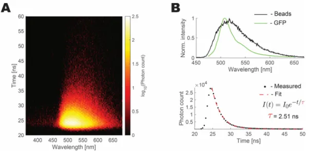
Figure 2.3 Volumetric imaging of beads with the WF-TeFo setup. ... 33
Figure 2.4 Fluorescent beads mimic the excitation spectrum and fluorescence lifetime of GFP. ... 34
Figure 2.5 Increased repetition rate results in proportional gains in fluorescence intensity. ... 37
Figure 2.6 Effect of increase in intensity and repetition rate on photobleaching time. ... 39
Figure 2.7 Temperature dissipation model. ... 45
Figure 2.8 Optimal combination of peak power density and repetition rate for the highest signal intensity within thermal constraints. ... 47
Figure 2.9 2.5D microfluidics device for animal immobilization. ... 49
Figure 2.10 Volumetric imaging of live animals at 100 kHz with WF-TeFo. ... 51
Figure 3.1 Expression of transgenic proteins in engineered animal strains. ... 59
Figure 3.2 Hardware components of the tracking system. ... 62
Figure 3.3 Characterization of the light source for optogenetic stimulation. ... 64
Figure 3.4 Stimulation repeatability and concentration dependence. ... 66
Figure 3.5 Software design flowchart diagram. ... 67
Figure 3.6 Experiment setup user interface. ... 68
Figure 3.7 Behavioral metric extraction from acquired frames. ... 70
Figure 3.8 Long-term recording analysis reveals principal worm eigenshapes. ... 72
Figure 3.9 The influence of diacetyl concentration on animal behavior. ... 74
Figure 3.10 Sensitization to repeated octanol pulses. ... 75
Figure 3.12 Velocity change as a function of optical stimulation power. ... 79 Figure 3.13 Integration of repeated pairing of olfactory and optogenetic stimuli of opposite valence. ... 81
List of Tables
Table 2.1 Modeling parameter and variable definitions ... 43 Table 3.1 Plasmids used for the preparation of transgenic animals ... 57 Table 3.2 Animal strains used in the study ... 60
Outline of the thesis
The main body of this thesis is structured into four parts:
1. In Chapter 1, I provide an introduction to the biology and behavior of C.
elegans. Optical techniques for investigation of neuronal networks are then reviewed.
2. In Chapter 2, I demonstrate how wide-field two-photon temporal focusing microscopy benefits from an increased repetition rate.
3. In Chapter 3, I describe a population tracking and stimulation system capable of dual-color optogenetic stimulation. I validate the system by demonstrating the behavioral implications of contradictory cues.
4. I conclude in Chapter 4.
1 Literature Review
Abstract
The overarching goal of neuroscience is to bridge the disconnect between observed animal behavior and the underlying ‘black-box’ of information flows in the complex neuronal networks. New optical and optogenetic methods have the potential to drastically accelerate the progress in this direction. Of the model systems used in neuroscience, Caehnorhabditis elegans is particularly well suited to address this question with optical means. The relative simplicity of its anatomy has motivated the research of all aspects of C. elegans biology. It is the only model system to date to be described with the annotated genome sequence and complete connectome. Despite the small nervous system composed of only 302 neurons, this animal exhibit complex behavior on short and long time scales, including complex navigation, decision making, learning and even social behaviors. In this chapter, I provide an overview of the C. elegans nervous system and highlight previous work dealing with optical behavioral tracking, optogenetic manipulation and neuronal imaging techniques aimed at dissection of function of neuronal networks.
1.1 Introduction
With the growing understanding of neuronal physiology and mechanisms of information transmission and integration, there is a mounting interest in the neuroscience community to shift focus toward the analysis of the function of more extensive networks (Friston 2011). For decades functional imaging using functional magnetic resonance imaging aimed to do just that; however, the indirect measure of blood oxygen level- dependent activity is a macro-scale measure and suffers from poor signal-to-noise ratios (Logothetis et al. 2001). Therefore more direct methods of measuring neuronal activity are in active development (Scanziani and Häusser 2009).
An established method for measuring and manipulating neuronal activity is the in vivo electrophysiology, which has redefined the 20th-century neuroscience (Bickle 2016).
The introduction of the electrophysiological techniques has, for the first time, enabled the dissection of mechanisms controlling the flow of electrical signals through neuronal tissue (Schwiening 2012). Additionally, the basic methodology was rapidly adapted for probing
and control of all conceivable neuronal cell types generating insights not only in the inner workings of neurons (Verkhratsky and Parpura 2014) but also the behavior of populations of neurons (Taketani and Baudry 2006).
Much like the introduction of electrophysiology, developments in optical imaging technology (J. N. D. Kerr and Denk 2008) and optogenetics (Fenno, Yizhar, and
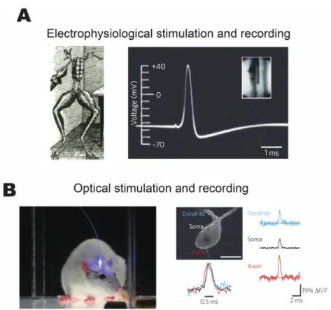
Deisseroth 2011) have sparked a modern revolution in neuroscience research. The former, coupled with genetically encoded activity probes, enables high-resolution imaging of neural activity over large volumes of neurons deep in the neural tissue and with minimal photo-damage (Ji, Freeman, and Smith 2016). The latter enables depolarization or inhibition of genetically targeted cells and cell populations. The combination of the two essentially recaptures an electrophysiological system (Hochbaum et al. 2014). Figure 1.1 presents a comparison between two families of techniques from a historical and practical perspective.
Figure 1.1 Historical comparison of electrophysiological and optical techniques for stimulation and recording. (A) The electrical stimulation of a frog nerve by Galvani revealing the electrical basis of neural information transmission and the first intracellularly recorded movement of action potential by Hodgkin and Huxley. (B) Optical stimulation of deep brain regions expressing optogenetic proteins and optical recording of action
potentials from multiple sites simultaneously. Figure adapted from (Scanziani and Häusser 2009).
Electrophysiological methods, in general, require substantial dissection of the specimen, and it is virtually impossible to reuse the animal for subsequent experiments.
Nevertheless, electrophysiology remains the ‘gold standard’ for analysis of neuronal activity, which is well illustrated by high signal-to-noise ratios of pioneering recordings of Hodgkin and Huxley (A. L. Hodgkin and Huxley 1939). On the other hand, optogenetic tools enable minimally invasive stimulation and recording of live, behaving animals. The same animal can be utilized for months after the installation of a cranial window (Goldey et al. 2014). The downsides, however, include the intrinsically noisy recordings and non-
specific labeling of neural targets (Scanziani and Häusser 2009). While
electrophysiological methods remain the most accurate methods for measurement and control, optical methods have far surpassed the former in experimental throughput as well as intrinsic non-invasiveness offered by the use of light. These features of light-based technologies are what make it such an attractive tool for study of network dynamics.
Among the established animal models, the nematode C. elegans is particularly interesting for the study of functional connectivity. Optically transparent bodies, as well as the small number of neurons, make it particularly tractable by optical means (R. A. Kerr 2006). C. elegans is a genetically flexible system, enabling the expression of optogenetic tools with relative ease (Husson, Gottschalk, and Leifer 2013). In fact, C. elegans was the first model organism to use the green fluorescent protein as a marker (Chalfie et al. 1994).
Additionally, its small size enables the study of both individual animal and population dynamics. A few pioneering studies have shown that these simple animals with 302 neurons are capable of unusually complex behaviors: long-term learning and adapting behavior to suit their environment (Morrison et al. 1999; Amano and Maruyama 2011; de Bono and Maricq 2005; Ghosh et al. 2016).
1.2 Overview of the nervous system of C. elegans
C. elegans is a small, detritivorous nematode found widely around the world (Kiontke and Sudhaus 2006). As a model organism, it was pioneered in the early 1960s by Sydney Brenner, who envisioned that it could be used for investigating developmental processes and nervous system functions (Brenner 1974). The biology of this species offers several advantages. Apart from fast generation time and simple cultivation conditions, they are of sufficient size to experiment on individual animals, but at the same time small enough to investigate a population without substantially changing the experimental setup.
The main strength of this model organism lies in its genetic amenability. High robustness under mutagenesis and hermaphroditic reproduction offer additional benefits in making and maintaining new lines. Over the years, many mutant strains have been identified by forward genetic screens giving insights into many critical life processes (Wood 1988). It was the first multicellular organism to have its full genome sequenced, and the sequence is well annotated (Harris et al. 2010). Additionally, the developmental fate of each cell has been traced, and a number of tissue and cell-specific promoters have been identified
(Riddle 1997). Transgenic animals can be made relatively quickly by direct injection of foreign DNA into the gonad (Fire 1986; Evans 2006).
From the perspective of neuroscience, it was the first animal to have its complete connectome mapped, revealing the connectivity of all 302 neurons (White et al. 1986). The nervous system of the animal can be roughly divided into four categories based on
information flow from inputs to observed behaviors: sensory neurons, interneurons, motor neurons, and polymodal neurons, which share the characteristics of two or more categories (polymodal). Others neurons still may be more important in the guidance of neuronal processes or maintenance than in information transmission. Neurons of the animal mostly exhibit monopolar or bipolar structure, with only a few showing higher degrees of
branching. Furthermore, the position of soma and processes are almost invariable between individuals, making it easier to identify specific neurons (Hall, Lints, and Altun 2005).
The nervous system of C. elegans shares features with those of higher animals at the molecular and cellular levels. Despite the available connectome, polarity and type have not been determined for the vast majority of synapses. Recent research indicates that, at least in the case of interneurons, the majority of synapses is likely to be inhibitory, and is optimized to receive excitatory input from sensory neurons (Rakowski et al. 2013). Despite the small size of its neurons, most electrophysiological techniques can be applied to C.
elegans (Goodman et al. 2012). Due to the lack of evidence for the presence of voltage- gated Na+ channels, it is assumed that most information coding in the neural network of the animal occurs via graded potentials. However, advances in electrophysiological recordings and the small size of the network and neurons themselves suggest that information may be represented digitally (Mellem et al. 2008). Also, there are hints that locomotion direction is coded as two possible states: reverse or forward (Lockery and Goodman 2009). One recent study indicates that some neurons are likely to function as digital gates, while others as analog modulators. AIY neurons form inhibitory synapses to AIZ neurons, which locks them in a low-activity state. Upon inhibition of AIY, AIZ switches from an inactive to an active state in an all-or-none manner, with a narrow dynamic range resulting in animal initiating a reversal in movement direction. AIY neurons also form excitatory connections with RIB neurons, which have a much wider dynamic range and have been shown to modulate locomotion speed in accordance with the magnitude of neuronal activity (Li et al.
2014).
Powerful genetic approaches available for C. elegans have helped to identify many protein candidates that play essential roles in all stages of synaptic transmission. These findings indicate that features of synaptic signaling are highly conserved throughout the animal kingdom (Richmond 2005). The expression pattern of glutamate transporter EAT-4 suggests that 38 out of 118 anatomical neuronal classes are glutamatergic (Serrano-Saiz et al. 2013). So far, ten putative ionotropic glutamate receptor subunits have been found in C.
elegans: 8 AMPA or kainate subfamilies and 2 of NMDA. Amino acid alignment with vertebrate homologs reveals many similarities. As in vertebrates, ionotropic glutamate receptors are implicated in long-term behavioral plasticity in C. elegans (Brockie and Maricq 2006). Furthermore, expression of the cAMP response element binding protein (CREB), required for long-term memory formation in animals ranging from sea slugs to mammals, is also essential for long-term memory in the nematode. More surprisingly, overexpression of C. elegans homolog of CREB, chr-1, results in enhanced long-term memory retention (Alcedo and Zhang 2013).
Considering that its nervous system is so small, an unexpected wealth of
complexity is observed in C. elegans from foraging to social interactions and other aspects of complex behavior (de Bono and Maricq 2005; Kiontke and Sudhaus 2006). These observations can be partly explained by several peculiar features of its nervous system.
First, the majority of neuronal processes can both receive and send potentials (Hall, Lints, and Altun 2005). Neurites of AIY interneurons have calcium transients that are
independent of the neuronal soma (Chalasani et al. 2007; Larsch et al. 2013). Second, compartmentalization of axons has been observed previously in RIA neurons, where it plays a role in monitoring the animal’s motion, accepting updates from its sensory system (Hendricks and Zhang 2013). It is not inconceivable that this feature of the nervous system is more prevalent. Secondly, many neurons are not strictly specialized but have evolved to perform different functions. Some neurons are capable of mechanosensation while
functioning as interneurons. AVL, for instance, is a motor neuron, which has interneuron features. ASH neurons incorporate a number of different sensory modalities (Hall, Lints, and Altun 2005). Third, neuromodulators can have a dramatic effect on the activity of particular neurons. For example, the level of NLP-12, a cholecystokinin homolog, has been shown to modulate motor responses in local search behavior. NLP-12 release is modulated by dopaminergic DVA neurons, which may have a role in the integration of sensory
information about food availability (Bhattacharya et al. 2014). Furthermore, serotonin and insulin-like peptides are necessary for aversive learning through modulation of
interneurons. Additional neuropeptides also are implicated in learning for other sensory modalities (McDiarmid, Ardiel, and Rankin 2015). These features, taken together, dramatically expand the complexity of the C. elegans nervous system beyond what might be expected from the connectome alone.
1.3 Olfaction and nociception in C. elegans
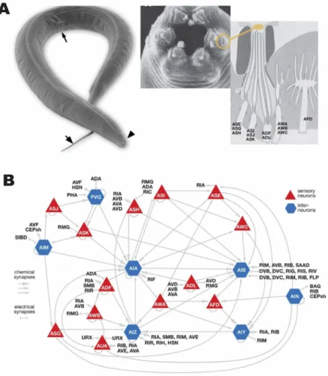
The chemosensory system of C. elegans is composed of 32 neurons found in the amphid, the phasmid, and neurons of the inner labia. Figure 1.2 shows the physiology and connectivity of sensory organs. These are exposed either directly or indirectly to the environment through pores in the cuticle made of socket and sheath cells. They are loosely specialized to respond to different classes of chemical cues, but also are observed to modulate sensitivity in a concerted manner (Cornelia I Bargmann 2006). Chemosensation in C. elegans is divided into two categories: gustatory and olfactory. Since gustatory cues are not volatile, while olfactory cues are, these two senses differ in the range over which their respective cues are detectable (C I Bargmann, Hartwieg, and Horvitz 1993; Cornelia I Bargmann 2006). Systematic behavioral screens have identified a number of substances that induce attraction or repulsion. C. elegans are observed to ascend gradients of cations, anions, cyclic nucleotides, and alkaline pH (Ward 1973) and show aversion to toxic cations (Sambongi et al. 1999). Similar experiments in olfactory preference have identified many volatile substances, including alcohols, amines, aldehydes, esters, organic acids, and heterocyclic compounds, to which specific preferences are exhibited by the animal (C I Bargmann, Hartwieg, and Horvitz 1993). In contrast to gustatory cues, the response to olfactory cues is more varied and is highly concentration-dependent. Individual neurons responsible for sensing many olfactory or gustatory cues have been identified: ASE, ADL, and ASH in the case of gustation and AWA, AWB, AWC, and ASH in olfaction (Cornelia I Bargmann 2006). More recently, it has been reported, however, that several different sensory neurons might respond to the same chemical cue as attractive or repulsive in a concentration-dependent manner (Yoshida et al. 2012; Taniguchi et al. 2014).
Figure 1.2. Olfactory and nociceptive system of C. elegans. (A) Electron microscopy image of the worm (arrows indicate the pharynx, vulva, and tail). Pharynx, the mouth of the animal, contains sensory amphid where neurons are exposed to the environment. (B) Connectivity diagram of different sensory neurons showing chemical and electrical connections. Adapted from (de Bono and Maricq 2005; Hall, Lints, and Altun 2005).
The ASH neuron mediates nociception in C. elegans. Exposure to high osmolality, a number of harmful compounds, such as heavy metals, detergents, acidic pH, and organic compounds produce an immediate reversal of direction, which requires ASH. Furthermore, the ASH neuron, in a similar manner, is necessary for withdrawal from a mechanical stimulation (Cornelia I Bargmann 2006). These observations establish the ASH neuron as a general sensor for damage detection, which, in conjunction with phasmid neurons in the
tail, transmit nociceptive signals to interneurons. The ASH forms connections with AVA, AVB, and AVD interneurons, which are responsible for the initiation of forward and backward locomotion (White et al. 1986). Direct activation of the ASH neuron is sufficient to induce a reversal response. It has been demonstrated by expressing the vertebrate TRPV1 channel in the ASH neuron and inducing avoidance behavior through the
administration of capsaicin, an antagonist of TRPV1 channels (Cornelia I Bargmann 2006;
Tobin et al. 2002). Additionally, a similar response has been observed through the
expression of channelrhodopsin-2 (ChR2) in ASH and inducing activation of the neuron by exposure to blue light (Faumont et al. 2011). Glutamate release at synaptic sites of ASH with interneurons transmits sensory information through activation of AMPA and NMDA type glutamate receptors (Mellem et al. 2002).
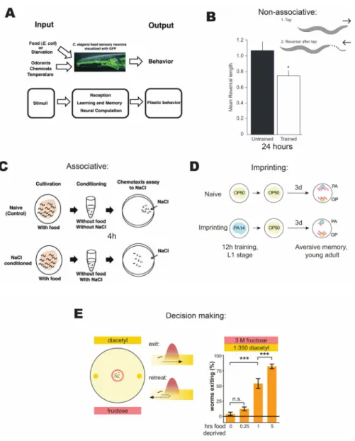
1.4 Long-term behavioral plasticity
Research efforts spanning over three decades has uncovered a vast repertoire of behavioral plasticity in C. elegans. These include observed plasticity in all sensory modalities, including learning achieved through major sensory systems: mechanosensory, thermosensory, and chemosensory (Giles, Rose, and Rankin 2005). Additionally,
behavioral plasticity has been shown in the application of sexual cues (Sakai et al. 2013) and as a response to changes in oxygen levels (Cheung et al. 2005) and humidity (Russell et al. 2014). Plasticity systems investigated thus far can be divided into several basic categories: non-associative learning (habituation), associative learning, imprinting (Ardiel and Rankin 2010), and decision making (see Figure 1.3). These studies, taken together, reveal unexpected behavioral plasticity in C. elegans despite its compact nervous system.
Figure 1.3 Relevant long time scale behavioral plasticity paradigms. (A) C. elegans neural system as an engine for studying plasticity of behavior. (B) Habituation is a form of non-associative plasticity, where a single sense loses sensitivity with repetition and can be retained for 24 hours. (C) Association of NaCl and starvation is a form of
associative plasticity that can last for 4 hours. (D) Imprinting is learning that occurs only at an early developmental stage and lasts for life. In this case, learning to avoid noxious bacteria. (E) Behavioral decision-making paradigms, where animals reevaluate the cost and benefit of experiencing pain to gain reward based on their satiety state. Adapted from (Sasakura et al. 2013; McEwan and Rankin 2013; Jin, Pokala, and Bargmann 2016;
Dipon Ghosh et al. 2016).
Mechanosensory stimulation induces reversal responses in C. elegans. If the animal is stimulated repeatedly, reversal responses reduce in magnitude, which serves as a
measure of sensory habituation. Interestingly, this can be replicated by activating ChR2 expressed in touch receptor neurons (Timbers et al. 2013). When presented at spaced intervals, this training paradigm induces long-term memory for habituation in C. elegans.
This effect is protein synthesis-dependent, as behavior is disrupted by heat shock, while training administered without resting periods is not. Furthermore, spaced training leads to decreased expression and changes in the distribution of glutamate receptor subunits on processes of interneurons in the posterior nerve cord (Rose et al., 2003). This change was later shown to be reversible in the case of heat shock delivery during reconsolidation (Rose and Rankin, 2006).
The study of associative learning in C. elegans traces its roots to the first report of animals showing preference to the temperature in which they were cultivated in an abundance of food (Hedgecock and Russell 1975). In the context of chemotaxis, initial evidence was provided by a study investigating behavioral plasticity towards Na+ and Cl- ions. Animals exposed to these ions in the presence or absence of food showed changes in preference on subsequent exposures (Wen et al. 1997). Later a similar paradigm was investigated, in which animals were left to starve in the presence and absence of NaCl for four hours. After conditioning, chemotaxis towards NaCl, as well as other soluble
compounds, decreased, indicating that animals learned to associate starvation with the presence of particular substances (Saeki, Yamamoto, and Iino 2001).
Moreover, plasticity of learned avoidance responses was dependent on the
expression of NMDA-type ionotropic glutamate receptor subunits NMR-1 and NMR-2, but not on GLR-1 or GLR-2, in contrast to long-term mechanosensory habituation memory.
Expression of NMR-1 and NMR-2 in RIM interneurons rescued defective memory retention (Kano et al. 2008). Animals cultivated at a particular NaCl concentration will migrate toward this concentration when placed on a gradient. This indicates that animals can associate concentration with cultivation conditions. The ability to follow the
concentration gradient was attributed to an ASE sensory neuron pair, which activates asymmetrically in response to salt concentrations below or above the preferred concentration (Suzuki et al. 2008). Recently it was shown with calcium imaging that memory of preferred conditions is stored directly in the ASE neuron, while the interneuron
network downstream of ASE only modulates motor output in accordance with a specific condition. This preference is plastic and can be changed within 4 hours (Luo, Wen, et al.
2014). Similar results were observed in the case of thermotaxis to a preferred temperature mediated by the AFD neuron (Luo, Cook, et al. 2014). These reports indicate that
associations can be stored directly in the sensory neurons themselves.
Adult animals exposed to pathogenic bacteria on growth plates for four hours showed increased avoidance of the smell of the same pathogenic bacteria (Zhang, Lu, and Bargmann 2005). The same condition results in imprinting, if applied in the early stages of development (Jin, Pokala, and Bargmann 2016). This response was plastic, and otherwise normal sensitivity was not affected. Furthermore, this plasticity was dependent on
serotonin modulation by ADF neurons (Zhang, Lu, and Bargmann 2005). Later the network for modulation of this aversive response was dissected through neuronal ablation and calcium imaging. Two neural circuits were identified for generation of naïve and trained aversion towards the pathogenic strain. The naïve response required AWC and AWB sensory-motor circuits, while ADF was necessary for trained responses through RIA interneurons and SMD motorneurons. ADF was also shown to play a role in the naïve response. Combined ablation of AIY and AIB interneurons, on the other hand, completely abolished aversive responses (Ha et al. 2010). Previously, animals have been compelled to climb artificial light gradients, when the AIY neuron was stimulated directly using
optogenetic stimulation with ChR2 (Kocabas et al. 2012). This observation demonstrates that the AIY acts as a chemosensory input hub, and its activation alone is sufficient to induce behavior.
The two circuit aversive olfactory learning network in C. elegans is reminiscent of the regulation of fear responses in mice, where behavior depends on the interplay between low and high fear neurons in the amygdala (Herry et al. 2008; Ha et al. 2010). A recent report used optogenetic stimulation of a group of cells expressing ChR2 in the amygdala of a mouse, combined with fear conditioning to induce artificial fear response memories.
Trained animals responded to optogenetic stimulation with an increased tendency to exhibit a freezing response, a hallmark of fear conditioning (Nabavi et al. 2014). When the functional similarities between fear response conditioning and aversive conditioning are taken into account, it is conceivable that a similar conditioning protocol, relying on optogenetic stimulation, could be implemented in the nematode as well.
An aversive olfactory conditioning paradigm was also reported to induce memory when animals were exposed to pure chemicals, rather than mixed stimuli (food). In this case, an initially attractive stimulus, diacetyl, was paired with an aversive acetic acid solution, which resulted in decreased chemotaxis toward diacetyl after training (Morrison et al. 1999). In this paradigm, mutant animals deficient in glutamate receptors did not show a reduction in attraction to diacetyl (Morrison and Van Der Kooy 2001). This paradigm was later modified to replace diacetyl with 1-propanol, a more neutral stimulus, which led to a pronounced difference in chemotaxis index before and after conditioning. When consecutive iterations of conditioning trials were presented with no or small delay, changes in chemotaxis persisted for only 3 hours. A memory that was induced using longer rests between consecutive iterations was shown to last for at least 12 hours after training.
Furthermore, induced memory was dependent on protein synthesis. Analysis of mutants deficient in GLR-1 and NMR-1 as well as CREB mutants did not show any signs of memory retention 6 hours after conditioning, indicating that long-term memory failed to form (Amano and Maruyama 2011).
In summary, published research shows that C. elegans is capable of behavioral plasticity between different sensory modalities and on time scales of days. Moreover, the mechanisms mediating plasticity share features with more complex animals.
1.5 Optogenetic toolkit
Due to its amenability to genetic manipulation, its small size, and transparent body, C. elegans is particularly suited for manipulation and imaging using a number of
optogenetic tools. Genetically expressed ChR2 has been widely used for the manipulation of neuronal activity with high temporal precision. This cationic ion channel is endogenous to the green alga, Chlamydomonas reinhardtii, where it functions in a photoreceptor
system. Blue light with an excitation peak at 450-460 nm induces a conformation change in the cofactor all-trans-retinal (ATR), which renders the channel permeable to cations. ATR is an essential cofactor for the function of ChR2 and is not produced by C. elegans (G.
Nagel et al. 2005). In physiological conditions, mostly Ca2+ are allowed to pass through ChR2, but K+ ions can also pass if they are present at high concentrations (E. Nagel et al.
2003). ChR2 was successfully expressed in muscles of C. elegans, where it caused
predictable contraction upon stimulation with blue light. Furthermore, expression in ALM,
PLM, AVM, and PVM mechanosensory neurons induces reversals in both wild type animals and mechanosensation-defective mutants (G. Nagel et al. 2005). ChR2 and its enhanced derivatives have become popular tools for analysis of synaptic transmission, as well as dissection of neuronal circuits in C. elegans (Husson, Gottschalk, and Leifer 2013).
Recently several red-shifted photoactivatable ion channels have been discovered and optimized. One such variant, a chimeric fusion of channelrhodopsin-1 and Volvox VChR1, containing point mutations at E122T and E162T (C1V1-ET/ET), has been shown to induce muscle contraction in response to green light (580 nm). The same authors further demonstrated that this protein could be used in conjunction with hypersensitized ChR2 (mutagenized at H134R and T159C, termed ChR2-HR/TC) to activate muscle contraction in a mutually exclusive manner by either near UV or green light. This approach, however, necessitates the use of low-intensity light stimulation as C1V1-ET/ET is also activated by high-intensity blue light (Erbguth et al. 2012). Another approach for engineering novel ChR2 variants through mutagenesis is to continue probing natural sources for such proteins. A recent study employed large scale algal transcriptome sequencing and discovered a novel channelrhodopsin variant, which can be reliably activated by the red light. Termed ChrimsonR (CrR), it was expressed in mouse cortical slices in conjunction with a new blue light-sensitive ChR2 variant and used to independently excite distinct neural populations with light of appropriate wavelengths (Klapoetke et al. 2014).
Photoinhibitory membrane proteins can be expressed in C. elegans for precise light-induced hyperpolarization of neurons (Husson, Gottschalk, and Leifer 2013). There are two widely used varieties: light-gated inward Cl-- pumps and outward-directed proton pumps (Chow et al. 2010). Halorodopsin (NpHR) belongs to the former class, while Archaerhodopsin-3 (Arch) and Mac belong to the latter. Furthermore, these protein classes differ significantly in spectral properties. Mac and Arch peak activation is blue-shifted compared to NpHR (Husson et al. 2012). NpHR is activated by yellow light (580 nm and longer), while Mac, for instance, can be switched on with light wavelengths falling in between 400 and 650 nm; however, activation peak is achieved at green-blue light region (Husson et al. 2012; Waschuk et al. 2005). All of the photoinhibition proteins mentioned above require ATR for normal function (Husson et al. 2012). These proteins promise a much higher degree of flexibility for the manipulation and analysis of neural circuits than just relying on natural stimuli.
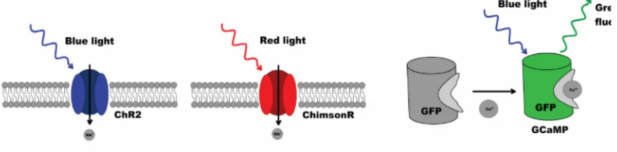
Activation of glutamate receptors produces an influx of Ca2+ into neuronal dendrites. This influx denotes the activation of a neuron and can be measured using genetically encoded calcium indicators. Often such proteins consist of a calcium-binding domain fused to a fluorescent protein. These proteins can generally be expressed readily in many model systems, including C. elegans. Furthermore, their color-type, dynamic range, and calcium sensitivity are being optimized continuously through mutagenesis (Tian et al.
2009; Zhao et al. 2011; Ohkura et al. 2012). A robust approach has been previously introduced for dissection of C. elegans neural networks and was used to examine reversal behaviors through stimulation of the ASH neuron and simultaneous imaging of ASH, AVA, AVD, and RIM interneurons. Calcium currents were imaged with then newly developed GCaMP2 calcium indicators. Using low-intensity blue illumination for imaging, coupled with periods of higher intensity for stimulation, authors were able to both image and excite predictably with a single wavelength of light (Guo, Hart, and Ramanathan 2009). Figure 1.4 shows an example of a toolkit for multineuronal stimulation and recording.
Figure 1.4 An example toolkit for two-color stimulation of neurons and readout of neuronal activity. Blue light-sensitive ChR2 and red light-sensitive ChrimsonR serve as actuators, while an intracellular calcium probe such as GCaMP allows for recording of neuronal activity.
1.6 Behavioral tracking of C. elegans
Animals dynamically adapt to the environment to increase the chances of survival.
How it is achieved is one of the central questions of neuroscience. For a long time, the nervous system was viewed as a center of command issuing, but it is becoming
increasingly apparent that the interaction between changing environment, the body, which is subject to laws of physics and animal brain is not a top-down hierarchy, but rather a feedback loop (Chiel and Beer 1997). It thus follows that understanding the dynamics of the nervous system requires taking into account all three levels of complexity.
Historically, relatively little attention was spared for the behavior of the nematode.
The view held by many researchers was that the animal, owing to its relatively simple structure, was not much more than a hardwired automaton. The focus fell more on unraveling developmental processes and biological structures. Nevertheless, this has changed with research work uncovering more sophisticated behaviors (Catharine H Rankin 2004) .
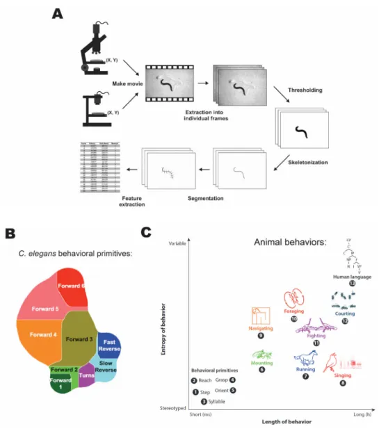
The earliest behavioral analysis for C. elegans was routinely performed in the context of mutant screens. The descriptions of the phenotype were broad and qualitative, such as the animal being uncoordinated or coordinated (Brenner 1974). The description list quickly expanded to include more coarse-grained descriptions such as kinky, coiling, sluggish, slow (J. Hodgkin 1983). The growing interest in understanding the sensory systems of the nematode motivated the introduction of the population assays. In this method, animals are allowed to move on the behavioral arena in response to a sensory cue for a set amount of time: temperature (Hedgecock and Russell 1975) and olfactory (C I Bargmann, Hartwieg, and Horvitz 1993). The distribution of animals relative to the cue was then determined manually. Recording of reversals of individual animals was used to study the properties of animal response to mechanical stimuli (C. H. Rankin, Beck, and Chiba 1990).
Automated behavioral tracking in the field had a slow start. The first system developed for tracking the movement of centroid of C. elegans was capable of monitoring
~ 25 animals at 1 Hz (Dusenbery 1985). Nevertheless, behavioral tracking was not adopted by the worm research community until the end of the 1990s. With cheaper computational costs and access to digital imaging equipment, the use of automated behavioral tracking has increased. A system capable of tracking the centroid position of approximately 50 animals was developed and used to investigate social aggregation behavior (de Bono and Bargmann 1998). Later a system was designed to track the movement speed and turning rate of individual animals during chemotaxis behavior using a motorized stage (Pierce- Shimomura, Morse, and Lockery 1999). The first standardized suit for behavioral tracking
of single animals was made available to the community in 2004, complete with software for feature extraction and classification (see Figure 1.5 A) (Feng et al. 2004; Cronin et al.
2005). This work and its derivatives have significantly increased the adoption of behavioral tracking in the C. elegans research community (Husson et al. 2018).
The revival of improved population tracking in the 2010s has dramatically increased the throughput of data acquisition. A system capable of collecting movement parameters of ~50 worms have been introduced as a tool for pharmacological analysis (Ramot et al. 2008). Later a multiworm tracking suit was developed for the use of researchers capable of simultaneous tracking of 120 animals and was used to analyze the spontaneous movement of animals on food in response to tapping stimulus. The software can collect animal outlines and centroid positions, while a supplemental analysis suite derives behavioral parameters (Swierczek et al. 2011).
Combining of optogenetic tools with behavioral tracking has been undertaken by several labs using different approaches. Single animal tracking and optogenetic stimulation were implemented using patterned illumination by digital micromirror devices (Leifer et al.
2011; Stirman et al. 2012) to enable stimulation of parts of the animal bodies expressing optogenetic proteins. Tracking and optogenetic control of the animal movement was demonstrated by stimulating AIY interneuron with shifting light gradients (Kocabas et al.
2012). Simultaneous tracking, optogenetic stimulation, and recording have been used to investigate the properties of the reversal response in individual animals (Faumont et al.
2011). Recently, the optogenetic stimulation with a single color of multiple animals has been shown as an extension of the multiworm tracking suit (Yu et al. 2019; Ardiel et al.
2016). Multiworm tracking has also been used with optogenetic analysis to uncover the landscape of short-term behaviors in response to random mechanosensory stimuli (see Figure 1.5 B). (Liu et al. 2018).
The behavior of C. elegans is fairly well understood on short timescales, owing to advances in behavioral tracking technology; however, ample research opportunities exist with multichannel stimulation on longer timescales (see Figure 1.5 C), which would require improved methods.
Figure 1.5 Principles of automated tracking and behavioral complexity. (A) Shows the key processes used in extracting behavioral data from animal recordings. (B) Short time scale behavioral repertoire of C. elegans uncovered with population tracking and optogenetic stimulation. (C) Breakdown of animal behaviors across different levels of complexity and time scales. Adapted from (Husson et al. 2018; Liu et al. 2018; Egnor and Branson 2016).
1.7 Optical microscopy for live imaging of neural activity
Capability to take into account the spatial and temporal dynamics of neuronal activity in cell populations with single-neuron resolution is essential to understanding how behavior adapts to the environment (Ji, Freeman, and Smith 2016). Recording of neuronal activity using electrodes still offers an unparalleled temporal resolution. Unfortunately, the spatial resolution, even using electrode arrays, is limited to a subset of nearby neurons (Henze et al. 2000). Neuronal activity recordings using optical imaging are significantly less intrusive and more comprehensive as light can access a vastly larger area and carry information to instruments that can be located away from the brain. Furthermore, genetically expressed calcium indicators evolve rapidly and provide an array of tools for extracting all sorts of metrics of interest, well beyond recording electrical signals (Wang, Kim, and Ting 2019). Optical methods enable the recording of neuronal communication over a large volume, which can reveal correlations, statistical properties, and large scale dynamics, that are vital to the understanding of adaptive brain function (Ji, Freeman, and Smith 2016).
The meticulous reconstruction of the whole connectome map of C. elegans neural system through electron microscopy (White et al. 1986) was posed to be a revolutionary step forward in understanding the nervous system function. However, decades later, its most significant achievement paradoxically was to highlight the additional complexity that is hidden behind mere connections between neurons. There are many features that are not obvious from the connectivity maps, which have been shown since to have crucial
importance to animal behavior (Cornelia I. Bargmann 2012). For instance, the knowledge of connectivity graph does not inform on the polarity of the individual synapses (Rakowski et al. 2013), which can quite drastically shape the interpretation of the roles of individual neurons. Then there are neuromodulators that often not only act in the synapse locally, but can diffuse to affect neighboring connections (Hobert 2016). Additionally, mechanisms operating entirely outside the scope of connectome can have profound implications for neural computation. These include monoamine neuromodulators and neuropeptides that diffuse over vast distances and change short and long-term neuronal functions
(McDiarmid, Ardiel, and Rankin 2015; Huang et al. 2017).
A way to take an integrative view of all the mechanisms that contribute to neuronal function is to determine the functional connectivity for investigated behaviors (DiLoreto et al. 2019), which could be accomplished by way of measuring the fluctuations in neuronal activity under different environmental conditions. In C. elegans, due to small size and transparent body as well as the relative ease of genetic manipulation, the most common way to measure neuronal activity is by using genetically encoded light-sensitive probes (Breimann, Preusser, and Preibisch 2019). Bioengineering efforts have resulted in many useful genetically encoded tools for the measurement of different aspects of neuronal signal transmission, of which the most broadly used are voltage and calcium sensors (Wang, Kim, and Ting 2019). The genetic engineering of animals carrying these proteins in the desired location has been simplified considerably by the introduction of CRISPR genome modification technology (El Mouridi et al. 2017). The introduction and refinement of these tools have greatly accelerated the progress in the neural imaging in this model system (Breimann, Preusser, and Preibisch 2019). Until recently, most published studies relied on imaging individual neurons (Zheng et al. 2012). Indeed, an impressive system, which can collect calcium dynamics from single neurons in populations of animals, while simultaneously acquiring behavioral features have been demonstrated (Larsch et al. 2015).
The findings of this study also indicate that variability in behavior is more pronounced than in the recordings of neural activity motivating the development of single animal recording systems that can image the whole brain of the animal (~180 neurons simultaneously).
Systems for the whole brain imaging of C. elegans are in active development. The first published system, showing near 70% activity for ~5-minute recordings of all head neurons, was based on wide-field temporal focusing two-photon microscopy (WF-TeFo) (Schrödel et al. 2013). This method, however, did not see wide adoption in the community due to relatively weak signal intensities that could be obtained with low repetition laser systems. Another intriguing approach was the use of light-field microscopy (Prevedel et al.
2014). The method is compelling in that it requires no scanning, but is limited by considerable post-processing required to deconvolute imaging data for volumetric
reconstruction. The more widespread approach in recent years is the use of a spinning disk confocal microscope (Nguyen et al. 2016; Venkatachalam et al. 2016). The main limitation of this approach is the use of single-photon excitation, which limits recording length in case of volumetric imaging. In spinning disk confocal microscopy, the sample is subjected
to a large number of spatially separated parallel beams scanning the imaging plane (Nakano 2002). Excitation is achieved in the one-photon regime, thus contributing to out- of-focus bleaching of the excitation volume and consequently reducing the viable imaging length (Diaspro, Chirico, and Collini 2005). The maximum reported length of recording is
~18 minutes of continuous volumetric, high-resolution imaging (Kato et al. 2015). It is conceivable that the application of two-photon spinning disk microscopy (Shimozawa et al.
2013) could resolve this problem, but the implementation of this technique is challenging.
The more recent technique applied to whole animal imaging employed light-sheet
microscopy (Ardiel et al. 2017). It is promising, but requires immobilization of the animals in a hydrogel to allow excitation by orthogonal plane, effectively isolating it from sensory stimuli. Out of the techniques mentioned, WF-TeFo is still the most attractive for achieving long-term high-resolution fast volumetric imaging as discussed in the following section.
1.8 Two-photon temporal focusing microscopy
The use of microscopy techniques based on two-photon excitation offers significant advantages for live imaging. Since two-photon excitation probability depends on the square of the intensity, a dramatic reduction in the background can be achieved, when compared to one-photon microscopy techniques. Additionally, tissues have lower absorption for infrared spectrum, which results in larger penetration depth and markedly reduced out of focus phototoxicity and photobleaching (Denk, Piston, and Webb 1995). In conventional two-photon microscopy, the axial confinement is primarily governed by spatial focusing, and the beam maintains a constant pulse width as it propagates along the optical axis. This is useful for scanning microscopy as the focused beam has a near- diffraction-limited spot size, which can be employed for obtaining high-resolution images.
Unfortunately, the scanning speed limits the rate at which readouts can be achieved. This is particularly problematic for volumetric imaging, where short pixel dwell times limit the dynamic range (Ji, Freeman, and Smith 2016). Operating in the wide-field two-photon configuration using soft focusing of the beam has been attempted and may have advantages for particular experiments, but suffers from poor axial resolution and weak signal
intensities (Amor et al. 2016).
Temporal focusing based two-photon microscopy introduces another degree of freedom, whereby the pulse duration is not fixed but depends on the distance away from
the focal plane (M E Durst, Zhu, and Xu 2008). The basic working principle is akin to that of an asymmetric 4f grating compressor (Oron, Tal, and Silberberg 2005). In ultrafast pulse compression using a double-pass grating compressor, a dispersive delay line is used to compensate for self-phase modulation (Weiner 2009). In temporal focusing microscopy, one of the gratings is replaced with an objective lens. Analogously, spectral components of an incoming pulse are spatially separated by the grating and recombined by the objective lens. According to Fermat's principle, the spatial and temporal overlap of components is realized only at the focal plane of the objective, while pulse duration outside the focal plane is subject to rapid broadening (M E Durst, Zhu, and Xu 2008).
This phenomenon significantly enhances the axial confinement offered by a
nonlinear power dependence in conventional two-photon microscopy (Yew, Sheppard, and So 2013). Spatial focusing using an ideal lens results in a diffraction-limited excitation spot, offering the highest axial resolution. In the case of temporal focusing, the
combination of grating and an objective, effectively create a temporal lens, whereby focusing is achieved in time rather than space. As a result, the resolution of temporal focusing microscopy is worse than point scanning microscope and is comparable to line focusing microscopy (Oron and Silberberg 2015). Intuitively, the axial resolution tends to ideal value, with shortening of excitation pulse duration. The reduction of the incoming pulse duration to sub-20 fs produces a depth of focus that is very close to 1.9 µm at 795 nm (Vaziri et al. 2008) and about twice that of point scanning two-photon microscope (Yew, Sheppard, and So 2013). Supplementing diffraction grating with an Echelle’s grating enables the generation of multifocal excitation that scans through the focal plane as the femtosecond pulse passes through the system. In this configuration, one can achieve an axial resolution comparable to point-scanning two-photon microscopy (Vaziri and Shank 2010), while retaining wide-field excitation.
While tissue exhibit low absorbance in the infrared, scattering presents significant problems for deep-tissue fluorescence imaging (Miller et al. 2017). Temporal focusing offers advantages for excitation of fluorophores inside the tissue due to inherent ‘self- healing’ properties. Owing to spatial separation of the spectral components, the beam acquires uncorrelated speckle patterns as it is propagating through turbid media. Moreover, even the adjacent components that are subject to correlated speckle maintain a spatial shift in speckle patterns resulting in anisotropic smoothing in the direction of the shift. The
combination of these phenomena enables wide-field excitation as deep as 500 µm in the scattering media. While it leads to the degradation of the axial resolution, the lateral sharpness is preserved (Papagiakoumou et al. 2013). A separate challenge is to capture the emitted fluorescence photons, that are subjected to scattering and absorption. The
independent readout of adjacent neurons (~10 µm apart) has been demonstrated to result in less than 7% crosstalk within 250 µm below tissue surface using a scanless temporal focusing approach (Bovetti et al. 2017). A novel approach has been implemented recently, enabling deep tissue imaging with wide-field temporal focusing using single-pixel
detection. The sample is excited using orthogonal Hadamard patterns imposed with an SLM. The spatial features are preserved due to the resilience of temporal focusing to scattering. The resulting excitation light intensity is collected for each pattern and is stored as a coefficient. The fluorescence image is reconstructed by adding all patterns weighed by respective coefficients. The authors demonstrated imaging through 400 µm of scattering medium with a lateral resolution of less than 5 µm (Escobet-Montalbán et al. 2018).
The ability of a two-photon temporal focusing microscope to excite large areas with high axial resolution, yet maintaining low out of focus phototoxicity characteristic of scanning two-photon microscopy, make it an ideal choice for fast volumetric imaging (Schrödel et al. 2013).
1.9 Problem statement and aims
This work focuses on advancing the toolkit for long-term observation and timed stimulation of live animals on behavior and neuronal levels. The overall goal is to provide the research community with better tools to perform long-term behavioral, optogenetic, and imaging experiments.
The specific aims of the project are as follows:
1. Develop and validate a neuronal imaging system capable of high throughput neuronal activity recording over long time scales.
2. Develop and validate a multicolor optogenetic stimulation system for analysis of behavior from individual to population level, capable of long- term recording.
A combination of these tools will enable the exploration of new scientific questions that were beyond the reach of current technologies.
2 Long-term neuronal activity recording with two-photon temporal microscopy
with high pulse repetition rates
Contributions: sample preparation, microfluidics manufacturing, data collection, modeling, and analysis were performed by me. I thank Prof. Bala Murali Krishna Mariserla, Chakradhar Sahoo and Reuben T. Harding for help with setup design and implementation. Also Dr.
Julien Madéo, Dr. E Laine Wong, and Dr. Michael K. L. Man for valuable advice on experiment design and data analysis.
Abstract
Neuronal recordings using fluorescent activity probes are routinely used in
neuroscience research to study the dynamics of networks. In C. elegans, such experiments are performed using high-speed imaging techniques such as spinning-disk confocal microscopy. One-photon imaging techniques are not suitable for detailed recordings over long periods due to photobleaching. Recently, the wide-field temporal focused two-photon microscopy (WF-TeFo) has been applied to whole-brain imaging of C. elegans. WF-TeFo can potentially achieve an order of magnitude increase in volumetric recording speed owing to the use of large planar excitation area. Achieving large excitation area requires the use of laser systems capable of providing high peak power densities, at least half a millijoule per pulse, and only low repetition lasers were capable of driving pulses at such energies. The use of low repetition sources results in fluorophore saturation due to high excitation fluence as well as photobleaching. The recent introduction of high-repetition, high-pulse-energy laser systems can reduce these drawbacks by providing increased signal intensities seen in traditional two-photon microscopes and reducing the negatives of high fluence as seen in WF-TeFo setups to date. Here I use a laser system with a 100 microjoule per pulse driven at 50-100 kHz to investigate the advantages in performance achieved in WF-TeFo microscope for imaging C. elegans neuronal activity. I characterize the increase in signal intensity with increase in repetition rate by imaging fluorescent beads compared with a 1 kHz system and find that there is no increase in photobleaching. Indeed,
increasing repetition rate and decreasing pulse intensity results in a proportional reduction
in photobleaching. As a proof-of-principle, I measure the activity of multiple neurons in live C. elegans over a period of 25 min in a custom-built microfluidic enclosure. Finally, I perform an analysis of steady-state temperature increase to find the optimal repetition rate.
I find that repetition rates between approximately 0.5 and 5 MHz limit the rise to 1 K, which is a soft limit for C. elegans experiments. These findings demonstrate that the availability of high repetition rate, high-pulse-energy fiber laser systems will significantly increase the scope of neuroimaging experiments allowing for imaging longer at higher speeds.
2.1 Introduction
The demand for a more holistic snapshot of neuronal activity in live and behaving animals has spurred the development of advanced bio-optical technologies that aim to increase both the available speed and duration of imaging experiments. Use of one-photon microscopy and in particular high-speed spinning disk confocal microscopy has achieved high-speed volumetric imaging of a large number of neurons (Nguyen et al. 2016; Nichols et al. 2017), but imaging duration is constrained by the intense photobleaching resulting from one-photon excitation. Out of focus photobleaching can be reduced by the use of two- photon microscopy (Denk, Strickler, and Webb 1990). This method is minimally invasive and offers diffraction-limited resolution; however, it is limited in speed at which scanning of large areas can be implemented (Ji, Freeman, and Smith 2016). Adaptive optics can be used to extend the usefulness of this technique by producing multifocal excitation (Qu et al. 2012) and thus potentially increasing the scan speed (Abrahamsson et al. 2016). Here I make use of temporal focusing phenomenon, which allows for large excitation area that can be recorded with each exposure and offers excellent axial resolution (Yew, Sheppard, and So 2013).
Wide-field temporal focusing microscopy (WF-TeFo) is a multiphoton far-field imaging method, which enables excitation of large sample area and therefore overcomes temporal drift of the acquired signal in the lateral imaging plane. Due to broad area excitation, it potentially enables an increase in recording rate of the volumetric
fluorescence signal. In this configuration, ultrashort laser pulses pass through a diffraction grating and become separated according to wavelength and then refocused in the focal plane of the objective lens. The result is the temporal focusing effect, where the shortest pulse duration is generated at the focal plane. The pulse rapidly broadens as it propagates